Primary vs Secondary Healing | Cellular Biology | Growth Factors | Mechanical Environment
- Primary healing requires absolute stability, no callus forms, cutting cones cross fracture
- Secondary healing is natural pathway with callus formation through four phases
- Strain theory (Perren): Under 2% = bone, 2-10% = cartilage, over 10% = fibrous tissue
- Diamond concept: Cells + Scaffold + Growth factors + Mechanical environment
- MSCs differentiate to osteoblasts under BMP signaling and mechanical stimulation
- “Primary healing is Haversian remodeling across fracture (cutting cones)
- “Secondary healing uses enchondral ossification (cartilage intermediate)
- “BMP-2 and BMP-7 are osteoinductive (induce MSC differentiation to osteoblasts)
- “VEGF is critical for angiogenesis during fracture healing
- “Smoking doubles nonunion risk, NSAIDs controversial but avoid long-term use
Overview and Epidemiology
Bone is the only tissue that heals by regeneration rather than by scar. What bridges a united fracture is bone, with the original structure and mechanical properties restored, and that capacity rests on the osteogenic cells held in periosteum and marrow, the osteoconductive matrix of bone itself, and a mechanical environment that guides remodelling (Wolff law).
Why the biology decides the operation. Choosing between absolute stability with a plate and relative stability with a nail is a choice of healing pathway, made before the incision. The same biology governs what else the patient needs - smoking cessation, adequate stability, and biological augmentation such as BMP in high-risk cases (smoking, diabetes, revision surgery) - and it is why delayed union and nonunion are looked for early, so that intervention is timely.
The numbers. Between 90 and 95% of closed fractures unite with appropriate treatment, and nonunion runs at 5-10% overall. The rate is bone-specific:
- Scaphoid 5-10%
- Femoral neck 10-30%
- Tibial shaft 5-15%
Who fails to unite. Smoking is present in 30-40% of nonunions, diabetes prevalence is increasing, and the population is ageing. Nonunion treatment consumes significant healthcare resources, and costs the patient prolonged disability and lost productivity.
Concepts and Mechanisms
Primary (direct) healing
Primary (direct) healing occurs only where the surgeon creates the conditions for it: fragments anatomically reduced and rigidly fixed with absolute stability, so that osteoclasts and osteoblasts cross the fracture directly and no intermediate callus forms.
What absolute stability requires:
- Anatomical reduction, with direct bone-to-bone contact
- Interfragmentary strain under 2%
- Compression between the fragments, from lag screws or compression plates
- Rigid fixation that prevents any motion
Cutting cones. Osteoclasts assemble on one side of the fracture, begin resorbing bone, and tunnel across the fracture line in a cone-shaped front. Osteoblasts follow behind them, depositing lamellar bone, so that each tunnel becomes a new osteon - a Haversian system crossing the fracture and bridging the gap directly. Multiple cutting cones do this, and together they restore continuity.
Contact healing (gap under 0.01mm)
With the fragments in direct contact, or separated by no more than 10 micrometres, lamellar bone forms directly across the fracture. Cutting cones tunnel perpendicular to the fracture line, the new Haversian systems are oriented longitudinally, and there is no intermediate woven bone stage at all.
Remodelling begins within 1-2 weeks, mechanical strength returns slowly over 3-6 months, and complete remodelling takes 6-12 months. No callus is visible on radiographs, and the fracture line may remain visible for months even though the bone is united. This is true primary healing, with direct osteonal reconstruction.
Where primary healing is the aim. A lag screw across a simple oblique or spiral fracture compresses it into absolute stability, which is the everyday application in the ankle malleoli, the forearm bones and the scaphoid. Dynamic compression plating does the same for the forearm and clavicle, compressing the near cortex while the far cortex is loaded in tension. Articular fractures are fixed this way for a different reason: the joint surface must be reduced anatomically, lag screws through the plate supply compression, and primary healing restores articular congruity.
Absence of callus on the radiograph after compression plating is what primary healing looks like. Do not mistake it for absence of healing.
What it buys and what it costs. The bone is restored anatomically, the implant supplies mechanical stability from the day of surgery, there is no bulky callus to interfere with the soft tissues, and a periarticular fracture can be moved early. Against that:
- It requires open reduction and internal fixation, with the surgical trauma that entails
- Extensive soft tissue dissection may impair the blood supply
- Early strength depends entirely on the implant
- Stress shielding by the plate may lead to bone resorption
- Biological healing is slow, taking months for full strength
Secondary (indirect) healing
Secondary (indirect) healing is the natural biological response to a fracture held with relative stability, where controlled interfragmentary motion is allowed. Callus forms around and between the fragments and bridges the gap by enchondral ossification, through a cartilage intermediate, in four distinct phases. It does not require anatomical reduction and is more biologically robust than primary healing.
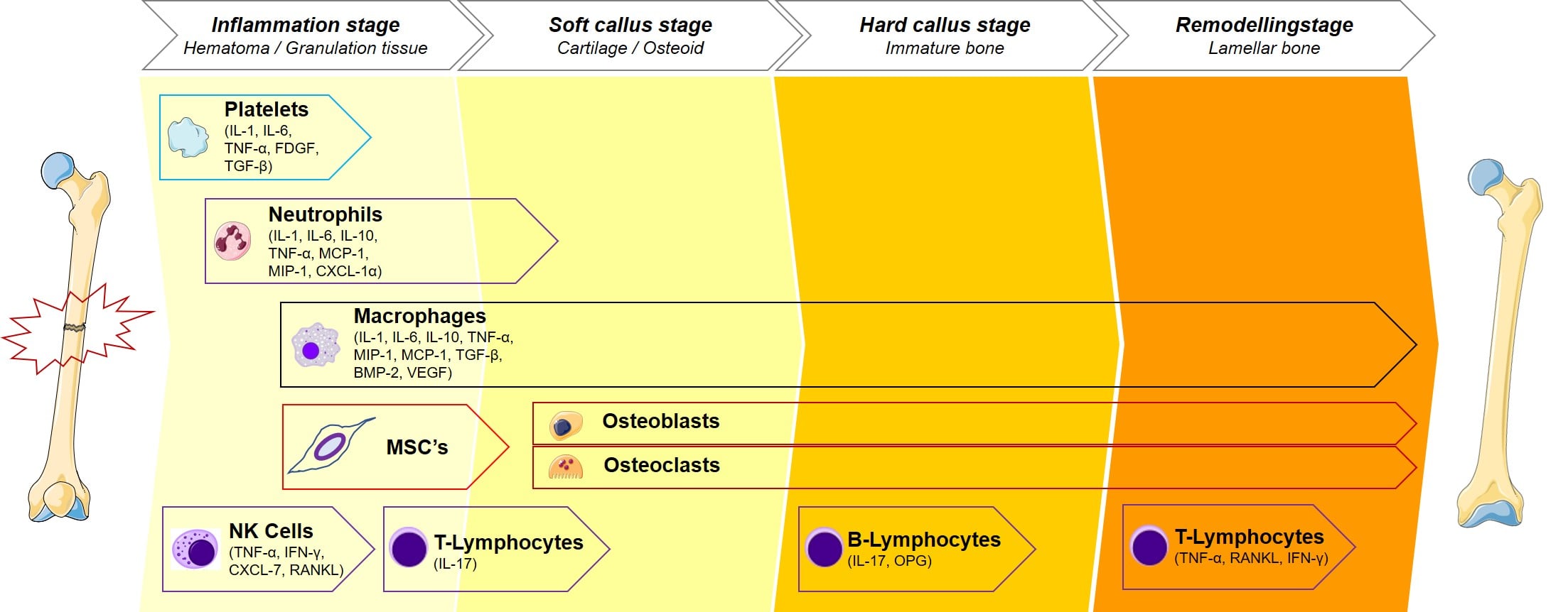
Secondary Bone Healing Timeline
Haematoma. Blood from torn vessels fills the fracture site and the fibrin clot forms the first scaffold.
Inflammatory response. Neutrophils arrive within 24-48 hours, macrophages after them, and between them they remove necrotic tissue and debris.
Cytokines. IL-1, IL-6 and TNF-α are released, recruiting mesenchymal stem cells and initiating angiogenesis.
Granulation tissue. Fibroblasts and new blood vessels form granulation tissue, the base on which callus is built.
Clinically, there is pain, swelling and heat at the fracture, and the haematoma is palpable initially.
MSC recruitment. Mesenchymal stem cells migrate to the fracture from periosteum, bone marrow and the surrounding tissues.
Chondrogenesis. Low oxygen tension at the fracture site promotes their differentiation into chondrocytes.
Fibrocartilaginous callus. Collagen type II and proteoglycans are produced, and the soft callus bridges the fracture.
Radiolucent. The callus is cartilage and fibrous tissue, not yet mineralised, so it appears radiolucent on radiographs.
Clinically, the soft callus provides limited stability, motion is still present at the fracture, and the pain is decreasing.
Enchondral ossification. Chondrocytes hypertrophy, calcify their matrix and undergo apoptosis, and blood vessels invade.
Osteoblast activity. Osteoblasts from periosteum and marrow lay down woven bone on the calcified cartilage scaffold.
Mineralisation. Hydroxyapatite deposition makes the callus radio-opaque and mechanically strong.
Periosteal and endosteal callus. Callus forms externally and internally, bridging the fracture from both sides.
Clinically, the fracture becomes stable, there is no motion on stress, pain is minimal and weight-bearing is possible.
Woven to lamellar bone. Osteoclasts resorb the woven bone and osteoblasts deposit organised lamellar bone in its place.
Callus resorption. Excess callus is gradually resorbed and the bone returns towards its original diameter.
Wolff law. Bone remodels along the lines of stress, and the trabeculae realign with mechanical loading.
Cortical restoration. The medullary canal is reconstituted and the cortical architecture restored.
Strength. Mechanical properties approach those of normal bone, which can take 6-18 months.
ISHRSecondary Healing Phases - ISHR
Hook:I See Hard Remodeling - the four phases of fracture healing in chronological order
The cells and the signals
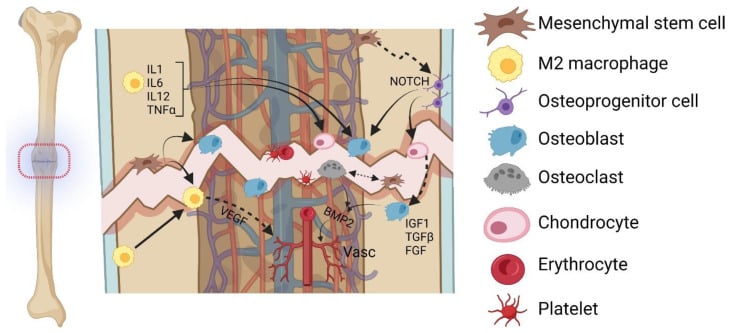
Key cells in fracture healing
The cells arrive in order, and the peak-activity column is the timetable of healing.
- Origin
- Blood
- Function in Healing
- Phagocytose debris, release cytokines
- Peak Activity
- 24-48 hours
- Origin
- Blood (monocytes)
- Function in Healing
- Phagocytose debris, release growth factors
- Peak Activity
- 3-7 days
- Origin
- Periosteum, marrow, soft tissue
- Function in Healing
- Differentiate to chondrocytes and osteoblasts
- Peak Activity
- 1-3 weeks
- Origin
- MSC differentiation
- Function in Healing
- Produce cartilage matrix in soft callus
- Peak Activity
- 1-3 weeks
- Origin
- MSC differentiation
- Function in Healing
- Deposit woven bone in hard callus
- Peak Activity
- 3-8 weeks
- Origin
- Monocyte fusion
- Function in Healing
- Resorb woven bone during remodeling
- Peak Activity
- 8+ weeks
MSCs differentiate according to the local environment. Low oxygen and mechanical instability favour chondrogenesis, giving cartilage in the soft callus; higher oxygen and stability favour osteoblastogenesis, giving bone in the hard callus. This is why soft callus forms first while the fracture is unstable, then converts to bone as stability improves.
Where secondary healing is expected
Intramedullary nailing of a femoral or tibial shaft fracture gives relative stability with controlled motion, and the large callus that follows provides biological bridging, with faster clinical union than plating. External fixation is used for open fractures, infected nonunions and distraction osteogenesis, and allows stability and compression to be adjusted, with the callus monitored and modulated as healing proceeds. Cast immobilisation treats most non-displaced or minimally displaced fractures along the same natural pathway, the callus supplying stability as it matures.
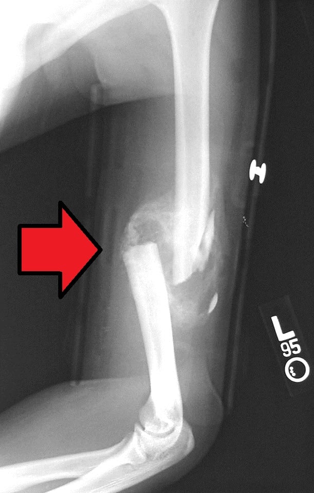
Both ossification routes run in the same callus. Intramembranous ossification occurs subperiosteally at the fracture peripheries, away from the gap, where the environment is relatively stable and well vascularised: osteoblasts lay down woven bone directly, with no cartilage intermediate, forming the peripheral hard callus. Enchondral ossification occurs in the central fracture gap, which is more mobile and hypoxic, where MSCs form a cartilage (soft callus) intermediate that calcifies and is then replaced by bone after vascular invasion. A single healing fracture therefore shows intramembranous bone at the edges and enchondral bone bridging the gap, and the proportions shift with the local strain and oxygen tension.
The blood supply reverses. Bone has a dual supply: the nutrient (medullary, endosteal) artery supplies the inner two-thirds of the cortex by centrifugal, outward flow, and the periosteal supply feeds the outer one-third by centripetal, inward flow. A fracture disrupts the medullary supply, so the periosteal and extraosseous supply becomes dominant during early healing, and the medullary supply is re-established as healing progresses. This is the biological reason to preserve the soft-tissue envelope and avoid periosteal stripping, and why reamed nailing transiently injures the endosteal supply while the periosteal supply compensates.
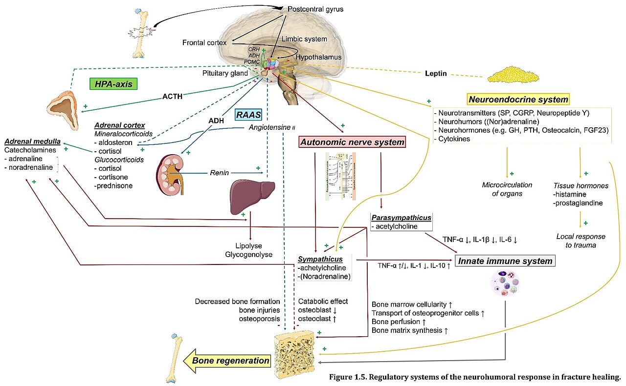
Management Algorithm
Clinical Relevance
Strain and the mechanical environment
Perren's interfragmentary strain theory. Strain is the change in the fracture gap divided by the original gap, ΔL / L, where ΔL is the change in gap width under load and L is the original gap width. Different tissues tolerate different amounts of strain, so the gap and the stiffness of the construct together decide what tissue can form in it.
- Tissue That Forms
- Bone (lamellar)
- Healing Type
- Primary healing
- Clinical Example
- Compression plating with anatomical reduction
- Tissue That Forms
- Cartilage then bone
- Healing Type
- Secondary healing with callus
- Clinical Example
- Intramedullary nail, external fixator, cast
- Tissue That Forms
- Fibrous tissue
- Healing Type
- Fibrous nonunion
- Clinical Example
- Inadequate fixation, large gap, infection
The surgeon sets the strain. A compression plate holds the fracture in the range where bone forms directly and no callus appears; an intramedullary nail leaves enough motion for a cartilage intermediate and callus; fixation inadequate for the pattern leaves the fracture in the fibrous range, and it does not unite.
Absolute stability is achieved by compression across the fracture, requires anatomical reduction, and produces primary healing with no callus. It is delivered by lag screws, compression plates, and an external fixator in compression mode.
Relative stability allows controlled interfragmentary motion, does not require anatomical reduction, and produces secondary healing with callus. It is delivered by:
- Intramedullary nails, locked or unlocked
- Bridge plating
- An external fixator in neutral or dynamic mode
- Cast immobilisation
Getting the mechanics right. The principles that govern the construct:
- Match the stability to the healing goal - articular fractures need anatomical reduction and primary healing, while secondary healing is acceptable for diaphyseal fractures
- Minimise the gap - large gaps delay or prevent healing
- Preserve the biology - minimise periosteal stripping, and use indirect reduction techniques where possible
- Correct length and alignment - malreduction impairs healing as well as function
The diamond concept
The Diamond Concept (Giannoudis et al., 2007) describes the four elements a fracture needs in order to heal: osteogenic cells, an osteoconductive scaffold, osteoinductive growth factors, and an appropriate mechanical environment. Deficiency in any one of them increases the risk of delayed union or nonunion, which is what makes it useful at the bedside - a nonunion is analysed by asking which element is missing.
Osteogenic cells
Where they come from.
- Periosteum - the richest source of MSCs and osteoprogenitor cells
- Bone marrow - haematopoietic and mesenchymal stem cells
- Endosteum - the inner lining of the medullary canal
- Surrounding soft tissues - muscle and fascia contain some MSCs
Numbers alone are not enough. The cells must be viable and present in adequate numbers, able to migrate to the fracture, capable of differentiating into bone-forming cells, and able to survive the local environment.
What depletes them.
- Extensive periosteal stripping during surgery
- Radiation therapy, which kills osteoprogenitor cells
- Severe open fractures with soft tissue loss
- Multiple operations, each with its own repeat trauma
How to replace them. Bone marrow aspirate harvested from the iliac crest and injected at the nonunion site brings MSCs and growth factors; cancellous bone graft brings cells, scaffold and growth factors together; demineralised bone matrix (DBM) brings growth factors that recruit the host's own cells.
Using the diamond on a nonunion. Ask which element is deficient: cells (radiation, smoking, excessive stripping), scaffold (a large gap, bone loss), growth factors (avascular tissue, diabetes), or mechanics (inadequate fixation, persistent motion). Then treat every deficient element rather than the most convenient one. An atrophic nonunion needs cells, scaffold and growth factors - bone graft, with BMP or DBM - alongside stable fixation; in a hypertrophic nonunion the mechanics are the likely problem, and the answer is compression and a reduced gap.
All four elements of the diamond must be present. Excellent fixation will not overcome absent cells or absent growth factors, and abundant growth factors cannot compensate for instability. It is why simply replating an infected nonunion often fails: the infection kills the cells, so the biology has to be addressed alongside the mechanics.
Differential diagnosis: impaired fracture healing
A fracture that is not progressing must be characterised precisely, because each pattern has a different deficient diamond element and therefore a different treatment. Delayed union and the nonunion subtypes are the core differential.
- Defining Feature
- Union slower than expected for site, but still progressing
- Typical Deficient Element
- Often mechanics or biology (modifiable)
- Radiographic Clue
- Some callus, fracture line persists
- Primary Treatment
- Optimise biology/stability, allow more time
- Defining Feature
- Abundant 'elephant-foot' callus, no bridging; biology intact
- Typical Deficient Element
- Mechanics (instability/motion)
- Radiographic Clue
- Exuberant callus, persistent gap
- Primary Treatment
- Improve stability/compression (e.g. nail, plate)
- Defining Feature
- Absent callus, sclerotic/resorbed ends; biology failed
- Typical Deficient Element
- Cells + scaffold + growth factors
- Radiographic Clue
- No callus, tapered/rounded bone ends
- Primary Treatment
- Biological augment (graft/BMP) plus stable fixation
- Defining Feature
- Nonunion with sepsis (sinus, raised inflammatory markers)
- Typical Deficient Element
- Cells (infection-impaired) + mechanics
- Radiographic Clue
- Lucency, sequestrum, loosening
- Primary Treatment
- Debridement, antibiotics, staged reconstruction
- Defining Feature
- Established false joint with synovial-lined cavity
- Typical Deficient Element
- Mechanics + biology (chronic)
- Radiographic Clue
- Smooth sclerotic ends, mobile gap
- Primary Treatment
- Resection of fibrous tissue, graft, stable fixation
What impairs healing
Patient factors. Systemic conditions and drugs change how a fracture heals, and each carries its own management step.
- Effect on Healing
- Doubles nonunion risk
- Mechanism
- Nicotine vasoconstriction impairs blood supply, CO decreases oxygen delivery
- Clinical Management
- Cessation essential - six to eight weeks before planned surgery
- Effect on Healing
- Delayed healing, increased infection
- Mechanism
- Hyperglycemia impairs cell function, neuropathy and vasculopathy
- Clinical Management
- Optimize glucose control (HbA1c under 7%)
- Effect on Healing
- Slower healing
- Mechanism
- Reduced cell number and function, comorbidities
- Clinical Management
- Optimize nutrition, consider augmentation
- Effect on Healing
- Impaired healing
- Mechanism
- Protein deficiency impairs collagen synthesis, vitamin deficiencies
- Clinical Management
- Nutritional supplementation, vitamin D, calcium
- Effect on Healing
- Inhibit healing
- Mechanism
- Suppress inflammation, reduce osteoblast function, increase osteoclast activity
- Clinical Management
- Minimize dose if possible, consider augmentation
- Effect on Healing
- Increased infection risk
- Mechanism
- Reduced immune surveillance, impaired inflammatory phase
- Clinical Management
- Prophylactic antibiotics, careful monitoring
- Effect on Healing
- Delayed healing
- Mechanism
- Reduced metabolic rate, decreased bone turnover
- Clinical Management
- Thyroid replacement therapy
Smoking roughly doubles the odds of nonunion - adjusted OR 2.32 (95% CI 1.76-3.06) in Scolaro's systematic review of 19 cohort studies, which is the card cited further down this page. Quote that figure, not a "Bhandari meta-analysis": no such paper exists, and the two 2012 JBJS papers with Bhandari as author are a tibial prognostic-factor analysis and a survey of how surgeons define nonunion.
Two things are commonly asserted here that the evidence does not support. Scolaro dichotomised smokers against non-smokers with no pack-year analysis, so a dose-response is biologically plausible but not demonstrated by that review; and no included study randomised cessation, so it establishes that smokers unite less often, not that stopping restores union. The review's own time-to-union difference (30.2 vs 24.1 weeks) did not reach significance (p = 0.18).
The cessation evidence that does exist is about wounds, not union. Møller's randomised trial (PMID 11809253) intervened 6 to 8 weeks before elective hip and knee replacement and cut wound complications from 31% to 5% (p = 0.001). That is the number to counsel with - and it is why the standard advice is six to eight weeks, not four. Still counsel every fracture patient to stop: the union risk is real even where the reversal evidence is thin.
Local factors. The fracture itself and its soft-tissue envelope carry risks of their own.
- Effect
- Higher nonunion and infection risk
- Why It Matters
- Soft tissue damage, contamination, impaired blood supply
- Management
- Debridement, antibiotics, staged fixation
- Effect
- Delayed union risk
- Why It Matters
- Bone loss, difficulty achieving stability, periosteal stripping
- Management
- Bridge plating, bone graft, consider IM nail
- Effect
- Nonunion if over 2-3mm
- Why It Matters
- Cells cannot bridge large gap, inadequate scaffold
- Management
- Bone graft, Masquelet technique, bone transport
- Effect
- Impaired healing
- Why It Matters
- Inadequate oxygen and nutrients, reduced cell delivery
- Management
- Preserve soft tissue, consider vascularized graft
- Effect
- Nonunion until controlled
- Why It Matters
- Inflammatory mediators inhibit healing, biofilm prevents antibiotics
- Management
- Debridement, antibiotics, staged reconstruction
- Effect
- Varies by bone
- Why It Matters
- Scaphoid, femoral neck have poor blood supply; tibia has limited soft tissue
- Management
- Consider biology when planning treatment
NSAIDs remain controversial. Animal studies show impaired healing, COX-2 being important for fracture healing, and human studies show mixed results, some showing delayed union and others no effect; the meta-analysis cited below found more delayed union and nonunion in adults, with no significant effect in children or with low-dose, short-duration exposure. The current consensus follows that split: avoid high-dose, long-term NSAIDs (especially COX-2 selective agents) in high-risk fractures, while short-term use of 1-2 weeks for pain is likely acceptable, with paracetamol, short-term opioids or regional anaesthesia as the alternatives.
Other drugs. Bisphosphonates reduce bone remodelling and may delay the remodelling phase, but they do not prevent healing and are continued for osteoporosis. Cytotoxic chemotherapy impairs cell proliferation, so elective surgery is delayed until after it where possible. Warfarin impairs the vitamin K-dependent proteins including osteocalcin, of unclear clinical significance, and is continued for thromboembolic disease.
Optimising the patient. Before surgery:
- Smoking cessation, six to eight weeks beforehand where the operation can be planned
- Glucose control in diabetes
- Nutritional supplementation with protein, vitamin D and calcium
- The lowest steroid dose the underlying disease allows, with biological augmentation considered where it cannot be reduced
At operation, minimise periosteal stripping, aim for stability that is adequate but not excessive, fill bone gaps with graft, and augment biologically with BMP or bone graft in high-risk cases. Afterwards:
- Avoid NSAIDs, or keep them short-term only
- Allow early weight-bearing if the fixation permits, since loading stimulates healing
- Monitor healing clinically and radiographically
- Intervene early where delayed union is suspected
Augmenting and rescuing healing
The diamond concept says what to optimise; a viva will also ask how to augment or rescue a slow-healing fracture actively. Three categories are high-yield.
Teriparatide (recombinant PTH 1-34). Intermittent low-dose PTH is anabolic, a net stimulus to osteoblasts, unlike the catabolic effect of continuous PTH. Its off-label uses are delayed union and nonunion (especially osteoporotic), pelvic and sacral insufficiency fractures, stress fractures and atypical femoral fractures, where impaired remodelling is the problem. Some randomised data, for instance in the distal radius, suggest faster healing, but the overall evidence is mixed, so it is an adjunct rather than a guaranteed therapy. Avoid it in the skeletally immature, in Paget disease, after prior skeletal radiation, or with an unexplained high alkaline phosphatase (the osteosarcoma signal in rodents), and it is typically limited to a lifetime course of about two years.
Biophysical stimulation. Low-intensity pulsed ultrasound (LIPUS) and pulsed electromagnetic fields or electrical stimulation (capacitive, inductive or direct current) are non-invasive adjuncts proposed for delayed union and nonunion. The evidence is mixed - the large TRUST randomised trial showed LIPUS did not improve tibial fracture healing - so they are selective adjuncts of uncertain benefit, never a substitute for correcting the deficient diamond element.
Surgical rescue. Exchange reamed intramedullary nailing suits a diaphyseal, especially hypertrophic, nonunion, the reaming providing autograft while the new nail is larger and stiffer. The Masquelet induced-membrane technique handles segmental defects in two stages: a cement spacer induces a vascular membrane, which is later opened and packed with bone graft. Bone transport, or distraction osteogenesis in the Ilizarov sense, is for large segmental defects.
Guidelines, Registries & Global Practice
Global Epidemiology of Fractures and Impaired Healing
Fractures are among the most common large-organ traumatic injuries worldwide, and approximately 10% fail to heal normally (Einhorn and Gerstenfeld, Nat Rev Rheumatol 2015, PMID 25266456). Global Burden of Disease modelling shows lower-limb fractures (patella, tibia/fibula, ankle) carry the highest age-standardised prevalence of all fracture sites, with falls the dominant mechanism and a projected continuing rise in fall-related fractures to 2035 (GBD 2019 analysis, PMID 39543488).
- Finding
- Approximately 10%
- Source
- Einhorn & Gerstenfeld 2015 (PMID 25266456)
- Finding
- Lower-limb (patella, tibia/fibula, ankle)
- Source
- GBD 2019 (PMID 39543488)
- Finding
- OR 2.32 (95% CI 1.76-3.06)
- Source
- Scolaro et al. 2014 (PMID 24740664)
- Finding
- OR 2.07 (95% CI 1.19-3.61)
- Source
- Wheatley et al. 2019 (PMID 30260913)
Guidance and Principles Across Jurisdictions
Bone-healing biology is governed by AO principles rather than disease-specific guidelines; the table below summarises how the major bodies frame stability and biology, with the underpinning evidence level.
- Core Guidance on Healing
- Absolute stability (lag screw/compression plate) for articular and simple fractures requiring anatomical reduction; relative stability (IM nail, bridge plating, external fixation) for comminuted/diaphyseal fractures
- Evidence Basis
- Mechanistic - Perren strain theory (PMID 376198)
- Core Guidance on Healing
- Optimise modifiable patient factors (smoking, glycaemia, nutrition); reserve osteobiologics (BMP) for high-risk/recalcitrant cases
- Evidence Basis
- RCT/meta-analysis (PMID 11314793, 24740664)
- Core Guidance on Healing
- Non-union and complex/open fractures managed in specialist (major trauma) centres; structured smoking cessation offered around surgery
- Evidence Basis
- Service standards + observational evidence
- Core Guidance on Healing
- Diamond-concept-guided management of nonunion - identify and correct the deficient element(s)
- Evidence Basis
- Expert consensus (PMID 18224731)
Registry and Surveillance Evidence
There is no dedicated international fracture-healing registry; population-level signal derives from trauma registries and Global Burden of Disease surveillance (PMID 39543488). Where biologics or implants are involved, device and adverse-event surveillance (e.g. national regulator pharmacovigilance) informs practice — for example, post-marketing signals shaped the cautious, lowest-effective-dose use of rhBMP-2/rhBMP-7 in spine and long-bone surgery.
Practice Variation
- Osteobiologic use varies markedly by funding and regulation. rhBMP use is constrained by cost in publicly funded systems; autograft remains the global default for biological augmentation.
- Smoking-cessation pathways differ but the principle (cessation around the time of surgery; structured support) is universal given the OR 2.32 nonunion signal (PMID 24740664).
- NSAID policy varies between centres given dose- and duration-dependent harm in adults but a reassuring profile in children and short courses (PMID 30260913).
- Underserved and remote populations carry compounded risk. Higher diabetes prevalence and barriers to surgery and follow-up access (including telehealth reach) worsen healing outcomes in Indigenous, remote-rural, and low-resource settings, making equitable access and culturally appropriate planning part of optimising healing globally.
Orthopaedic Examination Relevance
- Bone healing is a core basic science topic
- Primary vs secondary healing mechanisms
- Perren's strain theory (under 2%, 2-10%, over 10%)
- Diamond concept (cells, scaffold, growth factors, mechanics)
- Cutting cones and enchondral ossification
- Four phases of secondary healing (ISHR)
- Osteoinduction vs osteoconduction
- BMP and VEGF roles
- Smoking roughly doubles nonunion risk (OR 2.32)
- Fixation choice determines healing type via strain
MCQ Practice Points
Q: What is the interfragmentary strain threshold that determines whether primary or secondary bone healing occurs?
A: Under 2% strain = primary (direct) healing. 2-10% strain = secondary (indirect) healing with callus. Over 10% strain = only fibrous tissue survives (nonunion). This is Perren's strain theory: tissue survives only if local strain is less than its elongation at failure. Bone tolerates only 2%, cartilage tolerates 10%, fibrous tissue tolerates 100%.
Q: What are the four phases of secondary bone healing in correct order?
A: Inflammation → Soft callus → Hard callus → Remodeling (ISHR). Days 0-7: hematoma and inflammation. Weeks 1-3: fibrocartilaginous soft callus. Weeks 3-8: enchondral ossification to hard (woven bone) callus. Months to years: remodeling of woven to lamellar bone following Wolff's law.
Q: What is the mechanism of primary (direct) bone healing at the cellular level?
A: Cutting cones (osteoclast-led basic multicellular units) cross the fracture site directly. Osteoclasts at the leading edge resorb bone, followed by osteoblasts laying down new Haversian systems. This requires absolute stability (under 2% strain) achieved by compression plating or lag screws. No callus forms.
Q: Which growth factors are classified as osteoinductive and are used clinically to treat nonunion?
A: BMP-2 and BMP-7 (also called OP-1). They are osteoinductive because they induce mesenchymal stem cell differentiation into osteoblasts. BMP-2 is used in spinal fusion and tibial nonunion. VEGF, PDGF, and TGF-β are important for healing but are chemotactic/angiogenic rather than osteoinductive.
Q: What are the four components of the "Diamond Concept" for fracture healing?
A: (1) Osteogenic cells (MSCs, osteoblasts), (2) Osteoconductive scaffold (bone graft matrix), (3) Growth factors (BMPs, osteoinductive signals), (4) Mechanical environment (stability). All four are required for union. Nonunion management targets whichever element is deficient - atrophic needs biology, hypertrophic needs stability.
Basic Science Viva Scenarios
Practise clinical reasoning and management decisions out loud
“An examiner presents an X-ray of a tibial shaft fracture treated with intramedullary nail showing large callus, then an X-ray of a forearm fracture treated with compression plate showing no callus. They ask you to explain the difference.”
“A 55-year-old heavy smoker presents with atrophic nonunion of the tibia 9 months after open fracture treated with external fixator. The examiner asks how you would approach treatment using the Diamond Concept.”
“Explain the role of growth factors in fracture healing, focusing on BMPs and VEGF. What is the difference between osteoinduction and osteoconduction?”
Primary Healing (Direct)
- Absolute stability required (strain under 2%)
- Compression plating, lag screws achieve this
- No callus forms - cutting cones cross fracture
- Cutting cones = osteoclasts followed by osteoblasts
- Direct Haversian remodeling across fracture
- Slower biological healing, relies on implant early
Secondary Healing (Indirect)
- Relative stability (strain 2-10%), natural pathway
- Four phases: Inflammation (0-1wk), Soft callus (1-3wk), Hard callus (3-8wk), Remodeling (months-years)
- Inflammation: hematoma, cytokines, granulation tissue
- Soft callus: MSCs → chondrocytes, cartilage forms (radiolucent)
- Hard callus: enchondral ossification, woven bone (radio-opaque)
- Remodeling: woven to lamellar bone, Wolff law
Strain Theory (Perren)
- Strain = ΔL / L (gap change / original gap)
- Under 2% strain: bone forms (primary healing)
- 2-10% strain: cartilage then bone (secondary healing)
- Over 10% strain: fibrous tissue only (nonunion)
- Explains why fixation method determines healing type
Diamond Concept (All 4 Required)
- 1. Cells: MSCs, osteoblasts from periosteum and marrow
- 2. Scaffold: Osteoconductive matrix (bone graft, hematoma)
- 3. Growth factors: BMPs (osteoinductive), VEGF (angiogenesis), PDGF, TGF-β
- 4. Mechanics: Appropriate stability for healing type
Growth Factors (Know These)
- BMP-2, BMP-7: Osteoinductive (induce MSC → osteoblast)
- VEGF: Angiogenesis (critical for enchondral ossification)
- PDGF: Chemotactic for MSCs, proliferation
- TGF-β: Regulates cell proliferation, chondrogenesis
Factors Impairing Healing
- Smoking: doubles nonunion risk (nicotine, CO, impaired angiogenesis)
- Diabetes: hyperglycemia impairs cells, neuropathy/vasculopathy
- NSAIDs: controversial, avoid long-term especially COX-2
- Steroids: inhibit osteoblasts, increase osteoclasts
- Open fracture, infection, bone loss/gap, poor blood supply
Key Concepts for Viva
- Osteoinduction = inducing MSC differentiation to osteoblasts
- Osteoconduction = scaffold for existing cells to grow on
- Enchondral ossification = cartilage intermediate, requires vessels
- Cutting cones = osteoclasts then osteoblasts (primary healing)
- Autograft = cells + scaffold + growth factors (gold standard)
Evidence Base
Strain Theory and Tissue Differentiation
- Introduced interfragmentary strain theory (ΔL/L)
- Different tissues tolerate different strain levels
- Bone tolerates under 2% strain (primary healing possible)
- Cartilage forms at 2-10% strain (secondary healing)
- Over 10% strain leads to fibrous tissue (nonunion)
- Explained why different fixation methods produce different healing patterns
Diamond Concept of Fracture Healing
- Described four essential elements for healing: cells, scaffold, growth factors, mechanical environment
- Deficiency in any element can lead to nonunion
- Framework for analyzing nonunion etiology
- Guides augmentation strategies (which element is deficient?)
Cigarette Smoking Increases Fracture Complications
- Systematic review of 19 cohort studies (7 prospective, 12 retrospective) of smoking and long-bone fracture healing
- Adjusted odds ratio for nonunion in smokers vs non-smokers was 2.32 (95% CI 1.76-3.06, p under 0.001)
- Increased nonunion in smokers with tibial fractures (OR 2.16) and open fractures (OR 1.95)
- Mean time to union longer in smokers (30.2 vs 24.1 weeks)
- Non-significant trends toward more superficial and deep wound infection in smokers
rhBMP-7 (OP-1) for Tibial Nonunion
- Prospective randomised controlled trial (124 tibial nonunions) of rhOP-1 (BMP-7) with collagen carrier vs fresh autogenous bone graft, all with intramedullary fixation
- At 9 months, clinical success 81% (BMP-7) vs 85% (autograft) (p=0.524); radiographic union 75% vs 84% (p=0.218) - no significant difference
- Avoided donor-site morbidity: over 20% of autograft patients had chronic donor-site pain
- Supported regulatory approval of BMP-7 (OP-1) for recalcitrant tibial nonunion
Fracture Healing: Mechanisms and Interventions (Landmark Review)
- Authoritative review framing fracture repair as a postnatal regenerative process recapitulating embryonic skeletal development
- Approximately 10% of fractures do not heal normally
- Integrates the tissue, cellular and molecular sequence (inflammation, endochondral and intramembranous ossification, remodelling)
- Reviews the innate/adaptive immune contribution and the roles of fixation and vascularity
- Summarises biological interventions including rhBMPs and parathyroid hormone (teriparatide) therapy
Effect of NSAIDs on Bone Healing Rates (Meta-analysis)
- Systematic review and meta-analysis of NSAID exposure and bone healing (delayed union, nonunion, pseudarthrosis)
- NSAID exposure increased delayed union or nonunion overall (OR 2.07, 95% CI 1.19-3.61)
- No significant effect in paediatric patients (OR 0.58, 95% CI 0.27-1.21)
- No significant effect with low-dose / short-duration exposure (OR 1.68, 95% CI 0.63-4.46)
- Effect appears dose- and duration-dependent