Bone Signaling Pathways
RANK-RANKL-OPG sets resorption: RANKL (from osteoblast-lineage/osteocytes) drives osteoclasts, OPG is the decoy, and denosumab is anti-RANKL. The classic slip is saying OPG binds RANK - it binds RANKL (the ligand).
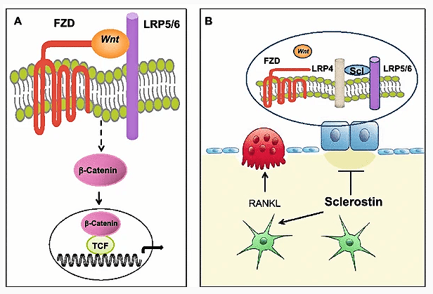
Wnt/β-catenin drives formation; sclerostin (osteocyte, SOST gene) is the brake, and romosozumab is anti-sclerostin. Don't confuse sclerostin (inhibits formation) with RANKL (drives resorption) - the two axes and their two antibodies are the commonest mix-up.
Overview & Introduction
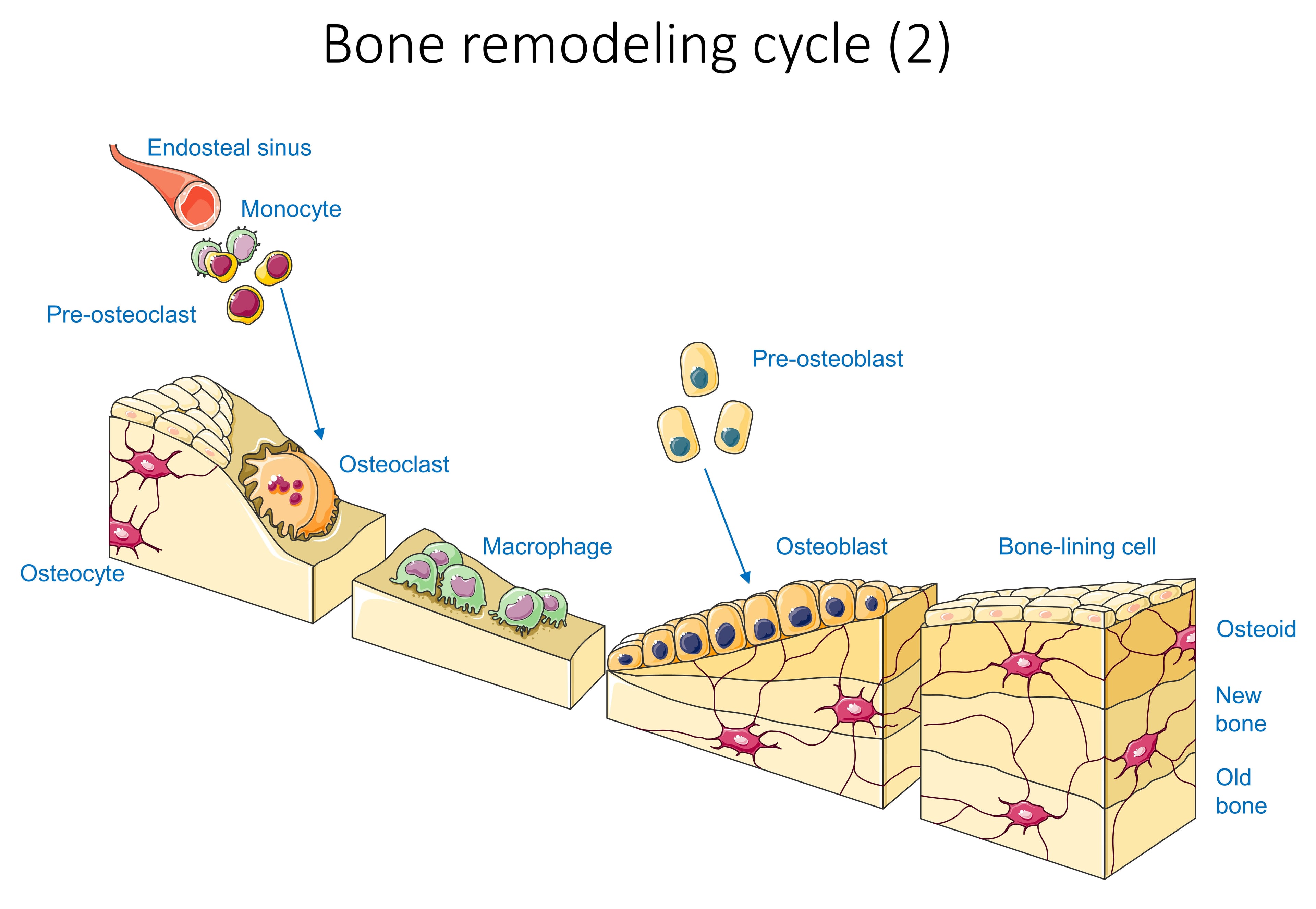
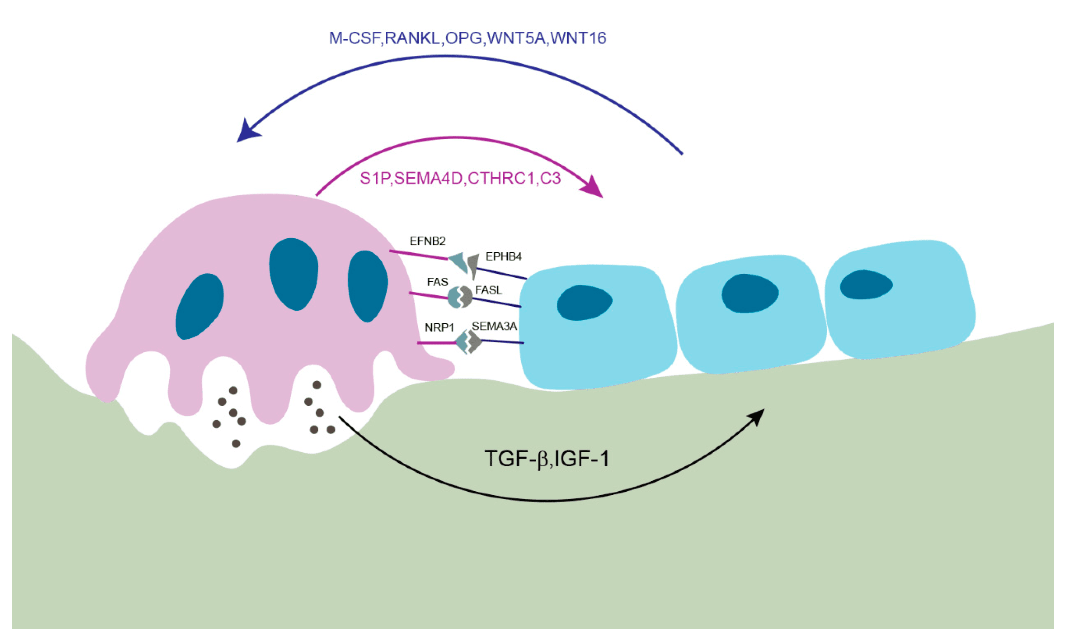
A handful of molecular pathways control every bone cell, and the exam distils them to two axes and two drugs. RANK-RANKL-OPG is the master switch for resorption (target: denosumab). Wnt/β-catenin is the switch for formation, restrained by the osteocyte brake sclerostin (target: romosozumab). Around these sit BMP/TGF-β (driving stem cells to osteoblasts; the basis of BMP-2), Indian hedgehog and PTHrP at the growth plate, and Notch as a contact-dependent modulator. Critically, the system is coupled: resorption itself releases the matrix-bound TGF-β and IGF-1 that recruit the osteoblasts to rebuild - which is why these are not independent switches but one integrated remodelling unit.
Concepts: RANK-RANKL-OPG (the Osteoclast Axis)
Osteoclasts are formed and activated by RANKL (Receptor Activator of Nuclear factor-κB Ligand), produced by osteoblast-lineage cells and osteocytes (and, in disease, by activated T cells). RANKL binds its receptor RANK on osteoclast precursors, which — together with M-CSF — drives their fusion, differentiation and bone-resorbing activity. The body's restraint is osteoprotegerin (OPG), a soluble decoy receptor secreted by osteoblasts that binds RANKL and prevents it reaching RANK. The RANKL:OPG ratio therefore sets the level of bone resorption: a high ratio means more osteoclasts and bone loss; a low ratio protects bone.
- Denosumab = a human monoclonal antibody to RANKL (mimics OPG) — a potent antiresorptive for osteoporosis and skeletal metastases.
- Oestrogen deficiency raises the RANKL:OPG ratio and increases IL-1/IL-6/TNF, accelerating postmenopausal bone loss.
- Many pathologies of bone loss (myeloma, inflammatory arthritis, metastasis) converge on up-regulated RANKL.
- Stopping denosumab causes a rebound in resorption with risk of multiple vertebral fractures — plan sequential therapy.
Wnt / β-catenin — the formation axis
In the canonical Wnt pathway, a Wnt ligand binds the Frizzled (FZD) receptor together with the co-receptor LRP5/6. This inhibits the destruction complex so that β-catenin is stabilised, enters the nucleus and partners with TCF/LEF to switch on osteoblastogenic genes — increasing osteoblast number and bone formation (and indirectly raising OPG, lowering resorption). The pathway is held in check by inhibitors: sclerostin (made by osteocytes from the SOST gene) and Dkk1, which bind LRP4/5/6 and switch Wnt off.
- LRP5 gain-of-function → high bone mass phenotype; LRP5 loss-of-function → osteoporosis-pseudoglioma syndrome (low bone mass). These human "experiments of nature" proved Wnt's central role.
- Romosozumab is a monoclonal antibody against sclerostin — it releases the Wnt brake and is a potent anabolic osteoporosis drug (dual effect: increases formation and decreases resorption).
- Mechanical loading lowers sclerostin (bone responds to load by building) — the molecular basis of Wolff's law.
BMP / TGF-β, Hedgehog & Notch
Bone morphogenetic proteins (BMPs) are members of the TGF-β superfamily. BMPs (especially BMP-2 and BMP-7) bind BMP receptors and signal through SMAD 1/5/8 to drive mesenchymal stem cells toward the osteoblast (and chondroblast) lineage, inducing Runx2/Osterix. TGF-β itself is abundant in bone matrix and, when released during resorption, recruits osteoblast precursors — helping couple formation to resorption. Recombinant BMP-2 is used clinically (e.g. spinal fusion, selected nonunions/open tibial fractures), though with dose and off-target considerations. BMP antagonists include noggin and gremlin.
These pathways do not act in isolation. Resorption and formation are coupled: osteoclastic resorption releases TGF-β and IGF-1 stored in bone matrix, which recruit osteoblasts to refill the cavity, while osteoblast-lineage cells reciprocally control osteoclasts through RANKL/OPG. Anabolic and antiresorptive drugs work by tipping this coupled system — which is why, for example, denosumab withdrawal produces a rebound in resorption.
Beyond sclerostin and RANKL, the osteocyte (and osteoblast) secretes FGF23, the master phosphate-regulating hormone. With its obligate co-receptor α-Klotho, FGF23 acts on the kidney to promote phosphate excretion (downregulating the NaPi-2a/2c transporters) and to suppress 1α-hydroxylase, lowering calcitriol. Excess FGF23 therefore causes hypophosphataemic rickets/osteomalacia: X-linked hypophosphataemia (XLH) from PHEX loss-of-function that raises FGF23, autosomal dominant hypophosphataemic rickets from FGF23 gain-of-function, and tumour-induced osteomalacia from an FGF23-secreting phosphaturic mesenchymal tumour. The targeted drug is burosumab, an anti-FGF23 monoclonal antibody now used in XLH and tumour-induced osteomalacia - a clean example of a bone-derived signalling molecule, its disease, and its antibody.
Clinical Relevance: Pathways & the Drugs That Target Them
- Net effect on bone
- RANKL → resorption; OPG protects
- Key inhibitor / brake
- OPG (decoy receptor)
- Therapeutic target
- Denosumab (anti-RANKL)
- Net effect on bone
- Formation (osteoblastogenesis)
- Key inhibitor / brake
- Sclerostin (SOST), Dkk1
- Therapeutic target
- Romosozumab (anti-sclerostin)
- Net effect on bone
- MSC → osteoblast; coupling
- Key inhibitor / brake
- Noggin, gremlin (BMP antagonists)
- Therapeutic target
- Recombinant BMP-2/7
- Net effect on bone
- Growth-plate & osteoblast development
- Key inhibitor / brake
- Patched/Gli regulation
- Therapeutic target
- Investigational
- Net effect on bone
- Progenitor proliferation/differentiation
- Key inhibitor / brake
- Context-dependent
- Therapeutic target
- Investigational
The two axes and two antibodies are not the only drug-bearing pathway. PTH and PTHrP act on the same receptor, PTH1R, on osteoblast-lineage cells - a Gs-coupled receptor signalling through cAMP/PKA. The examinable point is the paradox of timing: intermittent PTH is ANABOLIC - it increases osteoblast number and activity, reduces osteoblast apoptosis, and lowers sclerostin (de-repressing Wnt) - whereas continuous elevation (as in primary hyperparathyroidism, or a continuous infusion) is net CATABOLIC, because sustained signalling upregulates RANKL on osteoblasts and drives osteoclastic resorption. This is the basis of the anabolic drugs that complete the picture: teriparatide (recombinant PTH 1-34) and abaloparatide (a PTHrP analogue), given as a once-daily injection to exploit the intermittent-anabolic window. It also explains why hyperparathyroidism causes bone loss while a daily PTH injection builds it.
Mnemonics & Memory Aids
RODThe osteoclast axis
Hook:The osteoclast axis is a ROD: RANKL on, OPG off, Denosumab blocks.
WNTThe formation axis
Hook:WNT builds, sclerostin brakes, romosozumab releases the brake.
Evidence
WNT signaling in bone homeostasis and disease: from human mutations to treatments
- Rare human mutations affecting bone - osteoporosis-pseudoglioma (low mass), and high-bone-mass phenotype, sclerosteosis and Van Buchem disease (high mass) - all reside in components of canonical WNT signalling.
- Mouse genetics confirm that activating canonical Wnt increases, and inhibiting it decreases, bone mass and strength; GWAS link WNT loci to bone density in the population.
- The pathway is now a therapeutic target (sclerostin/Dkk1 inhibition) to restore bone strength - the rationale for romosozumab.
Osteoporosis Due to Hormone Imbalance: An Overview of the Effects of Estrogen Deficiency and Glucocorticoid Overuse on Bone Turnover
- Estrogen promotes OPG expression and suppresses RANKL, inhibiting osteoclast formation; it also activates Wnt/β-catenin to increase osteogenesis and upregulates BMP signalling to push MSCs toward osteoblasts.
- Estrogen deficiency increases IL-1, IL-6 and TNF and raises the RANKL:OPG ratio, accelerating resorption.
- Excess glucocorticoids interfere with BMP and inhibit Wnt, diverting MSCs to adipocytes, raise the RANKL:OPG ratio, and cause osteoblast/osteocyte apoptosis - explaining glucocorticoid-induced osteoporosis.
Relevance of Notch Signaling for Bone Metabolism and Regeneration
- Notch1-4 receptors control bone remodelling and regeneration - osteoblast differentiation/mineralisation, osteoclast recruitment/fusion, and progenitor proliferation - in a stage-dependent manner.
- Notch crosstalks with the Wnt/β-catenin, BMP and RANKL/OPG pathways that govern bone turnover.
- Mutations in Notch-pathway genes are associated with congenital skeletal disorders.
The Wnt-in-bone genetics and therapeutic rationale come from the Baron & Kneissel Nature Medicine review (DOI); the RANKL/OPG, Wnt and BMP relationships and their hormonal modulation from the Cheng et al. estrogen/glucocorticoid review (DOI); and the Notch biology and pathway crosstalk from the Ballhause et al. review (DOI). The growth-plate Ihh-PTHrP loop is the same axis described in our Physis topic, and the drug mechanisms (denosumab, romosozumab, rhBMP-2) are established pharmacology.