Bone Signaling Pathways
Overview & Introduction
These pathways are the mechanism; the pages around them are the consequences. The cycle they regulate is bone remodeling, run by osteoblasts and osteoclasts on the material described in bone composition and structure. Each axis has a drug that proves it: denosumab on RANKL, teriparatide and bisphosphonates on the two arms of turnover, and BMP-2 on the differentiation axis - which is why osteoporosis is treated by choosing which of these pathways to push, and why osteopetrosis shows what happens when the resorption arm cannot be pushed at all.
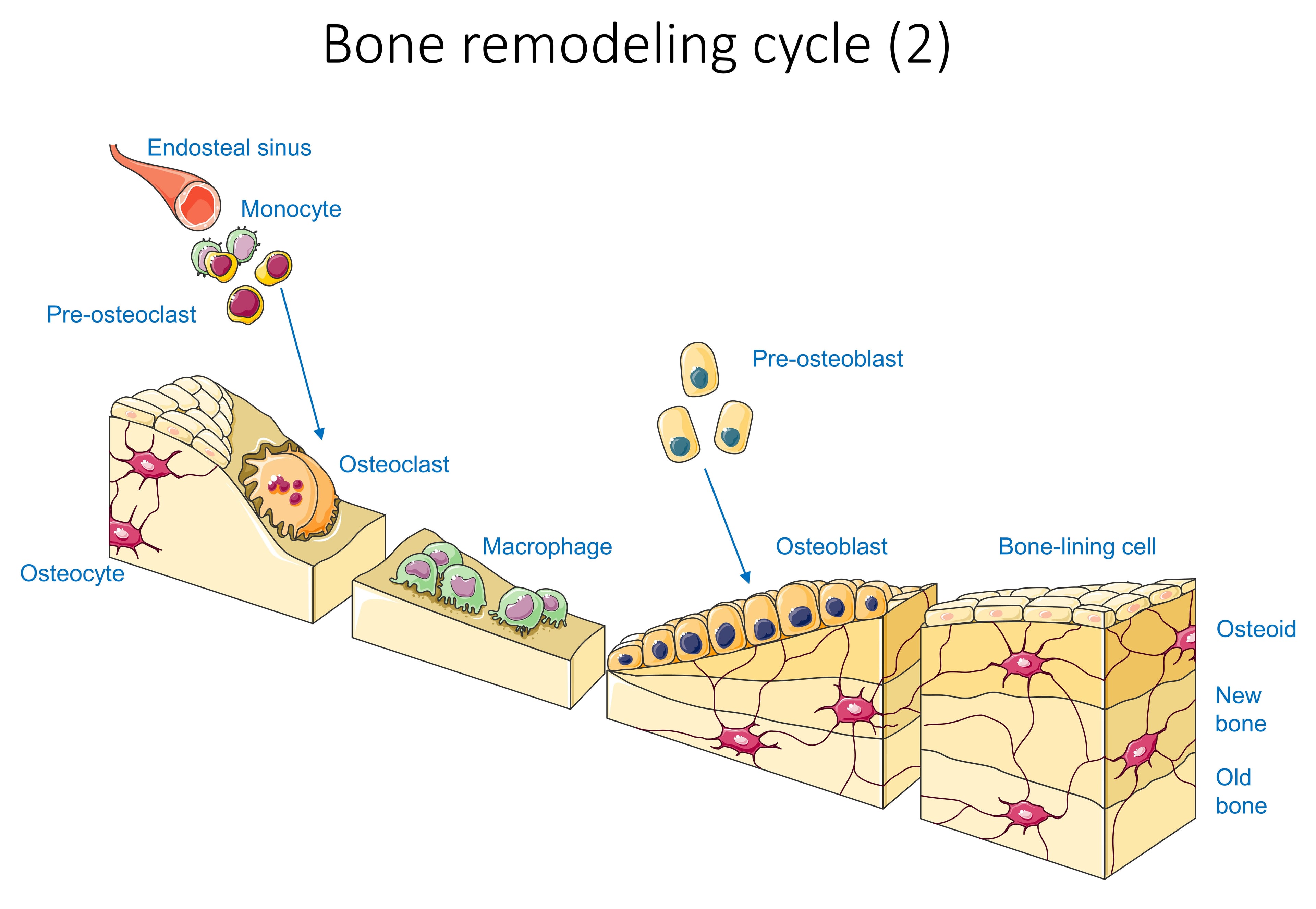
The stage these pathways act on. Remodelling runs as a basic multicellular unit: monocyte and pre-osteoclast precursors arrive from the endosteal sinus and become the resorbing osteoclast, and after a reversal phase pre-osteoblasts mature into osteoblasts that lay down osteoid and then new bone, finishing as flat bone-lining cells with osteocytes left embedded in the matrix. Every pathway in this topic acts somewhere along that sequence.
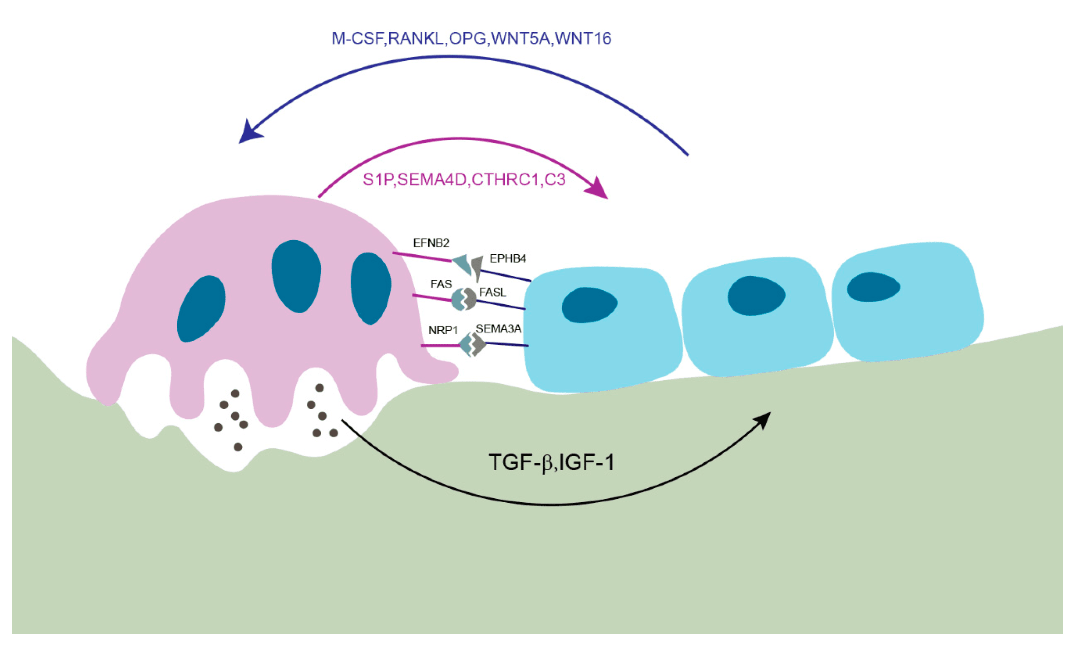
The map. RANK-RANKL-OPG drives the osteoclast, Wnt and BMP drive the osteoblast, and the TGF-β and IGF-1 released by resorption couple the two. Around them sit Indian hedgehog with PTHrP at the growth plate, and Notch as a contact-dependent modulator.
Concepts: RANK-RANKL-OPG (the Osteoclast Axis)
The switch. Osteoclasts are formed and activated by RANKL (Receptor Activator of Nuclear factor-κB Ligand), produced by osteoblast-lineage cells and osteocytes, and in disease by activated T cells. RANKL binds its receptor RANK on osteoclast precursors and, together with M-CSF, drives their fusion, differentiation and bone-resorbing activity.
The brake. The body's restraint is osteoprotegerin (OPG), a soluble decoy receptor secreted by osteoblasts that binds RANKL and prevents it reaching RANK. The RANKL:OPG ratio therefore sets the level of resorption: a high ratio means more osteoclasts and bone loss, a low ratio protects bone.
The drug. Denosumab is a human monoclonal antibody to RANKL, pharmacologically an extra supply of OPG, and a potent antiresorptive in osteoporosis and skeletal metastases.
Stopping it. Denosumab withdrawal causes a rebound in resorption with a risk of multiple vertebral fractures, so sequential therapy has to be planned.
Where disease enters the axis. Many pathologies of bone loss converge on up-regulated RANKL: myeloma, inflammatory arthritis and metastasis.
Wnt / β-catenin — the formation axis
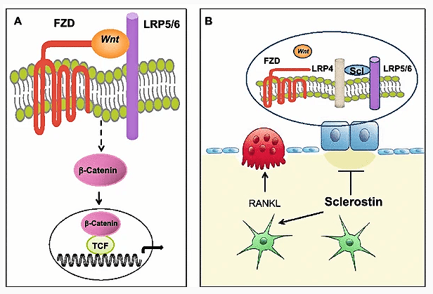
The pathway. In the canonical Wnt pathway a Wnt ligand binds the Frizzled (FZD) receptor together with the co-receptor LRP5/6. This inhibits the destruction complex, so β-catenin is stabilised, enters the nucleus and partners with TCF/LEF to switch on osteoblastogenic genes. Osteoblast number and bone formation rise, and OPG rises with them, so the same signal indirectly lowers resorption.
The brake. Two inhibitors bind LRP4/5/6 and switch Wnt off: sclerostin, made by osteocytes from the SOST gene, and Dkk1. Mechanical loading lowers sclerostin, so bone responds to load by building, which is the molecular basis of Wolff's law.
The genetics. LRP5 gain-of-function produces a high bone mass phenotype and loss-of-function produces osteoporosis-pseudoglioma syndrome, with low bone mass. These human experiments of nature proved Wnt's central role.
The drug. Romosozumab is a monoclonal antibody against sclerostin. Releasing the Wnt brake makes it a potent anabolic drug in osteoporosis, and the effect is dual: formation increases and resorption decreases.
The classic slip on the resorption axis is to say that OPG binds RANK. It binds RANKL, the ligand.
The classic slip across the axes is to swap the inhibitors or the antibodies. Sclerostin inhibits formation and romosozumab blocks sclerostin; RANKL drives resorption and denosumab blocks RANKL. These two axes and their two antibodies are the commonest mix-up.
BMP / TGF-β, Hedgehog & Notch
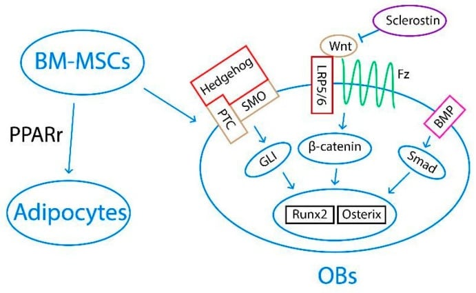
BMP and the TGF-β superfamily. Bone morphogenetic proteins are members of the TGF-β superfamily. BMPs, especially BMP-2 and BMP-7, bind BMP receptors and signal through SMAD 1/5/8 to drive mesenchymal stem cells toward the osteoblast and chondroblast lineages, inducing Runx2 and Osterix. The antagonists are noggin and gremlin.
The clinical use. Recombinant BMP-2 is used in spinal fusion and in selected nonunions and open tibial fractures, though with dose and off-target considerations. TGF-β itself is abundant in bone matrix, and what happens when resorption releases it is the coupling at the end of this section.
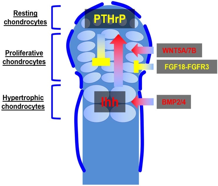
Indian hedgehog. Ihh, secreted by prehypertrophic chondrocytes, is central to endochondral ossification. It forms a feedback loop with PTHrP that paces chondrocyte proliferation against hypertrophy at the growth plate, and it is required for osteoblast differentiation in the developing bone collar. Dysregulated hedgehog signalling features in skeletal dysplasias and some bone tumours.
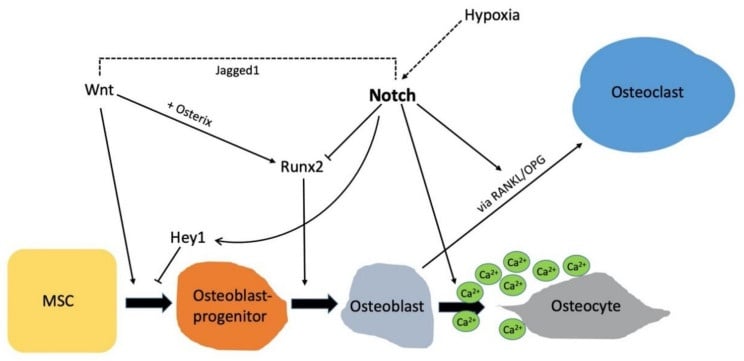
Notch. Notch signalling is contact-dependent, with receptors Notch1-4 and Jagged/Delta ligands. In bone it regulates osteoblast and osteoclast progenitor proliferation and differentiation and matrix mineralisation, with effects that are highly context- and stage-dependent. It crosstalks with Wnt/β-catenin, BMP and RANKL/OPG, and mutations in Notch-pathway genes cause congenital skeletal disorders.
Coupling. These pathways do not act in isolation. Osteoclastic resorption releases the TGF-β and IGF-1 stored in bone matrix, and those recruit osteoblasts to refill the cavity, while osteoblast-lineage cells reciprocally control osteoclasts through RANKL and OPG. Anabolic and antiresorptive drugs work by tipping this coupled system.
The osteocyte as an endocrine cell: FGF23, Klotho & phosphate
One cell, three outputs. From inside the matrix the osteocyte sets resorption through RANKL and OPG, restrains formation through sclerostin and DKK1, and controls phosphate through FGF23. Mechanically responsive mediators including prostaglandin E2 link loading to turnover, which is why the same cell can regulate resorption, formation and mineral metabolism.
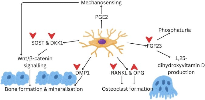
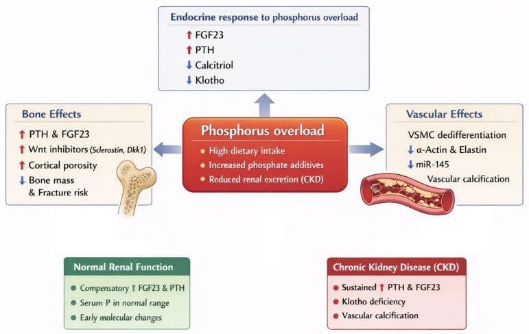
FGF23 and Klotho. The osteocyte and osteoblast secrete FGF23, the master phosphate-regulating hormone. With its obligate co-receptor α-Klotho it acts on the kidney to promote phosphate excretion, downregulating the NaPi-2a/2c transporters, and to suppress 1α-hydroxylase, which lowers calcitriol.
Too much of it. Excess FGF23 causes hypophosphataemic rickets and osteomalacia, in three forms worth naming:
- X-linked hypophosphataemia (XLH) - PHEX loss-of-function, which raises FGF23
- Autosomal dominant hypophosphataemic rickets - FGF23 gain-of-function
- Tumour-induced osteomalacia - an FGF23-secreting phosphaturic mesenchymal tumour
The drug. Burosumab, an anti-FGF23 monoclonal antibody, is used in XLH and in tumour-induced osteomalacia: a bone-derived signalling molecule, its disease and its antibody.
Clinical Relevance: Pathways & the Drugs That Target Them
- Net effect on bone
- RANKL → resorption; OPG protects
- Key inhibitor / brake
- OPG (decoy receptor)
- Therapeutic target
- Denosumab (anti-RANKL)
- Net effect on bone
- Formation (osteoblastogenesis)
- Key inhibitor / brake
- Sclerostin (SOST), Dkk1
- Therapeutic target
- Romosozumab (anti-sclerostin)
- Net effect on bone
- MSC → osteoblast; coupling
- Key inhibitor / brake
- Noggin, gremlin (BMP antagonists)
- Therapeutic target
- Recombinant BMP-2/7
- Net effect on bone
- Growth-plate & osteoblast development
- Key inhibitor / brake
- Patched/Gli regulation
- Therapeutic target
- Investigational
- Net effect on bone
- Progenitor proliferation/differentiation
- Key inhibitor / brake
- Context-dependent
- Therapeutic target
- Investigational
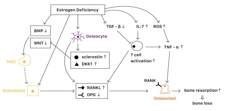
Oestrogen deficiency. Losing oestrogen raises the RANKL:OPG ratio and increases IL-1, IL-6 and TNF, accelerating bone loss after the menopause.
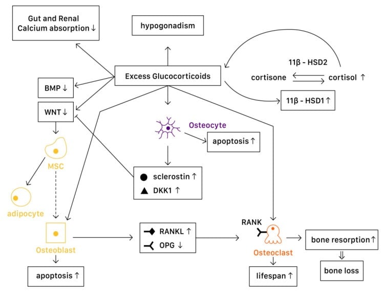
Glucocorticoids. Excess glucocorticoid interferes with BMP and inhibits Wnt, diverting mesenchymal stem cells to adipocytes, raises the RANKL:OPG ratio, and causes osteoblast and osteocyte apoptosis.
PTH and PTHrP share a receptor. The two axes and their two antibodies are not the only drug-bearing pathway in bone: PTH and PTHrP both act on PTH1R on osteoblast-lineage cells, a Gs-coupled receptor signalling through cAMP/PKA.
The paradox of timing. Intermittent PTH is anabolic: it increases osteoblast number and activity, reduces osteoblast apoptosis, and lowers sclerostin, de-repressing Wnt. Continuous elevation - in primary hyperparathyroidism, or from a continuous infusion - is net catabolic, because sustained signalling upregulates RANKL on osteoblasts and drives osteoclastic resorption.
The drugs that follow from it. Teriparatide (recombinant PTH 1-34) and abaloparatide (a PTHrP analogue) are given as a once-daily injection to exploit the intermittent-anabolic window. The same biology explains why hyperparathyroidism causes bone loss while a daily PTH injection builds bone.
Viva practice
Practise clinical reasoning and management decisions out loud
“Draw and explain the RANK-RANKL-OPG axis. Where does denosumab act, and how does oestrogen deficiency cause bone loss through this system?”
“How does the Wnt pathway regulate bone formation, what is the role of sclerostin, and which drug exploits this? Briefly add where BMPs fit.”
RANK-RANKL-OPG (resorption)
- RANKL (osteoblast/osteocyte) + RANK (precursor) + M-CSF to osteoclast formation/activity
- OPG = decoy receptor for RANKL; RANKL:OPG ratio sets resorption
- Denosumab = anti-RANKL; oestrogen raises OPG/lowers RANKL (loss = bone loss)
Wnt / β-catenin (formation)
- Wnt + FZD + LRP5/6 to β-catenin to TCF to osteoblastogenesis
- Sclerostin (SOST) & Dkk1 inhibit it; loading lowers sclerostin (Wolff's law)
- Romosozumab = anti-sclerostin (anabolic); LRP5 gain = high bone mass, loss = OPPG
BMP / TGF-β, Ihh, Notch
- BMP-2/7 (TGF-β superfamily, SMAD 1/5/8) to MSC to osteoblast; rhBMP-2 clinical
- TGF-β/IGF-1 released by resorption recruit osteoblasts (coupling)
- Ihh-PTHrP loop paces the growth plate; Notch (Jagged/Delta) tunes progenitors, crosstalks all
Clinical hooks
- Denosumab (anti-RANKL), romosozumab (anti-sclerostin), rhBMP-2 (fusion/nonunion)
- Glucocorticoids: inhibit Wnt/BMP, raise RANKL:OPG to osteoporosis
- Denosumab withdrawal to rebound resorption
Evidence
WNT signaling in bone homeostasis and disease: from human mutations to treatments
- Rare human mutations affecting bone - osteoporosis-pseudoglioma (low mass), and high-bone-mass phenotype, sclerosteosis and Van Buchem disease (high mass) - all reside in components of canonical WNT signalling.
- Mouse genetics confirm that activating canonical Wnt increases, and inhibiting it decreases, bone mass and strength; GWAS link WNT loci to bone density in the population.
- The pathway is now a therapeutic target (sclerostin/Dkk1 inhibition) to restore bone strength - the rationale for romosozumab.
Osteoporosis Due to Hormone Imbalance: An Overview of the Effects of Estrogen Deficiency and Glucocorticoid Overuse on Bone Turnover
- Estrogen promotes OPG expression and suppresses RANKL, inhibiting osteoclast formation; it also activates Wnt/β-catenin to increase osteogenesis and upregulates BMP signalling to push MSCs toward osteoblasts.
- Estrogen deficiency increases IL-1, IL-6 and TNF and raises the RANKL:OPG ratio, accelerating resorption.
- Excess glucocorticoids interfere with BMP and inhibit Wnt, diverting MSCs to adipocytes, raise the RANKL:OPG ratio, and cause osteoblast/osteocyte apoptosis - explaining glucocorticoid-induced osteoporosis.
Relevance of Notch Signaling for Bone Metabolism and Regeneration
- Notch1-4 receptors control bone remodelling and regeneration - osteoblast differentiation/mineralisation, osteoclast recruitment/fusion, and progenitor proliferation - in a stage-dependent manner.
- Notch crosstalks with the Wnt/β-catenin, BMP and RANKL/OPG pathways that govern bone turnover.
- Mutations in Notch-pathway genes are associated with congenital skeletal disorders.
The Wnt-in-bone genetics and therapeutic rationale come from the Baron & Kneissel Nature Medicine review (DOI); the RANKL/OPG, Wnt and BMP relationships and their hormonal modulation from the Cheng et al. estrogen/glucocorticoid review (DOI); and the Notch biology and pathway crosstalk from the Ballhause et al. review (DOI). The growth-plate Ihh-PTHrP loop is the same axis described in our Physis topic, and the drug mechanisms (denosumab, romosozumab, rhBMP-2) are established pharmacology.