Carpometacarpal Boss (Carpe Bossu)
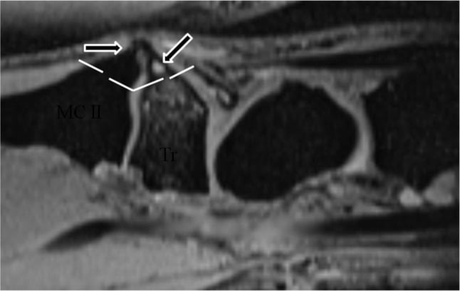
- The CARPAL BOSS (carpe bossu) is a symptomatic BONY PROMINENCE on the DORSUM of the wrist at the SECOND and/or THIRD CARPOMETACARPAL (CMC) joints - at the dorsal base of the index/middle metacarpal and the adjacent trapezoid and capitate - and represents a LOCALISED OSTEOARTHRITIS/osteophyte (exostosis) of these joints; an accessory ossicle (os styloideum) may be associated.
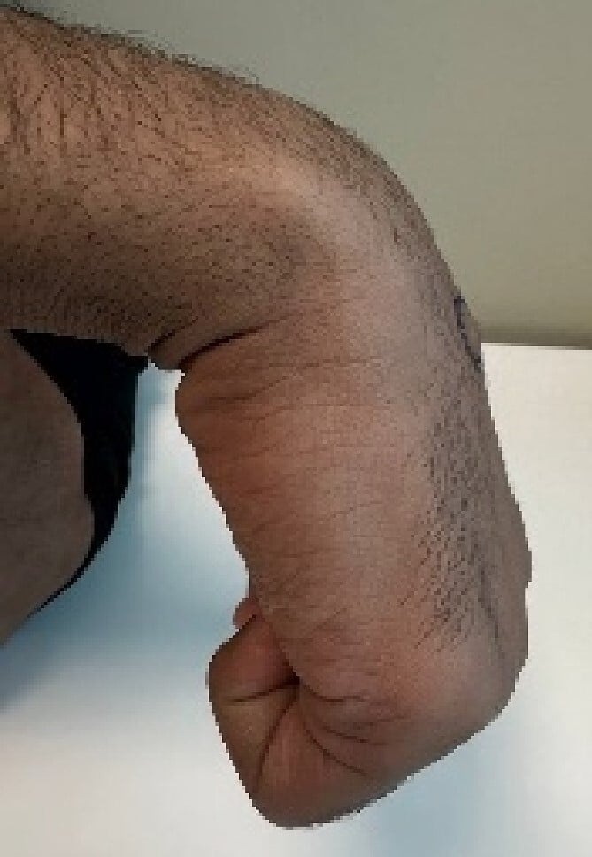
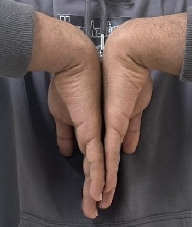
- It presents as a FIXED, HARD, non-mobile lump that is tender and may ache with activity or wrist extension; it becomes more prominent with the wrist FLEXED, and an overlying ganglion or extensor tendon irritation can coexist - it is most common in young to middle-aged adults.
- The KEY DIFFERENTIAL, and a classic exam point, is the DORSAL WRIST GANGLION: a ganglion is SOFT, MOBILE, often TRANSILLUMINATES, changes in size, and sits more proximally over the scapholunate area, whereas the carpal boss is HARD, FIXED and BONY at the CMC bases - palpation and the absence of transillumination usually separate them clinically.
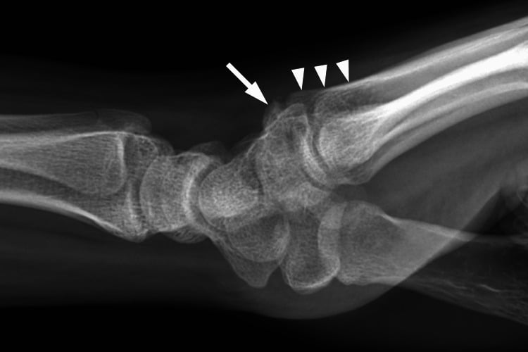
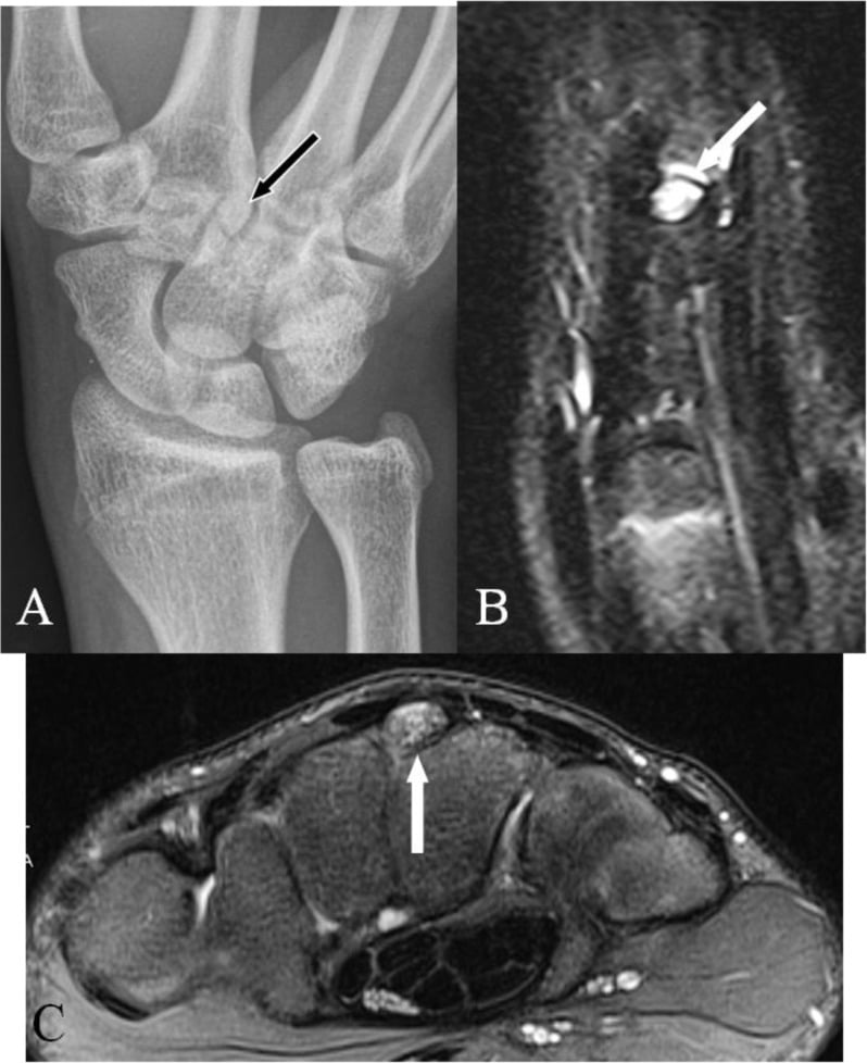
- DIAGNOSIS is clinical and confirmed on imaging: a dedicated 'CARPAL BOSS VIEW' (a lateral with the wrist about 30-40 degrees supinated and ulnar-deviated) profiles the dorsal CMC osteophyte; CT best demonstrates the bony prominence and the degenerative CMC joints when the diagnosis or surgical planning needs it.
- MANAGEMENT is NON-OPERATIVE first - reassurance, activity modification, a wrist splint, non-steroidal anti-inflammatories and a corticosteroid injection; the boss is benign, and the main reason to treat is persistent pain rather than the lump itself.
- SURGERY for refractory symptoms is SIMPLE EXCISION of the bony exostosis (with any associated ganglion), which gave no recurrences in 25 patients followed a mean of 8 years - a single-centre series, so read it as encouraging rather than as an established rate. The main pitfall is EXCESSIVE bone resection that destabilises the CMC joint; ARTHRODESIS (CMC fusion) is reserved for the rare secondary CMC INSTABILITY rather than as a primary procedure. Most bosses are ASYMPTOMATIC, so the indication is pain, never the lump.
- “Carpal boss = HARD, FIXED, BONY dorsal lump at the 2nd/3rd CMC bases (localised OA/osteophyte, +/- os styloideum). Contrast with a dorsal ganglion (SOFT, MOBILE, transilluminates, more proximal/scapholunate).
- “Confirm with the 'carpal boss view' (30-40 deg supinated, ulnar-deviated lateral) or CT - profiles the dorsal CMC osteophyte.
- “Most bosses are ASYMPTOMATIC - the indication is pain, not the lump. Non-operative first (splint/NSAIDs/injection aimed at the 3rd CMC joint under image guidance). Refractory → SIMPLE EXCISION (no recurrences in 25 patients at mean 8 years); avoid over-resection; fusion only for secondary CMC instability.
Definition and Anatomy
What it is. A carpal boss (carpe bossu) is a symptomatic bony prominence on the dorsum of the wrist at the second and/or third carpometacarpal (CMC) joints. It lies at the dorsal base of the index and middle metacarpals, where they meet the trapezoid and capitate. The prominence is an osteophyte, or exostosis, representing localised osteoarthritis of these joints, and an accessory ossicle, the os styloideum, may be associated. It is most common in young to middle-aged adults.
The extensor insertions. Extensor carpi radialis longus inserts on the base of the second metacarpal and extensor carpi radialis brevis on the base of the third, immediately over the boss. The prominence can irritate or catch these tendons, and an overlying ganglion may form, so a boss can present with dorsal tendon symptoms as well as a lump. Wrist flexion tightens the extensors over the ridge, which makes the boss more prominent and often more symptomatic.
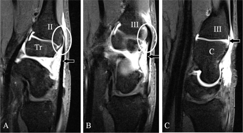
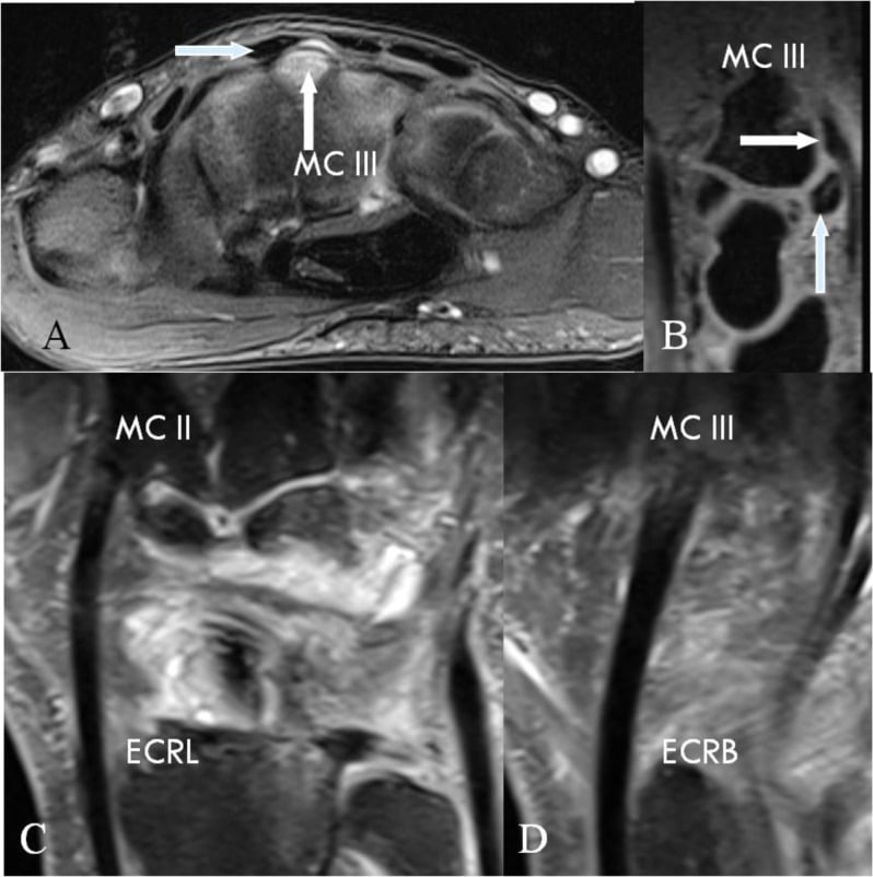
Pathogenesis
Dysplasia comes first. The primary abnormality is thought to be a developmental dysplasia at the second and third CMC bases: a dorsal ridge or prominence, or an os styloideum. It is not a generalised osteoarthritis.
Impingement makes it arthritic. During wrist and hand movement the dysplastic dorsal ridge impinges, and the resulting localised mechanical stress produces secondary osteoarthritis and osteophyte confined to the area of impingement. That is why the arthritis is localised rather than global, and why simple resection of the prominence works without treating a whole arthritic joint. Symptomatic cases occur more often in the dominant hand, which fits the impingement mechanism.
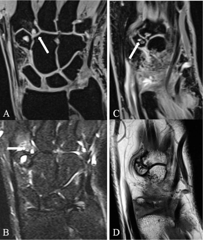
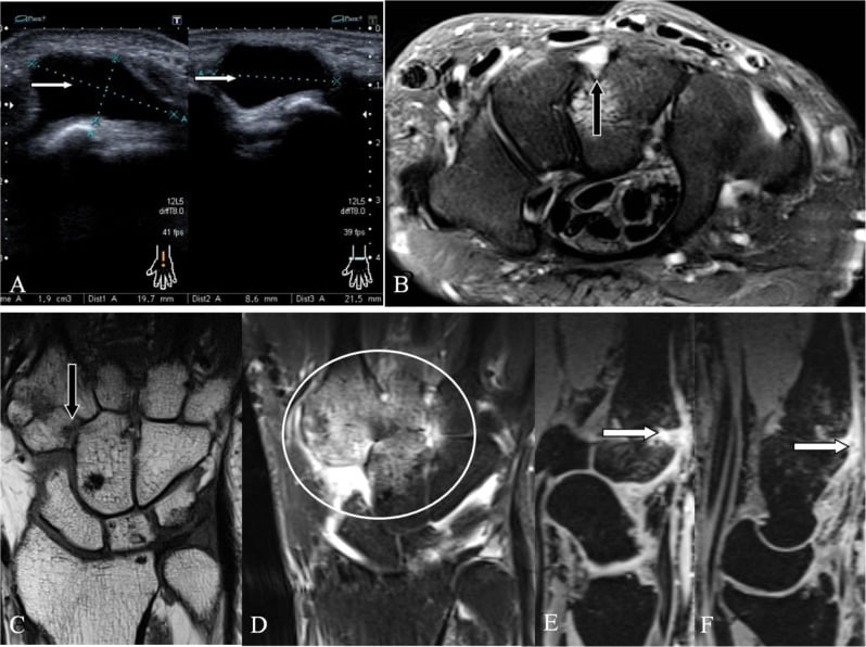
The os styloideum. An accessory ossicle at the dorsal base of the second and third metacarpals, where they meet the trapezoid and capitate: the exact site of the boss. It corresponds to the "styloid process of the third metacarpal" that projects dorsally, and is thought to arise from a separate, unfused ossification centre of the metacarpal base or from a post-traumatic ossicle. It is described as an anatomical variant that is normally asymptomatic and therefore often unrecognised.
How it becomes a boss. In many patients the os styloideum fuses to the metacarpal and carpal bases and, together with the associated dysplasia and secondary osteoarthritis, forms the bony prominence. In others it remains a discrete ossicle.
Clinical Presentation
Most are silent. A dorsal carpometacarpal prominence is usually symptom-free. A boss found on a radiograph taken for another reason is not a diagnosis of anything, and the argument for operating rests entirely on symptoms rather than on the size of the lump.
The symptomatic boss. It presents as a fixed, hard, non-mobile lump that is tender and may ache with activity or wrist extension. Pain is spontaneous or provoked by overuse and is aggravated by palmar flexion.
Subluxation of the extensor tendons overlying the boss is specifically described and is a different problem from tendons irritated as they pass over the prominence. A tendon that snaps across a bony ridge produces a painful catching sensation on wrist movement rather than simple tenderness over a lump, and it will not settle with a splint alone. Ask about catching or snapping, and examine the tendons through active wrist flexion and extension rather than only palpating the boss at rest.
The key differential. A dorsal wrist ganglion is the classic contrast. Palpation and the absence of transillumination usually separate the two clinically.
- On examination
- Hard, fixed, bony; more prominent in wrist flexion; does not transilluminate
- Site and nature
- 2nd/3rd CMC bases; a localised osteophyte/OA, +/- os styloideum
- On examination
- Soft, mobile, often transilluminates, fluctuates in size
- Site and nature
- More proximal, over the scapholunate region; a cyst, not bone
Investigations
The carpal boss view. The diagnosis is clinical and confirmed on imaging. The dedicated carpal boss view is a lateral with the wrist about 30-40 degrees supinated and ulnar-deviated, which profiles the dorsal CMC osteophyte.
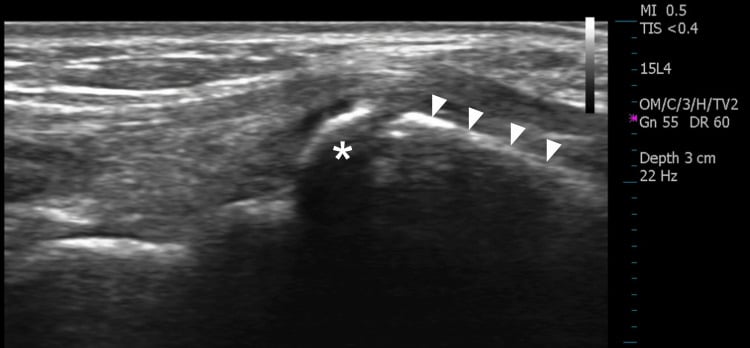
Os styloideum or fracture. On a radiograph, the carpal boss view included, a discrete os styloideum has smooth, corticated margins. An acute avulsion fracture of the metacarpal base has sharp, non-corticated edges and a matching donor site.
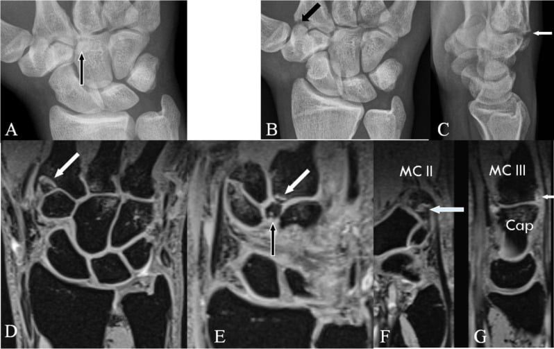
CT best demonstrates the bony prominence and the degenerative CMC joints when the diagnosis or surgical planning needs it.
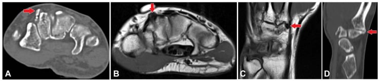
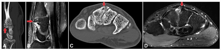
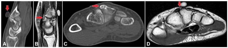
Ultrasound can demonstrate the ossicle as well as radiographs, and it can guide an injection in the same sitting.
Management
Non-operative first. Reassurance, activity modification, a wrist splint, non-steroidal anti-inflammatories and a corticosteroid injection. Most bosses are asymptomatic to begin with and the lump itself is harmless, so the main reason to treat is persistent pain rather than the lump. No cohort quantifying how many symptomatic bosses settle with conservative treatment, and no validated threshold for offering surgery, was retrieved.
Aim the injection. Inject the third carpometacarpal joint under image guidance, not the lump. Infiltration of that joint has been reported to resolve the pain of a symptomatic os styloideum completely, and early recognition of the entity is what allows conservative treatment instead of excision.
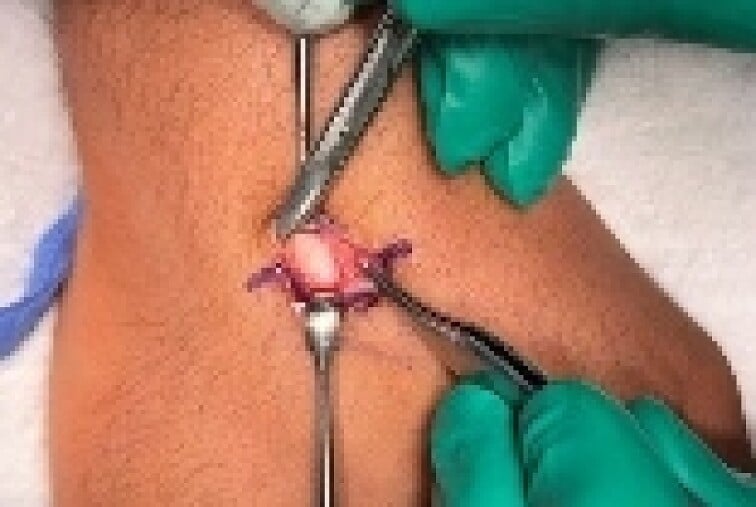
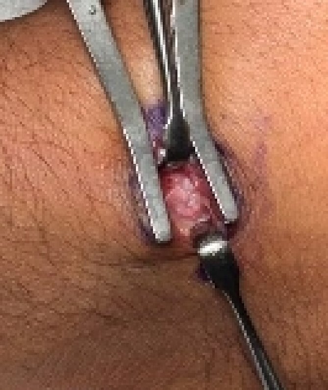
Simple excision for refractory pain. The indication is pain, never the lump. Before operating, confirm that the lump is bone and not a ganglion. The operation is simple excision of the bony exostosis, with any associated ganglion, and it gave no recurrences in 25 patients followed a mean of 8 years. That is a single-centre series, so read it as encouraging rather than as an established rate.
Over-resection is the main surgical pitfall. Taking excessive bone can destabilise the second/third carpometacarpal joint, and the resulting instability then requires a fusion, the very outcome to be avoided. Remove the prominence and preserve CMC stability; arthrodesis is reserved for the rare secondary CMC instability and is not a primary procedure.
Mnemonics & Memory Aids
BOSS
Hook:BOSS: Bony fixed CMC lump, Os styloideum, Separate from a ganglion, Simple excision.
Clinical Decision Scenarios
Practise clinical reasoning and management decisions out loud
“A young adult has a hard, fixed lump on the back of the wrist near the bases of the index and middle metacarpals. How do you assess and manage it?”
What it is
- Symptomatic bony prominence at the 2nd/3rd CMC joints (dorsal base of index/middle MC)
- Localised osteoarthritis/osteophyte (exostosis); may have an os styloideum
- Fixed, hard, non-mobile; more prominent in wrist flexion
Diagnosis
- Differentiate from a dorsal wrist ganglion (soft, mobile, transilluminates, more proximal)
- Carpal boss view (30-40 deg supinated, ulnar-deviated lateral)
- CT for bony detail / surgical planning
Management
- Non-operative first: splint, NSAIDs, corticosteroid injection
- Most are asymptomatic - operate for pain, not for the lump; ask about tendon catching (extensor subluxation)
- Refractory pain: simple excision of the exostosis (+ ganglion) - no recurrences in 25 patients at mean 8 years
- Avoid over-resection (CMC instability); fusion only for secondary instability
Evidence & Key Studies
Surgical treatment of carpal boss by simple resection - results at a mean of 8 years
- Carpal boss is a symptomatic bony protrusion on the dorsal surface of the wrist at the base of the 2nd and/or 3rd metacarpal; in all cases features of dysplasia were present, with secondary osteoarthritis limited to the area of impingement.
- 29 cases were treated between 1994 and 2014; 25 were assessed by TELEPHONE QUESTIONNAIRE at a mean of 8 years (range 1.1 to 20 years). There were no recurrences.
- The results are good but not silent: 15 patients (60%) considered themselves cured and 9 (36%) improved - but 8 of the 24 without fusion (33%) still reported moderate episodic pain (mean VAS 2.3, range 1 to 4).
- Range of motion improved in 8 (33%), was unchanged in 11 (46%) and DECREASED in 5 (21%). Twenty (83%) had no functional impairment; 4 were impaired during unusual hand movements.
- The single failure - carpometacarpal instability requiring fusion 5 years later - was attributed to excessive bone resection, so simple resection is sufficient and fusion should be reserved for that rare secondary instability.
The definition of the carpal boss (a symptomatic dorsal bony protrusion at the base of the 2nd/3rd metacarpal with localised secondary osteoarthritis), the reliable results of simple resection with no recurrence at a mean of 8 years, and the lesson that excessive resection can cause carpometacarpal instability (with fusion reserved for that rare event) come from the cited Roulet series. The differential with a dorsal wrist ganglion, the association with an os styloideum, and the dedicated carpal boss radiographic view are standard, well-established teaching. The Roulet series is 25 patients from one centre, so "no recurrence" means none in 25 at a mean of 8 years rather than a rate established across the literature, and its single failure is one patient. That the condition is usually symptom-free, that pain is aggravated by palmar flexion and that extensor tendon subluxation over the boss occurs come from Urban (Chir Narzadow Ruchu Ortop Pol 1996, PMID 8974848), a narrative account in Polish; the dominant-hand predilection, the resolution after fluoroscopically guided third-carpometacarpal corticosteroid infiltration and the utility of ultrasound come from Kaniewska and colleagues (J Radiol Case Rep 2017, PMID 29299098), a single case with literature review - so neither gives a proportion. No cohort quantifying how many symptomatic bosses settle with conservative treatment, and no validated threshold for offering surgery, was retrieved. See also ganglion cysts and thumb carpometacarpal arthritis; the library has no separate dorsal-wrist-ganglion or non-thumb carpometacarpal arthritis topic.