High Arch | Hindfoot Varus | CMT Most Common | Coleman Block Test Essential
- Coleman block test differentiates flexible vs fixed hindfoot varus - critical for surgical planning
- CMT (Charcot-Marie-Tooth) accounts for 66% of adult cavovarus - rule out neurological causes first
- Plantar-flexed 1st ray is the primary driver - forefoot pronates to get heel to ground, creating hindfoot varus
- Staged reconstruction: soft tissue balancing first, then bony correction if needed
- Posterior tibial tendon transfer for dynamic correction when peroneus brevis is weak
- “Viva question: Walk me through the Coleman block test - what does it tell you?
- “Distinguish idiopathic from neurological causes - CMT, spinal dysraphism, polio
- “Sequential surgery: plantar fascia release → 1st MT osteotomy → calcaneal osteotomy → midfoot osteotomy if needed
- “Complications: overcorrection to valgus, peroneal nerve injury, nonunion after calcaneal osteotomy
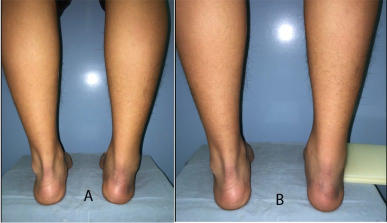
Patient stands on 1-inch block under lateral foot (1st and 2nd rays off edge). If hindfoot corrects to neutral or valgus = flexible (driven by forefoot). If hindfoot stays varus = fixed (needs calcaneal osteotomy). This single test dictates your surgical plan.
66% of adult cavovarus is Charcot-Marie-Tooth disease. Peroneal nerve dysfunction causes weak peroneals and overactive posterior tibialis. Always examine for wasting in anterior and lateral compartments, check family history, and consider nerve conduction studies.
Tripod effect: Plantar-flexed 1st metatarsal forces forefoot into pronation to get heel to ground. This creates functional hindfoot varus. Correcting the 1st ray often corrects the hindfoot without calcaneal osteotomy if flexible.
Stage 1: Plantar fascia release, 1st MT dorsiflexion osteotomy, peroneus longus to brevis transfer. Stage 2: Calcaneal lateralizing osteotomy if hindfoot remains varus. Stage 3: Midfoot osteotomy for severe cavus. Never correct everything at once - high complication rate.
- Coleman Block Result
- N/A - observation
- Primary Procedure
- Custom orthotics with lateral wedge
- Key Pearl
- Many patients never need surgery
- Coleman Block Result
- Hindfoot corrects to neutral on block
- Primary Procedure
- Plantar fascia release + 1st MT osteotomy
- Key Pearl
- Forefoot-driven deformity - fix the 1st ray
- Coleman Block Result
- Hindfoot stays varus on block
- Primary Procedure
- Add calcaneal lateralizing osteotomy
- Key Pearl
- Fixed deformity needs bone realignment
- Coleman Block Result
- Fixed hindfoot, weak eversion
- Primary Procedure
- Staged: soft tissue first, then calcaneal osteotomy + tendon transfers
- Key Pearl
- CMT pattern - transfer posterior tibial to dorsum or peroneals
CAVECavovarus Deformity Components - CAVE
Hook:CAVE: The patient's foot is stuck in a CAVE - deep arch, toes clawed, heel turned in!
LIFTColeman Block Test Interpretation - LIFT
Hook:LIFT the 1st ray off the block - does the hindfoot LIFT out of varus? If yes = flexible!
Overview and Epidemiology
Cavovarus foot is a complex three-dimensional deformity characterized by high medial longitudinal arch (cavus), hindfoot varus, and forefoot adduction. The most common cause in adults is Charcot-Marie-Tooth disease, a hereditary motor and sensory neuropathy. The deformity is progressive and leads to lateral ankle instability, peroneal tendinopathy, metatarsalgia, and stress fractures. Early recognition and staging prevents severe fixed deformity requiring triple arthrodesis.
- Neurological (75%): CMT, spinal dysraphism, polio residual, cerebral palsy
- Idiopathic (20%): No underlying cause identified
- Traumatic (5%): Compartment syndrome, malunion, neuroma
- CMT Type 1A: Most common subtype - autosomal dominant, PMP22 gene duplication
- Progressive deformity: Worsens over years as muscle imbalance continues
- Lateral ankle instability: Recurrent sprains from varus heel strike
- Metatarsalgia: Plantar-flexed 1st ray and claw toes concentrate pressure
- Arthritis: Midfoot and ankle joint degeneration by 5th-6th decade
Pathophysiology and Biomechanics
Muscle Imbalance Theory (CMT)
Peroneal nerve dysfunction in CMT causes selective weakness of peroneus brevis (eversion) and tibialis anterior (dorsiflexion). The posterior tibialis (tibial nerve innervated) remains strong and overpowers the weak peroneals, creating hindfoot varus. Intrinsic foot muscle weakness leads to claw toe deformity and elevated arch. This is a dynamic, progressive process.
- Innervation
- Superficial peroneal nerve
- CMT Status
- WEAK - overpowered
- Resultant Deformity
- Hindfoot varus (unopposed PT)
- Innervation
- Tibial nerve
- CMT Status
- STRONG - dominant
- Resultant Deformity
- Pulls hindfoot into varus
- Innervation
- Deep peroneal nerve
- CMT Status
- WEAK - drop foot risk
- Resultant Deformity
- Forefoot equinus, steppage gait
- Innervation
- Superficial peroneal nerve
- CMT Status
- WEAK but still fires
- Resultant Deformity
- Plantar-flexes 1st MT, worsens cavus
- Innervation
- Tibial and peroneal nerves
- CMT Status
- WEAK - atrophy
- Resultant Deformity
- Claw toes, loss of arch control
Tripod Effect and Forefoot-Driven Hindfoot Varus
Weak peroneals cannot resist peroneus longus pull. The 1st metatarsal plantar-flexes. When the patient tries to stand flat, the tripod of 1st MT head, 5th MT head, and heel creates a problem: the 1st MT is too low. The forefoot pronates (medial arch drops) to get the 1st MT to the ground. This forefoot pronation creates functional hindfoot varus.
By placing a block under the lateral foot, you allow the plantar-flexed 1st and 2nd rays to hang free. If the hindfoot corrects to neutral, it proves the varus was forefoot-driven and flexible. You only need to fix the 1st ray (dorsiflexion osteotomy). If the hindfoot stays in varus, there is a fixed component requiring calcaneal osteotomy.
The Coleman block test is the single most important clinical exam for cavovarus foot. It differentiates flexible (forefoot-driven) from fixed (structural) hindfoot varus. Flexible deformity corrects with soft tissue procedures and 1st MT osteotomy. Fixed deformity requires calcaneal lateralizing osteotomy. Getting this wrong leads to undercorrection (persistent varus) or overcorrection (iatrogenic valgus).
Classification Systems
Anatomical Classification by Location
- Clinical Findings
- Plantar-flexed 1st MT, claw toes
- Surgical Target
- Correct 1st ray position
- Procedure Options
- Dorsiflexion osteotomy, plantar fascia release
- Clinical Findings
- Heel inverted, lateral ankle instability
- Surgical Target
- Realign calcaneus under tibia
- Procedure Options
- Calcaneal lateralizing osteotomy, tendon transfer
- Clinical Findings
- Elevated longitudinal arch
- Surgical Target
- Reduce arch height
- Procedure Options
- Cole midfoot osteotomy if severe
- Clinical Findings
- Tight Achilles, limited dorsiflexion
- Surgical Target
- Lengthen posterior structures
- Procedure Options
- Gastrocnemius recession or TAL
In most cases, the plantar-flexed 1st ray is the primary driver. The hindfoot varus is secondary (functional) to the forefoot pronation. Correcting the 1st ray often resolves the hindfoot varus if flexible on Coleman block. Only fixed hindfoot varus requires calcaneal osteotomy.
Clinical Assessment
History
- Family history: CMT is autosomal dominant - parents, siblings affected?
- Onset and progression: Present since childhood or recent? Worsening?
- Ankle sprains: Recurrent lateral ankle instability very common
- Pain location: Lateral foot (peroneal tendinopathy), plantar forefoot (metatarsalgia), ankle arthritis
- Functional limitations: Difficulty with uneven ground, running, balance
- Numbness: Stocking-glove pattern in CMT (sensory neuropathy)
- Weakness: Anterior compartment weakness, foot drop, steppage gait
- Hand symptoms: CMT also affects hands - ask about fine motor difficulty
- Balance problems: Proprioception loss increases fall risk
- Footwear difficulty: Cannot fit standard shoes due to high arch
Physical Examination
Systematic Examination Sequence
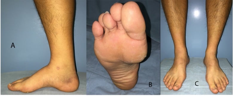
Alignment: Observe from behind for hindfoot varus, medial arch height, forefoot adduction. Coleman block test: Patient stands on 1-inch block under lateral foot - does hindfoot correct? Peek-a-boo heel sign: Can you see the toes peeking from behind medial side? (severe varus) Gait: Lateral foot strike, ankle instability, steppage gait if TA weak
Flexibility testing:
- Passively correct hindfoot varus - does it fully correct to neutral or beyond?
- Assess forefoot flexibility - can you dorsiflex 1st MT to neutral?
- Check ankle dorsiflexion with knee extended and flexed (Silverskiöld test for gastrocnemius tightness)
Muscle strength testing (grade 0-5):
- Peroneus brevis (eversion): Weak in CMT
- Posterior tibialis (inversion): Overactive in CMT
- Tibialis anterior (dorsiflexion): Weak in CMT, may have drop foot
- Gastrocnemius (plantarflexion): Usually normal
Motor: Anterior and lateral compartment weakness (peroneal nerve distribution) Sensory: Stocking-glove neuropathy in CMT Reflexes: Achilles and patellar reflexes often absent in CMT Inspection: Muscle wasting in anterior and lateral leg (stork leg), claw toes Upper extremity: Hand intrinsic wasting if CMT (ape hand deformity)
Coleman block test: Gold standard for flexibility assessment Silverskiöld test: Ankle dorsiflexion with knee straight vs bent - isolated gastrocnemius vs combined gastrocnemius-soleus tightness Ankle stability: Anterior drawer, talar tilt (chronic lateral instability common) Peek-a-boo heel sign: See toes from behind medial ankle = severe varus
Always examine the spine and perform neurological screening. Spinal dysraphism (tethered cord, diastematomyelia) can present as cavovarus foot. Look for midline skin stigmata (hairy patch, dimple, lipoma), asymmetric lower extremity, bladder dysfunction. MRI spine if any red flags. Missing this leads to progressive neurological deterioration.

Investigations
Imaging and Diagnostic Protocol
AP foot: Assess forefoot adduction, metatarsal break pattern, degenerative changes Lateral foot: Calcaneal pitch angle (normal 20 degrees, over 30 degrees = cavus), Hibbs angle (calcaneus-first metatarsal axis, reflects arch height), 1st MT plantar-flexion AP ankle: Assess ankle joint arthritis, talar tilt from chronic instability Hindfoot alignment view (Saltzman): Quantifies hindfoot varus (plumb line medial to heel = varus)
Key measurements:
- Calcaneal pitch greater than 30 degrees = cavus
- Hibbs angle (calcaneus-first metatarsal) less than 150 degrees = elevated arch (cavus)
- Talar-1st MT angle (Meary line) apex plantar = cavus
Assess peroneal tendons: Split tears, subluxation common with varus hindfoot Lateral ligament complex: Chronic ATFL/CFL injury from recurrent sprains Articular cartilage: Ankle joint degenerative changes Soft tissue balance: Plantar fascia, posterior tibial tendon quality
Nerve conduction studies: Reduced motor and sensory conduction velocities in CMT Type 1 (demyelinating) Electromyography: Denervation pattern in affected muscles Genetic testing: PMP22 duplication for CMT1A (70% of CMT cases) MRI spine: If any suspicion of spinal dysraphism or tethered cord
Pedobarography (pressure mat): Demonstrates lateral column overload, forefoot pressure concentration under 1st MT Gait analysis: Quantifies ankle instability, varus thrust, lateral foot contact pattern

Differential Diagnosis
- Key Distinguishing Features
- Bilateral, family history, peroneal-pattern wasting (stork leg), absent reflexes, hand involvement
- Decisive Test
- Nerve conduction studies + PMP22 genetic testing
- Key Distinguishing Features
- Unilateral or asymmetric, midline skin stigmata, bladder/bowel symptoms, rapid progression
- Decisive Test
- MRI of whole spine
- Key Distinguishing Features
- Mild, often unilateral, normal neurological exam, non-progressive
- Decisive Test
- Diagnosis of exclusion after full neurological work-up
- Key Distinguishing Features
- History of congenital clubfoot/treatment, midfoot cavus, internal rotation
- Decisive Test
- History + standing radiographs
- Key Distinguishing Features
- Unilateral, prior leg trauma or ischaemia, fixed contracture, normal contralateral foot
- Decisive Test
- History + MRI showing muscle fibrosis
- Key Distinguishing Features
- Recurrent sprains with mild hindfoot varus, no neuropathy
- Decisive Test
- Hindfoot alignment (Saltzman) view + stress radiographs
The first fork is neurological versus non-neurological. A bilateral, symmetric, progressive cavovarus with a family history is CMT until proven otherwise. A unilateral or rapidly progressive cavovarus mandates spinal imaging to exclude a tethered cord or intraspinal lesion before any foot surgery - missing this leads to relentless recurrence.
Non-Operative Management
- Mild deformity with minimal symptoms
- Patient unfit for surgery (medical comorbidities)
- Stable neurological condition (non-progressive)
- Patient refuses surgery or wants to delay
- Elderly low-demand patients with acceptable function
- Custom foot orthoses with lateral heel wedge (shifts weight medial, unloads lateral column)
- High-backed shoes or ankle braces for lateral ankle instability
- Accommodative padding for metatarsalgia (plantar MT pads)
- Rocker-bottom soles to reduce forefoot pressure
- AFO (ankle-foot orthosis) for severe drop foot in CMT
Non-Operative Protocols
Pain management: NSAIDs, activity modification, ice for acute exacerbations Orthotic fitting: Custom orthoses with lateral wedge, accommodative padding Footwear modification: Extra-depth shoes, wide toe box, rigid shank
Peroneal strengthening: Resistance band eversion exercises (won't reverse CMT but maintains function) Intrinsic foot strengthening: Towel curls, marble pickup to slow claw toe progression Ankle proprioception: Single-leg balance, wobble board
Annual reassessment: X-rays to monitor progression, clinical exam for worsening varus Orthotic adjustment: Replace or modify orthoses as deformity progresses Surgical timing: Offer surgery if deformity worsens, pain increases, or function declines significantly
Indications to proceed to surgery: Progressive deformity despite orthoses, recurrent ankle sprains (greater than 2 per year), severe metatarsalgia affecting walking, lateral foot pain from peroneal overload, ankle arthritis developing, patient unable to fit into shoes. Non-operative management is temporizing, not curative.
Management Algorithm
Forefoot-Driven Cavovarus (Hindfoot Corrects on Block)
Goal: Correct the plantar-flexed 1st ray to eliminate forefoot pronation and functional hindfoot varus.
Surgical Sequence for Flexible Cavovarus
Technique: Plantar medial or plantar lateral approach, release plantar fascia from calcaneus, release intrinsic origin. Effect: Drops the arch, reduces plantar 1st MT force. Caution: Do not over-release - can create flatfoot.
Dorsal closing wedge osteotomy at base of 1st MT (preferred) or Cotton osteotomy (opening wedge medial cuneiform). Fixation: Plate and screws or staples. Goal: Elevate 1st MT to neutral, eliminate tripod effect.
Rationale: Peroneus longus plantar-flexes 1st MT - removing this deforming force and augmenting brevis improves eversion strength. Technique: Divide peroneus longus tendon distally, weave into peroneus brevis. Benefit: Augments weak brevis (CMT pattern).
Flexor to extensor transfer (Girdlestone-Taylor): FDL or FHL transferred to dorsal hood to extend toes. IP joint fusion: If severe fixed clawing, fuse PIP or DIP joints.
Silverskiöld test positive: Ankle dorsiflexion less than 5 degrees with knee extended but improves with knee flexed = isolated gastrocnemius tightness. Strayer procedure: Recede gastrocnemius at musculotendinous junction.
By correcting the plantar-flexed 1st MT (Step 2), you eliminate the forefoot pronation that was creating functional hindfoot varus. The hindfoot spontaneously corrects to neutral without needing calcaneal osteotomy. This is proven by Coleman block test preoperatively. Soft tissue balancing alone is sufficient for flexible deformities.
Surgical Technique: Key Procedures
Pre-Operative Planning
- Infection: 2-3% superficial, 1% deep
- Nerve injury: Sural nerve (lateral approach), medial plantar nerve (plantar fascia release)
- Nonunion: 5-10% for calcaneal osteotomy, 10-15% for midfoot osteotomy
- Overcorrection to valgus: Difficult to salvage, may need revision
- Recurrence: 20-30% if underlying cause (CMT) is progressive
- Need for staged procedures: May require 2-3 surgeries for severe deformity
- Osteotomy instruments: Oscillating saw, osteotomes
- Fixation: Small fragment plates and screws for 1st MT, cannulated screws for calcaneus
- Tendon transfer instruments: Tendon passer, whip stitch sutures (FiberWire)
- Imaging: C-arm for intraoperative fluoroscopy (essential for bone cuts)
- K-wires: Temporary fixation during correction
Plantar Fascia Release Technique
Approach: Plantar medial (Steindler stripping) or plantar lateral (endoscopic).
Plantar Medial Approach (Steindler)
Position: Supine with bump under ipsilateral hip, foot externally rotated. Incision: 3-4 cm longitudinal incision over plantar medial foot, centered over medial calcaneal tuberosity. Landmarks: Palpate calcaneal tuberosity, stay medial to avoid lateral plantar nerve.
Identify plantar fascia (thick white band) and intrinsic muscle origins (abductor hallucis, flexor digitorum brevis). Release plantar fascia from calcaneal origin with sharp dissection. Release intrinsic muscle origins (Steindler stripping) to drop arch maximally. Caution: Stay on bone to avoid medial plantar nerve (runs deep to abductor hallucis).
Irrigate wound, close deep fascia with absorbable suture, skin with nylon. Compression dressing to prevent hematoma.
The medial plantar nerve runs deep to abductor hallucis muscle and is at risk during plantar medial approach. Stay on bone during release. Nerve injury causes permanent plantar numbness and painful neuroma. Test sensation postoperatively before discharge.
Plantar fascia release is the cornerstone of cavovarus soft tissue correction. It drops the arch and reduces tension on the plantar foot structures.
Intraoperative Troubleshooting
- Cause
- Fixed component underestimated, Coleman block misinterpreted
- Solution
- Add calcaneal lateralizing osteotomy - stage if necessary
- Cause
- Excessive lateral translation (greater than 10mm)
- Solution
- Remove screws, reduce translation to 8mm, re-fix with fluoroscopy confirmation
- Cause
- Inadequate fixation, osteoporotic bone
- Solution
- Add dorsal plate (not just screws), consider bone graft or substitute
- Cause
- Soft tissue tethering, incomplete osteotomy
- Solution
- Complete osteotomy with osteotome plantarly, release periosteum circumferentially
SOFT to BONEStaged Surgical Sequence - SOFT BONE
Hook:Fix SOFT tissues first, then BONE - never rush to cut bone when soft tissue release may suffice!
The Jones Procedure (Clawed Hallux Correction)
The Ward EvidenceCard's algorithm explicitly includes "transfer of the extensor hallucis longus to the neck of the first metatarsal", and the surgical section mentions "EHL/FHL transfers for clawing", but the Jones procedure and its cavovarus rationale are never described.
- The problem it solves. In cavovarus (especially CMT), a weak tibialis anterior recruits the extensor hallucis longus (EHL) to help dorsiflex the ankle. This over-recruited EHL cocks up the hallux (hyperextended MTP, flexed IP = a clawed great toe) and, because its distal pull acts on the toe rather than the metatarsal, indirectly depresses (plantarflexes) the first metatarsal, worsening the cavus.
- The operation (Robert Jones, 1916). The EHL is detached distally, transferred and fixed into the neck of the first metatarsal, and the hallux interphalangeal (IP) joint is fused (or tenodesed). This achieves three things at once: it removes the deforming plantarflexion force on the first ray; it converts the EHL into a first-metatarsal dorsiflexor (augmenting correction of the plantarflexed first ray); and it straightens the clawed hallux (the IP fusion prevents recurrent clawing).
- Where it fits. It is a soft-tissue adjunct performed with the plantar fascia release and first metatarsal osteotomy during the forefoot-correction stage - the hallux equivalent of the Girdlestone-Taylor flexor-to-extensor transfer used for the clawed lesser toes.
Q: What is the Jones procedure and why is it used in cavovarus foot? A: Transfer of the extensor hallucis longus to the neck of the first metatarsal, with fusion (or tenodesis) of the hallux IP joint. In cavovarus a weak tibialis anterior recruits the EHL to dorsiflex the ankle, which cocks up the hallux and depresses the first metatarsal. The Jones procedure removes that first-ray-plantarflexing force, turns the EHL into a first-metatarsal dorsiflexor (helping correct the plantarflexed first ray), and straightens the clawed great toe.
Radiographic Angles of the Cavus Foot
The topic names the calcaneal pitch, Meary's angle and Hibbs angle, but never sets out how each is measured or its cut-off in one place.
- Calcaneal pitch (calcaneal inclination angle). On the weight-bearing lateral, the angle between the plantar surface of the calcaneus and the supporting floor. Normal roughly 20-30 degrees; a pitch over about 30 degrees indicates a calcaneocavus (hindfoot-driven) high arch.
- Meary's angle (talo-first-metatarsal angle). On the lateral, the angle between the long axis of the talus and the long axis of the first metatarsal - normally colinear (about 0 degrees). In cavus the line breaks with the apex pointing dorsally, and the level of that apex (forefoot, midfoot, or a smooth curve) localises the deformity.
- Hibbs angle (calcaneal-first metatarsal angle). On the lateral, the angle between the long axis of the calcaneus and the long axis of the first metatarsal. Normal is roughly 150-175 degrees; a Hibbs angle under about 150 degrees reflects an elevated arch (cavus) - the more acute the angle, the higher the arch.
- Hindfoot alignment (Saltzman) view. A weight-bearing coronal view that quantifies the varus/valgus position of the calcaneus under the tibia - essential for planning and for confirming neutral alignment intraoperatively after a calcaneal osteotomy.
Q: Which radiographic angles quantify a cavus foot and what are their cut-offs? A: On the weight-bearing lateral: calcaneal pitch (calcaneus to floor; over about 30 degrees = calcaneocavus), Meary's angle (talus-first metatarsal; normally about 0 degrees, apex-dorsal in cavus and localising the deformity level) and the Hibbs angle (calcaneus-first metatarsal; normally about 150-175 degrees, under about 150 degrees = cavus). The Saltzman hindfoot-alignment view quantifies hindfoot varus and confirms neutral alignment after a calcaneal osteotomy.
Complications
- Incidence
- 5-10%
- Risk Factors
- Excessive calcaneal lateralization, weak PT tendon
- Management
- Difficult to salvage - may need medializing osteotomy, PT augmentation, or arthrodesis
- Incidence
- 10-15%
- Risk Factors
- Inadequate correction, fixed component underestimated
- Management
- Revision calcaneal osteotomy, add tendon transfer, consider triple arthrodesis
- Incidence
- 5-10% (calcaneal), 10-15% (midfoot)
- Risk Factors
- Smoking, osteoporosis, inadequate fixation
- Management
- ORIF with bone graft, revision fixation, consider bone stimulator
- Incidence
- 5-10%
- Risk Factors
- Lateral calcaneal approach, nerve not identified
- Management
- Numbness permanent, neuroma may require excision and nerve burial
- Incidence
- 5-8%
- Risk Factors
- Lateral foot surgery, thin skin, smoking
- Management
- Local wound care, VAC therapy if deep, flap coverage if severe
- Incidence
- 20-30% (CMT patients)
- Risk Factors
- Progressive neurological disease (CMT), inadequate initial correction
- Management
- Revision surgery, counsel about progression with CMT, may need multiple revisions over lifetime
Overcorrecting hindfoot to valgus is the most difficult complication to salvage. Patients develop medial ankle pain, medial column overload, posterior tibial tendon dysfunction, and progressive planovalgus deformity. Prevention: Use intraoperative fluoroscopy (Saltzman view) to confirm neutral or slight valgus alignment (not excessive valgus). Limit calcaneal translation to 8-10mm maximum. If in doubt, undercorrect slightly - easier to revise for varus than valgus.
Postoperative Care and Rehabilitation
Rehabilitation After Plantar Fascia Release and 1st MT Osteotomy
Recovery Timeline
Immobilization: Posterior splint or CAM boot, non-weight-bearing. Elevation: Leg elevated above heart to reduce swelling. DVT prophylaxis: Aspirin or LMWH if high risk. Wound check: Remove dressing at 2 weeks, assess for infection.
Weight-bearing: Progress to weight-bearing as tolerated in CAM boot. X-rays at 6 weeks: Check 1st MT osteotomy healing, alignment maintained. ROM exercises: Ankle pumps, toe curls (gentle). Precautions: No running, jumping, pivoting.
Transition to shoes: Wean from boot to supportive shoes with custom orthoses. Strengthening: Peroneal strengthening, intrinsic foot exercises, proprioception training. Return to low-impact activities: Walking, swimming, cycling.
X-rays at 3 months: Confirm union of osteotomy. Return to sports: Gradual return to running, jumping sports. Long-term orthoses: Continue custom orthoses indefinitely (especially CMT patients).
Full recovery from forefoot procedures takes 3-6 months. Patients can usually walk comfortably in shoes by 8-10 weeks.
Outcomes and Prognosis
- Success Rate
- 85-90% correction maintained at 5 years
- Patient Satisfaction
- High (greater than 80%)
- Common Residual Complaints
- Mild stiffness, continued need for orthoses
- Success Rate
- 75-85% correction at 5 years
- Patient Satisfaction
- Good (70-80%)
- Common Residual Complaints
- Lateral foot numbness (sural nerve), hindfoot stiffness
- Success Rate
- 90-95% pain relief, 100% correction
- Patient Satisfaction
- Good (75-85%)
- Common Residual Complaints
- Complete hindfoot stiffness, gait abnormality, ankle arthritis risk
- Success Rate
- 70-80% avoid arthrodesis long-term
- Patient Satisfaction
- Moderate (65-75%)
- Common Residual Complaints
- Prolonged recovery (12-18 months), multiple surgeries, recurrence risk
Predictors of Poor Outcome
- CMT Type 1A: Higher recurrence rate than idiopathic cavovarus
- Severe preoperative deformity: Calcaneal pitch over 40 degrees
- Fixed deformity: Inability to passively correct to neutral
- Age over 50: Lower activity demands but higher medical comorbidities
- Smoking: Doubles nonunion risk, triples wound complication risk
- Inadequate correction: Residual varus leads to recurrent lateral ankle instability
- Overcorrection to valgus: Difficult to salvage, creates new problems
- Single-stage correction of severe deformity: High complication rate vs staged approach
- Failure to address all components: E.g., correcting hindfoot but not 1st ray
- Nonunion of osteotomy: Requires revision, delays recovery
CMT patients need realistic expectations: Surgery delays progression and improves function but does not cure the underlying neuropathy. Recurrence occurs in 20-30% of CMT patients over 10-15 years. They may need revision surgery. Lifelong custom orthoses are essential. Despite this, most CMT patients report significant improvement in pain, ankle stability, and ability to wear shoes. Timing surgery when deformity is moderate (before severe fixed changes) gives best long-term results.
Guidelines, Registries & Global Practice
- CMT prevalence: approximately 1 in 2,500 worldwide - the commonest inherited neuromuscular disorder, and the dominant neurological cause of adult cavovarus
- CMT1A (PMP22 duplication) is the single most common genetic subtype globally
- Over half of CMT patients develop foot/ankle deformity, of which cavovarus is by far the commonest
- Idiopathic cavovarus and residual/overcorrected clubfoot account for most non-neurological cases
- Bilateral, slowly progressive presentation is typical of a hereditary cause; unilateral or rapidly progressive deformity should prompt spinal cord imaging
- High-resource settings: nerve conduction studies, genetic testing, gait/pedobarography, weight-bearing CT, and staged joint-sparing reconstruction are routine
- Limited-resource settings: diagnosis often clinical; tendon transfers and osteotomies favoured over implant-dependent techniques; custom orthoses may be unavailable
- Triple arthrodesis remains an important, durable salvage where staged reconstruction or long follow-up is not feasible
- Lifelong orthotic supply and neurology follow-up are the main access barriers for CMT patients globally
- Emphasis
- Identify neurological cause; flexibility-based algorithm
- Practical Recommendation
- Coleman block test drives surgery; joint-sparing osteotomy/tendon transfer before arthrodesis
- Emphasis
- Multidisciplinary work-up with neurology and genetics
- Practical Recommendation
- Exhaust orthotic management; stage soft tissue then bone; reserve fusion for rigid/arthritic feet
- Emphasis
- Deformity-component analysis and fixation principles
- Practical Recommendation
- Correct apex of deformity; protect sural and plantar nerves; confirm hindfoot alignment with fluoroscopy
- Emphasis
- Early intervention in flexible deformity
- Practical Recommendation
- Operate while deformity is flexible to delay fixed changes and arthritis, especially in progressive CMT
Document discussion of the following with every cavovarus reconstruction patient:
- Overcorrection to valgus - the hardest complication to salvage; must be specifically discussed
- Recurrence in CMT - radiographic recurrence of hindfoot varus is common over the long term because the underlying neuropathy is progressive; this is not a surgical failure
- Staged surgery - severe deformity often needs 2-3 procedures, planned not failed
- Nerve injury - sural nerve (lateral calcaneal approach) and medial plantar nerve (plantar fascia release), with risk of permanent numbness or neuroma
- Nonunion - higher for midfoot than calcaneal osteotomy; smoking markedly increases risk
- Prolonged recovery and impact on work - months of restricted weight-bearing after bony procedures
- CMT1A is autosomal dominant: roughly 50% transmission risk to each child - offer genetics/neurology referral for confirmed or suspected CMT and at-risk relatives
- Genetic confirmation (e.g. PMP22 duplication testing) supports diagnosis, prognostic counselling and family screening
- Lifelong multidisciplinary follow-up: neurology, orthotics/AFO provision, physiotherapy for gait and falls prevention, and orthopaedic surveillance for deformity progression
Controversies and Areas of Uncertainty
The classic teaching - plantarflexed first ray driving hindfoot varus via the tripod effect - underpins the Coleman block test. Recent dynamic pedobarography (Ferguson et al., 2024) found first-ray-first contact in only ~40% of varus CMT feet, suggesting many feet are pre-positioned in varus by progressive muscle imbalance rather than purely forefoot-driven. The block test remains useful but may oversimplify a multifactorial deformity.
No high-level trial defines the optimal hindfoot osteotomy. Lateral translation/slide and the Z (Malerba) osteotomy are now generally preferred over the classic Dwyer lateral closing wedge because they correct alignment without shortening the calcaneus or altering the Achilles moment arm. Comparative outcome data remain limited and largely retrospective.
Long-term data favour joint-sparing surgery in flexible deformity (lower arthritis/reoperation than historical fusion series), but the threshold of rigidity and arthritis at which to abandon reconstruction for triple arthrodesis is not precisely defined and remains a judgement call.
Whether to operate early in flexible CMT to delay fixed changes, and which tendon transfers best balance a progressive neuropathy, are debated. Because CMT continues to progress, no transfer fully prevents recurrence, and revision over a lifetime is common.
CAMPCharcot-Marie-Tooth Foot Deformity Pattern - CAMP
Hook:CMT patients end up at CAMP - their legs look like stork legs at summer camp!
MCQ Practice Points
Q: What is the most common cause of cavovarus foot deformity in adults? A: Charcot-Marie-Tooth disease (CMT) accounts for 66% of adult cavovarus cases. CMT is a hereditary motor and sensory neuropathy causing peroneal nerve dysfunction, leading to weak peroneals (eversion) and overactive posterior tibialis (inversion), creating hindfoot varus. Idiopathic cavovarus accounts for 20%, with the remainder due to spinal dysraphism, polio, or trauma.
Q: What does the Coleman block test assess, and how do you interpret it? A: The Coleman block test differentiates flexible (forefoot-driven) from fixed (structural) hindfoot varus. The patient stands on a 1-inch block under the lateral foot (4th and 5th MTs), allowing the plantar-flexed 1st and 2nd rays to hang free. Interpretation: If the hindfoot corrects to neutral or valgus, the varus is flexible and forefoot-driven - soft tissue procedures and 1st MT osteotomy are sufficient. If the hindfoot stays in varus, there is a fixed component requiring calcaneal lateralizing osteotomy. This test is the gold standard for surgical planning.
Q: Explain the tripod effect and how it creates hindfoot varus in cavovarus foot. A: The foot normally bears weight on three points: 1st MT head, 5th MT head, and calcaneus (tripod). In cavovarus, the plantar-flexed 1st ray creates an imbalance - the 1st MT head is too low. When the patient stands, the forefoot pronates (medial arch drops) to get the 1st MT to the ground and maintain the tripod. This forefoot pronation forces the hindfoot into functional varus alignment. By correcting the plantar-flexed 1st ray (dorsiflexion osteotomy), you eliminate the forefoot pronation and the hindfoot spontaneously corrects to neutral - proven by Coleman block test.
Q: What is the most common complication of calcaneal lateralizing osteotomy and how do you prevent it? A: The most common nerve complication is sural nerve injury (5-10% incidence), causing permanent lateral foot numbness and potentially painful neuroma. The sural nerve runs along the lateral border of Achilles and courses behind the lateral malleolus - at high risk during lateral calcaneal approach. Prevention: Identify the sural nerve early in the dissection, protect it with retraction, and stay anterior to the nerve during periosteal elevation. The most devastating surgical complication is overcorrection to valgus from excessive lateral translation, which is very difficult to salvage. Prevention: Use intraoperative fluoroscopy (Saltzman view), limit translation to 8-10mm maximum, aim for neutral alignment not valgus.
Q: Why is staged surgical reconstruction preferred over single-stage correction for severe cavovarus foot? A: Staged reconstruction reduces complications and allows assessment after each stage to avoid overcorrection. Stage 1: Soft tissue procedures (plantar fascia release, peroneus longus to brevis transfer) and 1st MT dorsiflexion osteotomy to correct the forefoot. Reassess at 3-6 months with weight-bearing X-rays. Stage 2 (if needed): Calcaneal lateralizing osteotomy if hindfoot varus persists. Stage 3 (rare): Midfoot dorsal closing wedge osteotomy for severe residual cavus. Single-stage correction of severe deformity has high rates of overcorrection, undercorrection, wound complications, and nonunion. Staged approach takes longer (12-18 months total) but has more predictable outcomes.
Q: What is the recurrence rate of cavovarus deformity after surgical correction in CMT patients, and why does it occur? A: Recurrence of deformity is common in CMT - long-term series (e.g. Ward et al., JBJS 2008) show that most feet develop some radiographic recurrence of hindfoot varus over 10-26 years, far more than in idiopathic cavovarus. Reason: CMT is a progressive neurological disease - the muscle imbalance (weak peroneals, overactive posterior tibialis) continues to worsen over time despite surgical correction. Surgery delays progression and improves function but does not cure the underlying neuropathy. Management: Counsel patients preoperatively about recurrence risk, lifelong custom orthoses, and possible need for revision surgery in the future. Timing surgery when deformity is moderate (before severe fixed changes) gives best long-term results.
Exam Viva Scenarios
Practise clinical reasoning and management decisions out loud
“A 28-year-old woman presents with bilateral high-arched feet, recurrent lateral ankle sprains (3-4 per year), and difficulty finding comfortable shoes. Her father has similar foot shape. On examination, you note high medial arches, hindfoot varus, and muscle wasting in the anterior and lateral compartments of both legs. What is your assessment and how do you proceed?”
“You have performed a Coleman block test on a patient with moderate cavovarus foot deformity. The hindfoot corrects to neutral when the 1st and 2nd rays are allowed to hang free off the block. The patient has failed orthotic management and wishes to proceed with surgery. Walk me through your surgical plan and technique for the primary procedure.”
“You performed a calcaneal lateralizing osteotomy for fixed hindfoot varus in a cavovarus foot patient 6 months ago. The patient returns complaining of new medial ankle pain and feeling like they are walking on the inside of their foot. On examination, the hindfoot appears to be in valgus alignment. X-rays show the calcaneal osteotomy has healed with 15mm of lateral translation. How do you manage this?”
Key Pathophysiology
- CMT 66% of adult cavovarus - peroneal nerve dysfunction, weak peroneals, overactive PT = varus
- Plantar-flexed 1st ray = primary deformity, forefoot pronates to get heel down = functional hindfoot varus
- Coleman block test = gold standard for flexibility: corrects on block = flexible (1st MT osteotomy sufficient), stays varus = fixed (needs calcaneal osteotomy)
- Tripod effect: 1st MT too low → forefoot pronates → hindfoot varus to compensate
Clinical Assessment Essentials
- Coleman block test: Stand on 1-inch block under lateral foot (1st/2nd rays off) - does hindfoot correct?
- Peek-a-boo heel sign: See toes from behind medial ankle = severe varus
- Muscle testing: Weak peroneus brevis (eversion), weak TA (dorsiflexion), strong PT (inversion)
- Stork leg appearance: Muscle wasting anterior/lateral leg in CMT
- Calcaneal pitch angle: greater than 30 degrees = cavus (normal 20 degrees)
Surgical Algorithm
- Flexible hindfoot (Coleman negative): Plantar fascia release + 1st MT dorsiflexion osteotomy + PL to PB transfer
- Fixed hindfoot (Coleman positive): Stage 1 forefoot correction, Stage 2 calcaneal lateralizing osteotomy if varus persists
- Severe deformity: Staged approach over 12-18 months (soft tissue → bone → midfoot if needed)
- End-stage (arthritis): Triple arthrodesis salvage
Surgical Pearls
- Plantar fascia release: Stay on bone to protect medial plantar nerve (runs deep to abductor hallucis)
- 1st MT osteotomy: 5-8mm dorsal closing wedge at base, plate fixation
- Calcaneal osteotomy: Limit lateral translation to 8-10mm max, use fluoroscopy (Saltzman view) to avoid overcorrection
- PL to PB transfer: Removes 1st MT plantar flexion force, augments weak eversion
Complications and Management
- Overcorrection to valgus (5-10%): Worst complication, needs calcaneal medializing revision, hard to salvage
- Sural nerve injury (5-10%): Lateral calcaneal approach, permanent lateral foot numbness
- Nonunion (5-10% calcaneal, 10-15% midfoot): Revision ORIF with bone graft
- Recurrence in CMT (20-30%): Progressive neuropathy, may need revision at 10-15 years
Evidence Base and Key Studies
Coleman & Chesnut: The Original Hindfoot Flexibility Test (Landmark)
- Original description of the block test for assessing hindfoot flexibility in the cavovarus foot
- Patient stands on a block under the lateral border of the foot, allowing the plantarflexed first ray to drop free
- If the heel varus corrects, the hindfoot is flexible and the deformity is forefoot-driven
- If the heel remains in varus, a fixed hindfoot component is present requiring bony hindfoot correction
- Established the principle that surgical planning hinges on whether hindfoot varus is flexible or fixed
Ward et al: 26-Year Outcomes of Flexible Cavovarus Reconstruction in CMT
- Retrospective study of 25 patients (41 feet) with CMT-related flexible cavovarus, operated 1970-1994
- Algorithmic reconstruction: first metatarsal dorsiflexion osteotomy, peroneus longus to brevis transfer, plantar fascia release, EHL transfer, selective tibialis anterior transfer
- Mean follow-up 26.1 years (mean age 41.5 years at review)
- Cavus correction well maintained, though most feet showed some radiographic recurrence of hindfoot varus
- No patient required a triple arthrodesis; 7 patients underwent 11 subsequent foot/ankle operations; smokers had significantly worse Foot Function Index pain and disability scores
Ferguson et al: Is the Plantarflexed First Ray Really the Primary Deformity?
- Dynamic pedobarographic analysis of 118 feet in 60 children with CMT (68 varus vs 50 non-varus feet)
- Tested the classic forefoot-driven (tripod) hypothesis underlying the Coleman block test
- First ray contacted the ground before the fifth ray in only 39.7% of varus feet vs 34.0% of non-varus feet (P=0.526, not significant)
- About 60% of varus feet landed in varus BEFORE the first ray made contact, suggesting the foot is pre-positioned in varus
- Challenges the assumption that first ray plantarflexion is always the single primary driver of CMT hindfoot varus
Chen et al: Calcaneal Z-Osteotomy for Hindfoot Cavovarus
- Retrospective series of 14 feet in 12 adults treated with the Malerba calcaneal Z-osteotomy for hindfoot varus
- At mean 80 months, VAS pain improved 7.86 to 1.64, Foot Function Index 57.78% to 18.11%, AOFAS 39.57 to 80.71 (all statistically significant)
- Hindfoot alignment corrected from 19.83 degrees varus to 8.50 degrees varus; Meary angle and calcaneal pitch also improved
- Complications: 1 nonunion and 1 sural nerve neuralgia, both ultimately doing well; no tarsal tunnel syndrome
- Every patient stated they would undergo the procedure again
Saltzman et al: 25- and 44-Year Outcomes of Triple Arthrodesis (Landmark)
- 67 feet in 57 young patients reviewed at average 25 and 44 years after triple arthrodesis for hindfoot deformity
- Most common indication was neuromuscular imbalance (poliomyelitis 55%, CMT 9%, spinal cord 6%, cerebral palsy 4%)
- By the second follow-up, ALL ankles had developed degenerative changes and 55% of feet/ankles were painful
- Pseudarthrosis occurred in 13 feet; need for walking support rose from 32% to 68% over time
- Despite progressive adjacent-joint arthritis, 54 patients (95%) remained satisfied with the operation
Rosenbaum et al: The Cavus Foot - Contemporary Review
- Narrative review framing cavus deformity as a consequence of muscle imbalance presenting in childhood or adulthood
- Neurologic, traumatic, idiopathic and residual clubfoot causes identified; CMT emphasized as the dominant neurologic cause
- Stresses thorough history and examination (Coleman block test, peek-a-boo sign, Meary angle) to identify the underlying cause
- Conservative measures (orthoses, bracing) used first, with surgery reserved for refractory cases
- Surgical goal is to rebalance muscle forces via tendon transfers and osteotomies, with fusion reserved for the most severe deformity