Extensor Mechanism Disruption | Boutonniere Deformity Risk | Zone III Injury
- Central slip inserts onto middle phalanx base dorsally
- Injury leads to volar migration of lateral bands = boutonniere deformity
- Elson test: weak PIP extension with a RIGID, fixed-extended DIP = central slip rupture (a FLOPPY DIP means the slip is intact)
- Splint PIP in full extension for 6-8 weeks, allow DIP flexion
- Chronic boutonniere may require tendon reconstruction or arthroplasty
- “Boutonniere = PIP flexion + DIP hyperextension
- “Lateral bands migrate volar to PIP axis when central slip fails
- “Elson test is pathognomonic for central slip injury
- “Never delay diagnosis - early splinting prevents deformity
Overview and Epidemiology
The central slip is the middle band of the extensor mechanism, and its injury at the PIP joint is the zone III extensor injury. It is frequently missed in the acute setting because the intact lateral bands often preserve PIP extension at first. Left alone, the finger develops a boutonniere deformity insidiously over 3-6 weeks; early recognition and splinting prevent it.
Mechanism. The central slip is torn by force, a blade or a dislocation:
- Forced flexion - ball catching, the jammed finger
- Direct laceration - a dorsal PIP wound
- Crush - industrial accidents
- Volar PIP dislocation - reduction can leave the central slip torn even though the joint is now congruent
What it costs the patient. Loss of PIP extension power weakens grip. The deformity is conspicuous and the usual reason for referral, but an established boutonniere is often more disfiguring than disabling, because PIP flexion and the ability to make a fist are preserved. Chronic cases develop secondary PIP arthritis, and hyperextension of the adjacent DIP joint causes pain.
Pathophysiology
The extensor mechanism at the PIP. Over the proximal phalanx the extensor mechanism divides into three slips: the central slip and two lateral bands. The central slip inserts on the dorsal base of the middle phalanx and is the primary PIP extensor. The lateral bands assist PIP extension and continue distally to join as the terminal tendon, which extends the DIP; the triangular ligament connects them dorsally and prevents them separating.
How the boutonniere forms. When the central slip ruptures, the lateral bands lose their dorsal tether, the triangular ligament attenuates and the bands migrate volar. Once they lie volar to the PIP axis they change from PIP extensors into PIP flexors and exert increased extension force on the DIP, producing PIP flexion with DIP hyperextension. The migration runs to a timetable:
- Weeks 1-2 - the lateral bands begin to migrate volar
- Weeks 3-6 - they cross volar to the PIP axis
- Later - a fixed contracture develops over weeks, and once fixed the deformity is difficult to correct without surgery
Why extension survives at first. In the acute injury the lateral bands are still dorsal to the PIP axis, so the patient can extend the PIP through them, weakly. Extension is lost over the following weeks as they migrate, so a finger that extends on the day of injury has not had a central slip rupture excluded.

Classification and Types
Injuries are classified by timing from injury, which predicts the outcome of conservative treatment, and by mechanism.
- Clinical Features
- Swelling, weak extension, positive Elson
- Treatment
- Extension splinting 6-8 weeks
- Prognosis
- Excellent with early splinting
- Clinical Features
- Early boutonniere, mild PIP contracture
- Treatment
- Splinting trial, may need serial casting
- Prognosis
- Good if deformity correctable
- Clinical Features
- Fixed PIP flexion, DIP hyperextension
- Treatment
- Tendon reconstruction or arthroplasty
- Prognosis
- Variable, depends on PIP stiffness
Clinical Assessment
History. Establish the mechanism, forced flexion or laceration, and whether the presentation is acute or delayed. The patient reports difficulty extending the finger, grip weakness, and pain over the dorsum of the PIP that is worse with flexion.
Examination. Inspect for PIP swelling, an open wound and the resting posture. Active PIP extension is weak or absent, although it can be near normal acutely through the lateral bands. Passive extension is full in the acute injury and limited by contracture in the chronic one, and DIP function checks the integrity of the terminal tendon.
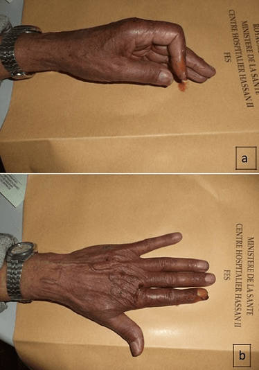
The Elson test. The patient's PIP is flexed to 90° over the edge of a table, and the examiner resists attempted PIP extension while palpating the DIP. What the DIP does gives the answer:
- Intact central slip - strong PIP extension force, and the DIP stays floppy because the lateral bands are slack in PIP flexion
- Ruptured central slip - weak PIP extension, and the DIP goes rigid in extension because all the extensor force is transmitted through the lateral bands to the DIP, bypassing the PIP
The rigid, fixed-extended DIP is the positive finding; a floppy DIP is the normal result. The test is regarded as pathognomonic, but two cautions come from the Evidence Base: the original description reported no accuracy data, and in a painful acute finger the test should be performed under digital block.
- Technique
- PIP at 90°, resist extension, palpate DIP
- Positive Finding
- Weak PIP extension + RIGID DIP
- Comment
- Reference clinical test; no published accuracy data
- Technique
- Ask patient to extend PIP from flexion
- Positive Finding
- Inability to fully extend (lag)
- Comment
- Can be normal acutely (lateral bands)
- Technique
- Passively extend PIP fully, release
- Positive Finding
- PIP drops into flexion immediately
- Comment
- Less specific than Elson
- Joint posture
- PIP flexion + DIP hyperextension
- Discriminating finding
- Weak PIP extension with a RIGID DIP
- Key test / imaging
- Positive Elson test; X-ray for avulsion
- Joint posture
- DIP flexion (drop) + neutral PIP
- Discriminating finding
- Loss of active DIP extension, PIP normal
- Key test / imaging
- Lateral X-ray for dorsal distal phalanx avulsion
- Joint posture
- PIP hyperextension + DIP flexion (opposite)
- Discriminating finding
- Volar plate / FDS laxity, not central slip loss
- Key test / imaging
- Bunnell test; assess intrinsic tightness
- Joint posture
- Painful PIP, possible hyperextension instability
- Discriminating finding
- Tenderness volar, stable extensor mechanism
- Key test / imaging
- Stress views; Elson test negative
- Joint posture
- Fixed PIP flexion, DIP normal (no hyperextension)
- Discriminating finding
- No central slip rupture; flexion contracture from volar scar
- Key test / imaging
- Elson test negative; history of hyperextension injury
- Joint posture
- Globally swollen, hot, painful PIP
- Discriminating finding
- Constitutional signs, no clear extensor deficit
- Key test / imaging
- Bloods, joint aspiration if infection suspected
Pseudo-boutonniere is the trap. It follows a hyperextension injury with volar plate damage and produces a fixed PIP flexion contracture, but the DIP is normal and the Elson test is negative. Mistaking it for a true boutonniere leads to extension splinting that fails.
Investigations
The diagnosis of a central slip injury is clinical. Radiographs are primarily to rule out a bony avulsion and to assess PIP congruity, and treatment does not wait for an MRI: if the Elson test is positive, splint immediately.
Radiographs. PA, lateral and oblique views of the affected finger are first line. Most central slip injuries are purely soft tissue and the films are normal; what they are looking for is:
- An avulsion fracture of the dorsal base of the middle phalanx
- PIP dislocation
- PIP arthritis, in chronic cases
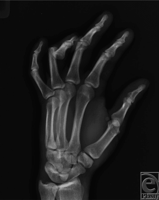
CT is added when a fracture is seen. It measures the size and displacement of the fragment against the fixation threshold below and plans the fixation, screws or a tension band.
MRI has its place in the chronic boutonniere, where it assesses tendon quality and joint cartilage: a scarred central slip, volar lateral bands and arthritic change at the PIP. It helps decide between repair, reconstruction and arthroplasty.
Bony Central Slip Avulsion: the Dorsal Middle-Phalanx-Base Fracture
The bony equivalent of a central slip rupture is managed differently from the pure soft-tissue tear, and deserves its own description.
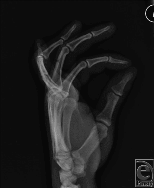
What it is. Instead of tearing in mid-substance, the central slip avulses a fragment of dorsal cortex from the base of the middle phalanx. The fragment marks the detached central slip insertion, and the injury carries the same boutonniere risk as the closed rupture.
How it is seen. The true lateral radiograph is the key view: look for a small dorsal flake off the middle-phalanx base, analogous to the dorsal distal-phalanx flake of a bony mallet. A subtle fragment is easily missed on the AP, so a dedicated lateral is mandatory in any PIP dorsal injury.
When to fix. A small, minimally displaced, congruent fragment is treated exactly like the soft-tissue injury, with PIP extension splinting and the DIP free, because reducing the extended PIP reduces the fragment. Operative fixation is reserved for:
- A large fragment, commonly quoted as more than about a quarter (over 25%) of the articular surface
- Significant displacement
- PIP joint incongruity or subluxation
The techniques include a small screw, a pull-out suture, or a dorsal tension band or anchor.
The pitfall. Do not mistake it for a stable chip. An unrecognised displaced dorsal-base avulsion produces the same fixed boutonniere as a missed soft-tissue rupture, plus articular incongruity and secondary PIP arthritis.
Management Algorithm
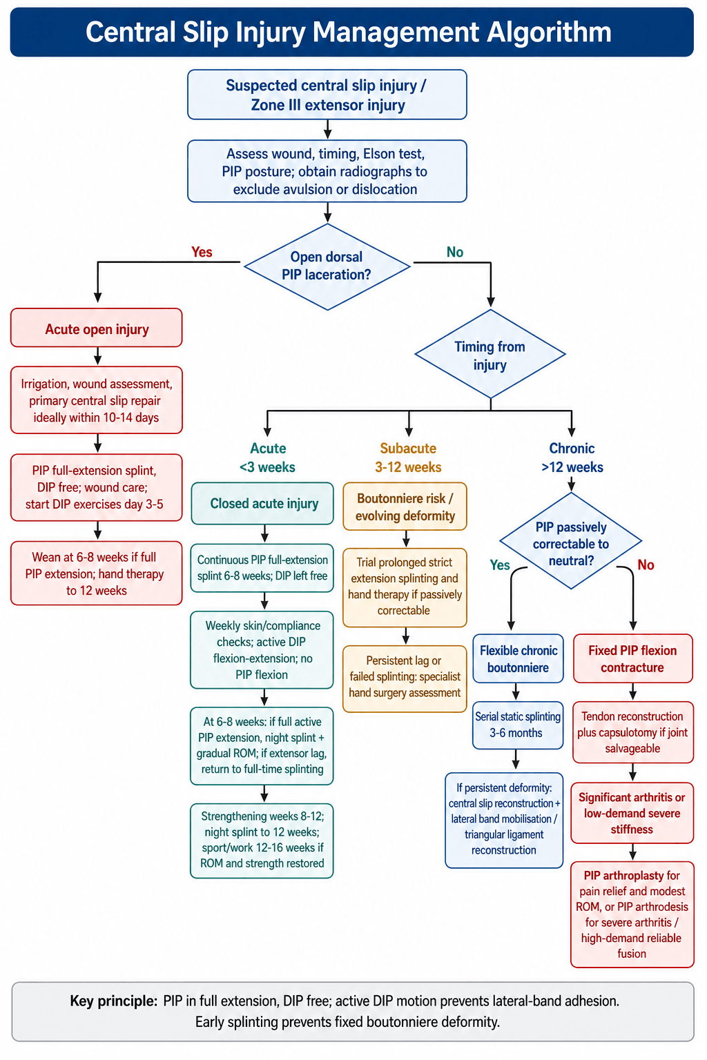
Acute closed injury (under 3 weeks). The goal is to prevent the boutonniere by holding the PIP in extension while the lateral bands are still in their anatomical position. Splint immediately: delay beyond 3 weeks risks a fixed boutonniere.
Treatment Protocol
PIP in full (0°) extension with the DIP free, in a dorsal aluminium splint, a custom thermoplastic splint or a stack splint. The splint is worn continuously and removed only for hygiene, under supervision. Active DIP flexion and extension exercises are started to prevent lateral band adhesion.
Full-time PIP extension splinting continues, with weekly checks of skin integrity, compliance and any developing deformity. DIP range of motion is essential to maintain lateral band excursion. No PIP flexion is allowed, because even passive flexion risks lateral band migration.
Full active PIP extension must be achieved before weaning. The splint then becomes a night splint, with gradual PIP range-of-motion exercises and buddy taping for protection during daytime activities. Any extensor lag means a return to full-time splinting.
Progressive resistance with grip strengthening and putty exercises. The night splint continues for 12 weeks in total, and return to sport is at 12-16 weeks if full range and strength are restored.
The key is PIP extension with DIP free. If you immobilise the DIP in extension, the lateral bands will adhere dorsally and lose excursion. Active DIP flexion during PIP extension splinting maintains lateral band mobility and prevents both boutonniere and swan neck deformities.
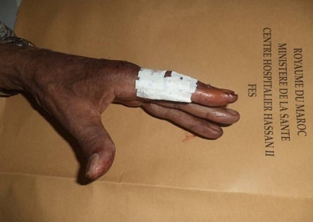
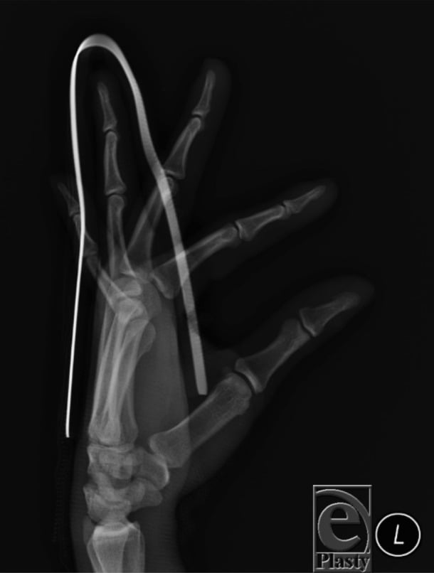
Relative-Motion Extension Splinting: Early Controlled Motion for Zone III
The relative-motion extension orthosis is an increasingly used alternative to rigid static splinting.
The principle. A small yoke across the dorsum holds the injured finger's MCP joint in slightly more extension than the adjacent MCPs, roughly 15-20° more. Altering the relative MCP posture offloads the healing central slip, through the quadriga/juncturae linkage, while still permitting active PIP and DIP motion, so the finger is not rigidly immobilised.
Why it is attractive. Early controlled active motion during healing can reduce PIP stiffness and adhesion and lets the patient keep using the hand. The splint is generally better tolerated and easier to comply with than 6-8 weeks of continuous rigid PIP extension, and compliance is the main determinant of outcome.
How it is applied. In zone III it is typically used as a relative-motion extension orthosis, often with a short additional PIP extension component early, and progressed by a hand therapist. It suits selected acute closed injuries and rehabilitation after repair rather than fixed chronic deformity.
The honest caveat. Systematic-review evidence shows no clear long-term superiority of relative-motion or dynamic regimens over static extension splinting for zone III. The choice is pragmatic and therapist-led, an option rather than a mandate, and rigid static splinting remains a perfectly acceptable default.
Complications
- Incidence
- Near 100 percent if untreated over 6 weeks
- Risk Factors
- Delayed diagnosis, poor compliance
- Management
- Prevention via early splinting best strategy
- Incidence
- 20-30 percent despite treatment
- Risk Factors
- Late presentation, inadequate splinting
- Management
- Serial splinting or surgical release
- Incidence
- 10-15 percent in chronic cases
- Risk Factors
- Overcorrection, DIP immobilisation during splint
- Management
- DIP flexion exercises, splint DIP if severe
- Incidence
- Common after surgical repair
- Risk Factors
- Prolonged immobilisation, scarring
- Management
- Early DIP motion, hand therapy at 6 weeks
- Incidence
- 5-10 percent after repair
- Risk Factors
- Early return to activity, inadequate splint time
- Management
- Return to splinting if caught early
Prevention is the best treatment. The most important complication to prevent is the boutonniere deformity itself. Once established it is difficult to correct and often results in permanent functional impairment, even though, as above, it is often more disfiguring than disabling. Prevention rests on a high index of suspicion in any PIP injury, an Elson test, and splinting started immediately and continued for 6-8 weeks.
Outcomes and Prognosis
- Treatment
- Extension splinting 6-8 weeks
- Expected Outcome
- 90 percent full ROM, rare extensor lag
- Long-term Function
- Excellent return to sport and work
- Treatment
- Prolonged splinting or surgery
- Expected Outcome
- 70-80 percent good ROM, mild lag common
- Long-term Function
- Good for daily activities, grip weaker
- Treatment
- Reconstruction or arthroplasty
- Expected Outcome
- 50-70 percent useful ROM, variable lag
- Long-term Function
- Functional improvement but rarely normal
Prognostic factors. The best prognosis belongs to an acute injury in a compliant patient, splinted full time for 6-8 weeks with early DIP mobilisation. The poorest follows diagnosis delayed beyond 6 weeks, a fixed PIP contracture, PIP arthritis on imaging, or non-compliance. The threshold to remember is 3 weeks from injury: outcomes decline significantly after it.
Guidelines, Registries & Global Practice
- Zone III extensor injuries are among the more common extensor tendon injuries seen in hand units worldwide
- Closed central slip rupture typically follows ball-sport jamming, falls, or forced PIP flexion across all regions
- Open lacerations dominate in occupational and assault settings (knife, glass, machinery)
- Volar PIP dislocation is an underrecognised cause and a frequent source of missed diagnosis
- High-resource: certified hand therapists fabricate custom thermoplastic or relative-motion splints with weekly review
- Limited-resource: low-cost aluminium/foam splints and buddy strapping are effective if applied early
- Universal principle: outcome depends far more on prompt diagnosis (Elson test) and splint compliance than on splint cost
- Surgery: chronic reconstruction is concentrated in specialist hand centres globally
- Diagnosis emphasis
- Elson test, ideally under digital block, on every PIP dorsal injury
- Acute treatment
- Continuous PIP extension splint 6 weeks, DIP free for active flexion
- Surgery / reconstruction
- Anatomic repair (suture anchor) for established supple deformity
- Diagnosis emphasis
- Clinical diagnosis; X-ray to exclude avulsion, not to confirm
- Acute treatment
- Orthosis-led conservative care via hand therapy; relative-motion splints increasingly used
- Surgery / reconstruction
- Reserved for failed conservative care or open injury
- Diagnosis emphasis
- High index of suspicion; tenodesis and Elson testing
- Acute treatment
- Static extension splinting; dynamic regimens optional (no proven superiority)
- Surgery / reconstruction
- Repair, lateral band relocation, or tendon graft by deformity type
- Diagnosis emphasis
- Assess for bony central slip avulsion at middle phalanx base
- Acute treatment
- Splint if soft tissue; ORIF if displaced avulsion involving the joint
- Surgery / reconstruction
- Fragment fixation principles for bony avulsions
There is no dedicated arthroplasty/implant registry for central slip injuries, as most are soft-tissue and treated non-operatively. The evidence base is dominated by small case series and operative-technique reviews rather than randomised trials, so guidance is principle-based: diagnose early, splint the PIP in extension with the DIP free, and reserve surgery for established or open injuries.
Record in every acute PIP injury:
- Elson test performed and its result (positive / negative)
- Splinting instructions given (PIP extension, DIP free, duration 6-8 weeks)
- Patient counselled on boutonniere risk if non-compliant
- Follow-up arranged within one week to assess compliance
A missed central slip diagnosis leading to a fixed boutonniere is a recurring source of complaints and claims worldwide. Always perform and document the Elson test on any PIP dorsal injury, even when the initial X-ray is normal.
Controversies & Areas of Uncertainty
Relative-motion extension orthoses allow early controlled motion and may reduce stiffness, but systematic-review evidence shows no clear long-term superiority over static extension splinting for zone III injuries. Choice is often pragmatic and therapist-led.
Six to eight weeks is conventional, but the precise minimum is not defined by high-quality trials. Most guidance favours at least six weeks of continuous PIP extension, balancing deformity prevention against stiffness from over-immobilisation.
A functional but cosmetically displeasing boutonniere may be best left alone: surgery to gain PIP extension can cost PIP flexion. Reconstruction is offered cautiously, mainly for supple, passively correctable deformities or significant functional limitation.
Anatomic anchor-augmented repair, lateral band relocation, and crossed palmaris longus grafting all report fair-to-good results in small series, with no head-to-head trials. Technique is dictated by tissue quality and surgeon experience rather than firm evidence.
MCQ Practice Points
Q: What is the insertion of the central slip of the extensor mechanism? A: Dorsal base of the middle phalanx. The extensor mechanism divides into three slips at the proximal phalanx level: the central slip (middle band) inserts on the middle phalanx base, while the two lateral bands continue distally to form the terminal tendon inserting on the distal phalanx.
Q: What is the Elson test and what does a positive test indicate? A: Elson test diagnoses central slip rupture. The patient's PIP is flexed to 90 degrees over the table edge. The examiner resists attempted PIP extension while palpating the DIP. A positive test shows weak PIP extension force and a RIGID, fixed-extended DIP (not floppy). This indicates central slip rupture - all extensor force is transmitted through the lateral bands to the DIP, bypassing the PIP, which is exactly why the DIP stiffens into extension.
Q: What is a boutonniere deformity and how does it develop? A: Boutonniere deformity is PIP flexion with DIP hyperextension. It develops when the central slip ruptures and the lateral bands gradually migrate volar to the PIP joint axis over 3-6 weeks. Once volar, the lateral bands act as PIP flexors (instead of extensors) and DIP hyperextensors, creating the classic deformity pattern.
Q: What is the splinting protocol for acute central slip injury? A: PIP in full (0 degrees) extension for 6-8 weeks continuously, with DIP free. The key is maintaining PIP extension to prevent lateral band volar migration while allowing DIP flexion exercises to maintain lateral band excursion and prevent adhesion. Splinting both PIP and DIP risks swan neck deformity.
Q: What is the critical time window for preventing boutonniere deformity? A: Within 3 weeks of injury. If extension splinting is started within 3 weeks, 90 percent of patients avoid boutonniere deformity. After 3-6 weeks, the lateral bands have migrated volar and conservative treatment success drops significantly, often requiring surgical reconstruction.
Q: When is surgical reconstruction indicated for boutonniere deformity? A: Chronic fixed deformity over 12 weeks that does not passively correct to neutral. Acute injuries (under 3 weeks) are treated with splinting. Subacute (3-12 weeks) can trial prolonged splinting. Surgery is needed when the PIP has a fixed flexion contracture, requiring capsular release, lateral band relocation, and central slip reconstruction or arthroplasty.
Exam Viva Scenarios
Practise clinical reasoning and management decisions out loud
“A 28-year-old basketball player presents 2 days after jamming his left middle finger catching a ball. He has swelling over the PIP joint and difficulty extending the finger. X-rays show no fracture. On examination, he has weak PIP extension and a positive Elson test. What is your diagnosis and management?”
“A 45-year-old presents with chronic PIP flexion deformity of the ring finger following an untreated injury 6 months ago. He has fixed 40-degree PIP flexion contracture and 20-degree DIP hyperextension. PIP can be passively extended to 20 degrees flexion but not neutral. X-rays show early PIP joint space narrowing. How would you manage this?”
“A 35-year-old sustains a 2cm laceration over the dorsum of the PIP joint of his index finger from a kitchen knife. In the emergency department, you can see the central slip is completely divided. The wound is clean and occurred 6 hours ago. How would you manage this?”
Key Anatomy
- Central slip = middle band of extensor mechanism, inserts dorsal base middle phalanx
- Lateral bands = continue to DIP as terminal tendon
- Central slip rupture allows lateral bands to migrate volar to PIP axis
- Volar lateral bands become PIP flexors and DIP hyperextensors = boutonniere
Diagnosis
- Elson test = pathognomonic (PIP at 90°, weak extension + RIGID DIP; floppy DIP = intact)
- Acute injury may have near-normal PIP extension (via lateral bands)
- X-ray: check for avulsion fracture middle phalanx base
- High index of suspicion on any PIP dorsal injury or forced flexion mechanism
Treatment Algorithm
- Acute (under 3 weeks) = PIP extension splint 6-8 weeks, DIP free
- Acute open = primary repair + splinting (same duration)
- Subacute (3-12 weeks) = trial prolonged splinting vs surgery
- Chronic fixed (over 12 weeks) = reconstruction or arthroplasty
Splinting Pearls
- PIP in full (0 degrees) extension continuously 6-8 weeks
- DIP must be free - active DIP flexion prevents lateral band adhesion
- Never splint PIP and DIP together (risks swan neck)
- Wean at 6-8 weeks: night splint + gradual ROM, monitor for lag
Complications
- Boutonniere deformity = near 100 percent if untreated over 6 weeks
- PIP flexion contracture = 20-30 percent even with treatment
- Re-rupture = 5-10 percent if return to activity too early
- Stiffness = common after surgery or prolonged immobilization
Evidence Base and Key Trials
Rupture of the central slip of the extensor hood: a test for early diagnosis
- Original description of the Elson test for early central slip rupture
- From 90 degrees of PIP flexion over a table edge, the patient extends against resistance
- Absent PIP extension force plus fixed (rigid) DIP extension indicates complete central slip rupture
- Boyes test turns positive only late, once boutonniere deformity is established
Managing swan neck and boutonniere deformities
- Boutonniere arises from loss of coordination between central slip and lateral bands with altered IP joint force balance
- Boutonniere fingers remain functional (PIP flexion and fist preserved); the deformity is largely aesthetic
- Surgical correction must be cautious: gaining PIP extension can impede PIP flexion and worsen function
- Corrective splinting is first line; operative reconstruction is reserved and counselled carefully
Anatomic repair of the central slip with anchor suture augmentation for established boutonniere deformity
- Retrospective series of 13 chronic, supple (Burton stage I) post-traumatic boutonniere deformities
- Direct end-to-end central slip repair augmented with a suture anchor
- Mean PIP extension lag improved from 43.5 degrees by an average of 21.9 degrees
- Excellent or good results in 77 percent (10 of 13) and 69.2 percent good by Souter criteria; 3 patients had recurrence during ROM exercises. Of the 10 patients reached by phone, 2 were very satisfied and 3 satisfied, but 3 rated the result average, 1 poor and 1 very poor
Chronic boutonniere deformity: cross-lateral band technique using palmaris longus autograft
- Seven flexible chronic boutonniere deformities with lateral bands deficient or beyond repair
- Palmaris longus grafts reconstruct both lateral bands, crossing over the middle phalanx
- Mean PIP extension deficit reduced from 54 degrees to 7 degrees
- DIP hyperextension reduced from 9 degrees to 2 degrees with no DIP flexion contracture
Long-term results of extensor tendon repair
- Retrospective review of 101 digits in 62 patients with extensor tendon injury (mostly static splinting)
- Good or excellent results in 64 percent of fingers with no associated injury
- Distal zones (I-IV, including central slip zone III) did significantly worse than proximal zones (V-VIII)
- Loss of flexion was more frequent and greater than loss of extension after repair
Extensor tendon repair (operative technique review)
- Zone III repairs use one to two core sutures plus a running suture; the Elson test should be performed under digital block
- Suture anchors or bone tunnels are used when no distal tendon remains for repair
- Systematic reviews suggest dynamic rehabilitation gives no superior long-term benefit over static splinting
- Running interlocking horizontal mattress repair is stiffer and faster than alternatives