From functional assessment to multilevel reconstruction
- Start with function and goals: walking distance, aids, falls, pain, fatigue, stairs, transfers, school participation and family priorities.
- Classify the child after assessment: distribution of involvement, movement disorder, GMFCS level, gait pattern and lever-arm status.
- Separate dynamic spasticity from fixed contracture, weakness, poor selective control, torsion and compensation before selecting treatment.
- The key sagittal patterns in spastic diplegia are true equinus, jump gait, apparent equinus and crouch gait.
- SEMLS is not multiple releases on one day; it is gait-analysis-informed correction of the whole kinetic chain with a long rehabilitation plan.
- “GMFCS is assigned from observed and reported functional mobility; it is not a single history question.
- “Toe walking may be true equinus, jump gait, apparent equinus, dystonia, short-limb compensation or balance strategy.
- “Crouch gait is multifactorial: weak plantarflexors, patella alta, knee flexion contracture, planovalgus, torsion and previous overlengthening may matter more than hamstrings.
- “A gait lab report supports clinical reasoning. It is not an operation list.
Overview and Epidemiology
Cerebral palsy gait is a dynamic, multiplanar orthopaedic problem. The brain lesion is non-progressive, but the musculoskeletal consequences progress because the child grows through abnormal tone, muscle imbalance, weakness, impaired motor control and altered loading. A toddler with dynamic equinus may become a school-aged child with fixed calf contracture, tibial torsion, planovalgus, knee flexion contracture, patella alta and increasing energy cost.
Rarely one tight muscle. The recurring message of the modern literature is that cerebral palsy gait is a whole-limb mechanical problem rather than a list of tight muscles. A visible abnormality may be the primary pathology, a compensation, or the only mechanism keeping the child upright, which is why the assessment follows a sequence rather than becoming a list of operations:
- Define the functional problem.
- Classify function and gait pattern.
- Identify dynamic tone, fixed contracture, weakness, torsion, foot deformity and compensation.
- Decide whether non-operative care, tone management, orthoses, surgery or surveillance best matches the goal.
- If surgery is required, plan the whole limb and the rehabilitation pathway.
Who is in front of you. Cerebral palsy is the most common cause of lifelong physical disability beginning in childhood, and orthopaedic gait surgery mainly concerns the ambulant child.
The common error in cerebral palsy gait is to operate on the most obvious deformity instead of the primary driver. Achilles lengthening for apparent equinus and isolated hamstring lengthening for multifactorial crouch are classic examples.
Definition of SEMLS
Single-event multilevel surgery is the planned correction of clinically important musculoskeletal deformities at multiple anatomical levels during one operative episode. In ambulant CP, it is usually planned from clinical examination, radiographs, rotational assessment and instrumented gait analysis.
SEMLS is a gait-analysis-informed single-stage strategy for ambulant children with cerebral palsy, designed to correct fixed soft-tissue contractures and bony lever-arm dysfunctions at the hip, knee, tibia, ankle and foot, with one coordinated rehabilitation pathway.
Why it exists. Treating equinus one year, hamstrings another year and femoral anteversion later can create sequential imbalance and repeated rehabilitation cycles. SEMLS corrects the clinically important fixed deformities and lever-arm dysfunctions together, once the gait pattern is sufficiently understood and the child can participate in rehabilitation.
What "single-event" does not mean. It does not mean every abnormality must be treated. The clinically relevant fixed deformities that drive the gait problem are corrected together where this is safer and more logical than serial isolated surgery.
SEMLS is not a random package of releases. It is not a response to a gait-lab printout alone. It is not appropriate for every child with CP. It is a selected reconstruction plan for a selected child with a defined functional goal.
SEMLS evidence at a glance
- Pooled analysis showed multilevel surgery improved gait (Gait Profile Score; SMD -0.80, 95% CI -0.95 to -0.65) but did not change gross motor function (SMD 0.38, 95% CI -0.25 to 1.01) or gait speed (SMD 0.12, 95% CI -0.01 to 0.25).
- Only one of 74 included studies was a randomised controlled trial; the remainder were cohort studies.
- Participation, quality of life and adverse events were inadequately reported in most studies.
Essential Gait Biomechanics
The normal cycle. Gait divides into stance and swing. Stance occupies roughly 60 percent of the cycle - initial contact, loading response, midstance, terminal stance and pre-swing - and swing the remaining 40 percent, as initial swing, mid-swing and terminal swing.
Efficient gait depends on a set of mechanical requirements, and cerebral palsy can disturb any of them:
- Heel strike or controlled initial contact
- Controlled tibial progression over a stable foot
- Adequate stance-phase knee extension without recurvatum or collapse
- Hip extension in terminal stance
- Heel rise and plantarflexion power in terminal stance and pre-swing
- Adequate knee flexion in swing for foot clearance
- Controlled terminal swing knee extension
- Balanced pelvic motion with limited compensatory trunk movement
Why the pattern breaks down. The child with CP loses gait efficiency when tone, weakness, contracture and skeletal alignment disturb that sequence, and a deformity at one level changes the apparent problem elsewhere. The table reads in both directions: from a driver to what it does, and from a visible abnormality back to what may be producing it.
- Effect on gait
- Poor push-off and reduced stance-phase knee extension moment.
- Treatment implication or trap
- Avoid overlengthening the calf and worsening crouch.
- Effect on gait
- Limits knee extension.
- Treatment implication or trap
- Lengthen only when contracture is a true driver, not a compensatory stabiliser.
- Effect on gait
- Absent heel strike.
- Treatment implication or trap
- Misdiagnosing apparent equinus as true calf equinus.
- Effect on gait
- Anterior pelvic tilt and lumbar lordosis.
- Treatment implication or trap
- Missing the proximal driver while focusing on the ankle.
- Effect on gait
- Hip internal rotation and altered knee progression.
- Treatment implication or trap
- Consider femoral derotation when gait analysis and examination agree.
- Effect on gait
- Foot lever points away from the line of progression; poor push-off and a crouch tendency.
- Treatment implication or trap
- Tibial derotation may be needed when lever-arm failure drives crouch; blaming hamstrings alone is the trap.
- Effect on gait
- Foot cannot act as a stable lever; weak push-off and midfoot break.
- Treatment implication or trap
- Foot stabilisation or an orthotic strategy may be needed before gait improves; treating calf weakness without restoring the foot lever fails.
- Effect on gait
- Extensor lag and crouch.
- Treatment implication or trap
- Restore the extensor mechanism rather than relying on hamstring lengthening.
- Effect on gait
- Reduced swing-phase knee flexion.
- Treatment implication or trap
- Stiff knee gait is not corrected by hamstring surgery.
Natural History of Gait Deterioration and Timing of SEMLS
The brain lesion is static; the gait is not. It deteriorates because the child grows through abnormal tone, weakness and lever-arm dysfunction, and that trajectory is what determines when to intervene.
The young child. Spasticity is still largely dynamic - dynamic equinus is the usual example - and the gait pattern is still evolving. Non-operative tone management with botulinum toxin, serial casting and orthoses carries this period: gait analysis is less reliable and soft-tissue lengthenings recur with growth, so early irreversible surgery is avoided.
School age, about 7 to 10 years. The gait pattern matures and becomes reproducible, and fixed contracture and lever-arm dysfunction declare themselves. This is the usual window for gait-analysis-informed SEMLS, once the deformity is fixed and functionally important.
The pubertal growth spurt. Rising body mass and longer levers outstrip plantarflexor and quadriceps strength, and a toe-walking or jump-gait child can collapse into high-energy crouch - the crouch crisis. It is the high-risk window: act before crouch and bony deformity become fixed and irreversible.
Adolescence and adulthood. Crouch is expensive to walk in, and a proportion of GMFCS II-III walkers lose efficient or independent ambulation. Preserving ambulation and energy efficiency is a central goal of well-timed surgery.
Pathophysiology
CP gait abnormalities arise from neurological and musculoskeletal factors acting together.
The neurological contribution. The lesion produces abnormal tone, weakness, poor selective control and impaired balance, and each of these has a different treatment logic.
- Spasticity - velocity-dependent resistance to stretch.
- Dystonia - involuntary fluctuating postures or movements.
- Co-contraction - simultaneous activation of antagonists, reducing smooth motion.
- Poor selective motor control - inability to isolate hip, knee, ankle and foot movement.
- Weakness - particularly hip extensors, hip abductors, quadriceps, dorsiflexors and plantarflexors.
- Impaired balance - abnormal trunk and pelvic strategies.
- Motor learning differences - persistence of inefficient movement patterns after deformity correction.
The musculoskeletal contribution. These accumulate as the child grows through the abnormal pattern.
- Muscle-tendon contracture.
- Joint contracture.
- Femoral anteversion.
- Tibial torsion.
- Patella alta and extensor mechanism insufficiency.
- Planovalgus or equinovarus foot deformity.
- Hip displacement and pelvic obliquity.
- Scoliosis or spinal imbalance.
Lever-arm dysfunction. Muscles need stable skeletal levers, and even strong muscles cannot work efficiently if the lever is malaligned. Femoral anteversion changes hip rotation and reduces efficient abductor mechanics, while external tibial torsion rotates the foot lever outwards and weakens the plantarflexion-knee-extension couple. Planovalgus shortens and destabilises the foot lever, a midfoot break allows collapse rather than push-off, and patella alta reduces the effectiveness of the quadriceps mechanism and contributes to extensor lag. This is why bony correction and foot reconstruction may be more important than additional soft-tissue lengthening in selected children.
T-C-W-LDrivers of Abnormal Gait
Hook:T-C-W-L asks why the gait pattern exists before deciding what to treat.
Sagittal Gait Patterns in Spastic Diplegia
The Rodda and Graham sagittal classification is useful because it links observed gait to treatment reasoning. It should not replace full assessment. Classify each limb separately. Asymmetry between the two sides is common in diplegia - a child may be in jump gait on one side and crouch on the other - and the operative plan then differs limb by limb. Assigning a single pattern to the whole child is a recognised way to end up doing the wrong operation on one leg.
- Hip
- Neutral or extended
- Knee
- Neutral or recurvatum
- Ankle/foot
- Plantarflexed
- Common driver
- Gastrocsoleus spasticity or contracture
- Treatment principle
- Treat calf only when true equinus is confirmed
- Hip
- Flexed
- Knee
- Flexed
- Ankle/foot
- Equinus
- Common driver
- Hip flexor, hamstring and calf involvement
- Treatment principle
- Multilevel assessment and selected correction
- Hip
- Flexed
- Knee
- Flexed
- Ankle/foot
- Neutral or dorsiflexed
- Common driver
- Proximal flexion pattern
- Treatment principle
- Do not lengthen Achilles unless true equinus coexists
- Hip
- Flexed
- Knee
- Flexed
- Ankle/foot
- Dorsiflexed or calcaneus
- Common driver
- Weakness, lever-arm failure, patella alta or contracture
- Treatment principle
- Correct all drivers; avoid reflex hamstring or calf lengthening
- Hip
- Variable
- Knee
- Reduced swing flexion
- Ankle/foot
- Toe drag or compensation
- Common driver
- Rectus femoris overactivity or poor clearance mechanics
- Treatment principle
- Rectus transfer only if confirmed
- Hip
- Variable
- Knee
- Crouch tendency
- Ankle/foot
- Valgus and midfoot break
- Common driver
- Ineffective foot lever
- Treatment principle
- Restore a stable plantigrade foot
What it is. Ankle plantarflexion during stance. The ankle is plantarflexed relative to the tibia, the forefoot commonly contacts first, and the heel may not reach the ground.
Drivers. Dynamic gastrocnemius or soleus spasticity, fixed gastrocnemius contracture, fixed gastrocsoleus contracture, an equinovarus foot, or dystonia and poor motor control.
Assessment. Examine passive ankle dorsiflexion with the knee flexed and extended, perform the Silfverskiold test, assess hindfoot position and check for a midfoot break. Decide whether the equinus is dynamic or fixed, gastrocnemius-only or gastrocsoleus, unilateral or bilateral, and whether the child needs it for stance stability.
Treatment logic. Dynamic equinus may respond to therapy, orthoses, botulinum toxin or serial casting. Fixed gastrocnemius contracture may need gastrocnemius recession. Fixed gastrocsoleus contracture may need tendo-Achilles lengthening in selected cases, but overlengthening can cause calcaneus and crouch.
Stiff-knee gait. The problem is reduced or delayed swing-phase knee flexion, with toe drag and poor clearance. Rectus femoris transfer is considered only when the gait-lab and clinical pattern support inappropriate rectus activity, because poor push-off, weakness and poor selective control can also reduce swing knee flexion. Circumduction, hip hiking and vaulting may be compensations for poor clearance rather than problems in their own right.
Rotational gait. Internal rotation gait may come from femoral anteversion, internal tibial torsion, foot deformity or compensation. Derotation decisions should match the functional gait findings rather than a single static measurement.
Scissoring. Assess adductor tone, hip abduction range, hip displacement risk, balance and selective control. Do not treat scissoring in isolation without understanding the hip and the functional goal.
Hemiplegic Gait Patterns
Hemiplegic gait is commonly described using the Winters, Gage and Hicks pattern. The value is not memorising labels; it is linking the involved levels to treatment.
- Main abnormality
- Drop foot in swing
- Typical features
- Ankle is usually acceptable in stance but dorsiflexion is inadequate in swing
- Treatment logic
- AFO, strengthening, functional electrical stimulation in selected cases
- Main abnormality
- Equinus in stance and swing
- Typical features
- Forefoot contact, possible recurvatum in type IIB
- Treatment logic
- Calf procedure only if fixed contracture and clinically indicated
- Main abnormality
- Equinus plus stiff knee
- Typical features
- Reduced swing knee flexion, toe drag, rectus femoris overactivity may contribute
- Treatment logic
- Calf correction plus rectus femoris transfer only when gait analysis supports it
- Main abnormality
- Multilevel involvement
- Typical features
- Hip flexion, adduction/internal rotation, stiff knee and equinus
- Treatment logic
- Often needs multilevel assessment and selected SEMLS
Functional Classification
Describe the child, not the operation. Begin with the distribution of involvement and the movement disorder.
- Spastic hemiplegia - one side predominantly involved, often with equinus or equinovarus, drop foot in swing, stiff knee gait or internal rotation gait.
- Spastic diplegia - both lower limbs involved, usually with relative upper limb sparing, commonly showing equinus, jump gait, apparent equinus, crouch, scissoring, internal rotation and planovalgus.
- Quadriplegia - all four limbs involved; gait surgery is less often the primary goal and orthopaedic care commonly focuses on hips, sitting, spine, hygiene, comfort and transfers.
- Dyskinetic or dystonic CP - variable posture and tone may make SEMLS outcomes less predictable if dystonia is dominant.
GMFCS comes from function. The level is assigned or confirmed from observed and reported function, based on what the child does in daily life, with emphasis on sitting, transfers and mobility. Build it from functional questions: does the child walk indoors, outdoors and at school; are stairs possible, and is a rail required; does the child use AFOs, crutches, a walker or a wheelchair; how far can the child walk before fatigue; does the child fall; can the child keep up with peers; are transfers independent; has function declined with growth; and is pain limiting walking?
- GMFCS I - walks without major limitations, but speed, balance, running and jumping may be impaired.
- GMFCS II - walks without handheld mobility devices but has limitations with distance, uneven ground, crowds, inclines or speed.
- GMFCS III - walks with a handheld mobility device and often uses wheeled mobility for longer distances.
- GMFCS IV - self-mobility is limited; powered mobility or substantial assistance is often required.
- GMFCS V - transported in a manual wheelchair, with severe limitations in head and trunk control.
Who the gait operation is for. The common gait-improvement SEMLS group is ambulant CP, especially GMFCS I to III, and particularly spastic diplegia and hemiplegia. GMFCS IV and V children may still need orthopaedic surgery, but the priorities usually shift to hip surveillance, sitting, hygiene, pain, skin protection, hip containment, standing transfers and care rather than efficient community walking.
Clinical Assessment
History. The history should explain why the child is presenting now: tripping, fatigue, pain, loss of walking, brace intolerance, cosmetic concern, hygiene, transfers or school participation. Ask what the child actually does - indoor walking, outdoor walking, stairs, playground, sport, self-care, endurance and community mobility - and what they use to do it, including AFO type, walker, crutches, wheelchair, night splints, how well each is tolerated and whether it leaves pressure areas. Falls deserve detail (frequency, direction, triggers and injuries), as do fatigue (distance before stopping, end-of-day deterioration and recovery time) and pain (hip, knee, foot, back, brace pressure or after activity).
The background that changes the plan. Take the developmental history: prematurity, neonatal course, milestones and any cognitive or communication needs. Ask about seizures, dystonia, medication, selective control concerns and tone management, and about every previous treatment - physiotherapy, strengthening, botulinum toxin, serial casting, orthoses, baclofen, intrathecal baclofen, selective dorsal rhizotomy and prior orthopaedic surgery. Ask too about recent growth spurt, worsening contracture or new functional decline.
Goals and rehabilitation reality. Ask what change would make treatment worthwhile, then ask whether it can be delivered: school support, therapy access, transport, home setup and family capacity.
Observation. Observe before touching the limb. Watch transfers, standing balance, shoes, braces and spontaneous walking, then observe barefoot and braced gait from the side, front and behind, recording:
- Walking speed and endurance
- Initial contact: heel, flat foot or toe
- Knee position in stance
- Hip flexion and pelvic tilt
- Foot progression angle
- Step length and symmetry
- Base width and scissoring
- Trunk lean, arm posture and balance
- Compensations: vaulting, circumduction, hip hiking and excessive pelvic rotation
Physical examination. The examination should explain the gait, not merely document range.
- What to assess
- Velocity-dependent catch, clonus, dystonia and co-contraction
- Why it matters
- Dynamic tone may respond to non-operative tone management
- What to assess
- Hip flexors, adductors, hamstrings, gastrocnemius and soleus
- Why it matters
- Fixed contracture changes operative planning
- What to assess
- Hip abductors/extensors, quadriceps, dorsiflexors and plantarflexors
- Why it matters
- Weakness limits lengthening and predicts crouch risk
- What to assess
- Ability to isolate hip, knee, ankle and foot movement
- Why it matters
- Poor control limits expected surgical gain
- What to assess
- Hip rotation, thigh-foot axis, transmalleolar axis and foot progression
- Why it matters
- Identifies lever-arm dysfunction
- What to assess
- Planovalgus, equinovarus, midfoot break, callosities and braceability
- Why it matters
- A poor foot lever weakens push-off
The tests that decide the plan. Each answers a specific question rather than filling a proforma:
- Thomas test - hip flexion contracture
- Hip abduction in flexion and extension - adductor tightness, and relevant to hip surveillance
- Popliteal angle - hamstring length
- Duncan-Ely or prone rectus test - rectus femoris tightness, and a clue to overactivity
- Silfverskiold test - gastrocnemius versus soleus contribution to equinus
- Patellar height and extensor lag - extensor mechanism insufficiency
- Rotational profile - femoral anteversion and tibial torsion
- Foot assessment - hindfoot, midfoot, forefoot, subtalar flexibility and braceability
- Strength and selective motor control - essential before any lengthening decision
Investigations and Gait Analysis
Plain radiographs. Request them to answer a question the examination has already raised. Gait planning sits inside the wider CP orthopaedic assessment of hips, spine, contractures, torsion, foot shape and functional goals.
- AP pelvis - migration percentage, acetabular index, femoral head shape, neck-shaft angle, hip subluxation, pelvic obliquity and pain source.
- Standing long-leg radiographs - coronal alignment and mechanical axis when deformity is suspected.
- Foot radiographs - weight-bearing AP and lateral views for planovalgus, cavovarus, midfoot break, talonavicular coverage and calcaneal pitch.
- Spine radiographs - scoliosis or pelvic obliquity when clinically relevant.
- Patellar height imaging - useful when crouch, extensor lag or anterior knee pain is present.
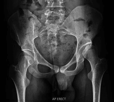
CT and MRI. CT may help quantify femoral anteversion or tibial torsion when clinical and gait findings are unclear; it should support the decision rather than replace functional assessment. MRI brain is not routinely required for established CP gait planning, but may be appropriate if the neurological diagnosis is unclear, progressive or atypical.
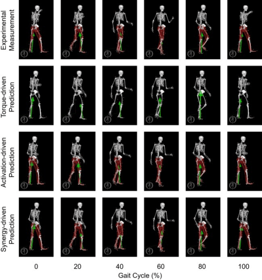
Instrumented gait analysis. It is most useful in complex ambulant CP, especially before SEMLS, and combines video, kinematics, kinetics, dynamic EMG, plantar pressure and temporospatial measures.
- What it shows
- Visual pattern, symmetry, orthotic effect and compensations
- Clinical use
- Correlates the data with what the clinician sees
- What it shows
- Speed, cadence, step length, stride length and support time
- Clinical use
- Quantifies function and response to treatment
- What it shows
- Joint angles across the gait cycle
- Clinical use
- Defines hip, knee, ankle, foot and pelvic motion
- What it shows
- Joint moments, power and ground reaction force
- Clinical use
- Shows whether muscles generate useful support and push-off
- What it shows
- Timing of muscle activity
- Clinical use
- Helps identify inappropriate rectus femoris, hamstring or calf activity
- What it shows
- Loading distribution and foot lever quality
- Clinical use
- Guides orthoses and foot reconstruction
- What it shows
- Oxygen cost or physiological cost index
- Clinical use
- Frames endurance and efficiency
What the gait lab does not replace. It cannot decide whether the family can manage rehabilitation, whether pain is the main complaint, whether school planning is realistic, or whether surgery matches the child's priorities. The report has to be reconciled with the examination, the radiographs and a shared decision.
Quantifying Gait: Summary Indices
SEMLS outcomes are reported as the Gait Profile Score, so it is worth knowing what these summary indices are. They turn a gait-laboratory dataset into a single comparable number, and they are how gait quality and treatment response are reported and compared.
- What it measures
- Root-mean-square deviation of nine key kinematic variables (the Gait Variable Scores) from typical data, in degrees
- Direction and use
- Lower is better (less deviation); fell after SEMLS in the meta-analysis; the Movement Analysis Profile displays the component scores
- What it measures
- Scaled multivariate index of kinematic normality
- Direction and use
- 100 represents unimpaired gait and each 10 points below 100 is about one standard deviation from typical; higher is better
- What it measures
- Structured observational (video) scoring of gait events at foot, knee, hip, pelvis and trunk
- Direction and use
- Usable without a full gait laboratory - valuable where instrumented analysis is unavailable
- What it measures
- Validated measure of gross motor capacity, distinct from the GMFCS classification
- Direction and use
- Tracks change in gross motor function over time
A summary index standardises outcome reporting and improves after SEMLS, but it cannot tell you which level to treat: the individual kinematic curves and the Movement Analysis Profile do that. Hold on to the dissociation in the evidence - SEMLS improves the gait indices but not the Gross Motor Function Measure or walking speed.
Differential Diagnosis
A non-progressive brain lesion is the defining feature of cerebral palsy. Before committing a child to a gait-correction pathway, exclude conditions that mimic spastic diplegia but progress, are inherited, or are treatable in a completely different way. A worsening, regressing or strongly familial picture, or an unusually pure dystonia, should prompt neurology review rather than orthopaedic surgery.
- Discriminating features
- Non-progressive lesion, often with prematurity or perinatal insult; static motor disorder that worsens mechanically only through growth
- Why it changes management
- Standard gait-pattern and SEMLS framework applies
- Discriminating features
- Slowly progressive, symmetrical lower-limb spasticity, frequent positive family history, often normal birth history
- Why it changes management
- Progression and inheritance change prognosis and counselling; surgery gives only temporary benefit
- Discriminating features
- Diurnal fluctuation worse in evening, dystonic posturing, dramatic response to a levodopa trial
- Why it changes management
- A levodopa trial can avoid unnecessary surgery; missing it is a classic trap
- Discriminating features
- Bilateral toe-walking, normal tone and neurology, normal development, able to stand flat-footed on request
- Why it changes management
- Often non-operative; not part of the CP spectrum
- Discriminating features
- Boys, calf pseudohypertrophy, Gowers sign, proximal weakness, very high creatine kinase, progressive
- Why it changes management
- Lengthening surgery is contraindicated reasoning; weakness, not spasticity, drives gait
- Discriminating features
- Mixed upper and lower motor neuron signs, bladder symptoms, back or foot changes, progression
- Why it changes management
- Requires neurosurgical assessment before any limb surgery
- Discriminating features
- Cavovarus feet, distal wasting, areflexia, sensory signs, family history
- Why it changes management
- Foot deformity is neuropathic and progressive; plan differs from spastic foot
Loss of previously acquired skills, marked diurnal variation, a strong family history, pure dystonia without spasticity, sensory level or bladder involvement should all trigger neurological reassessment before gait surgery is planned.
Management Principles
Management is matched to function, age, gait pattern, impairments, goals and rehabilitation capacity.
Physiotherapy and strengthening. Therapy targets strength, balance, endurance, motor control and gait retraining. Strengthening of hip extensors, hip abductors, quadriceps and plantarflexors is particularly important in crouch and after SEMLS.
Orthoses. Orthoses should match the gait pattern. A hinged AFO, solid AFO, ground-reaction AFO or supramalleolar orthosis can help or harm depending on the deformity, strength and foot lever. A ground-reaction AFO may help selected crouch by influencing the ground reaction force, but it requires adequate passive correction, a braceable foot and enough tolerance to wear it.
Tone management. Botulinum toxin and serial casting may help younger children with dynamic focal spasticity, especially dynamic equinus. They do not correct fixed bony torsion, established lever-arm dysfunction or severe contracture.
Surveillance. Growth changes everything. Reassess hip displacement, pain, contracture progression, brace tolerance, function and walking endurance over time.
- Reasonable direction
- Physiotherapy, AFO optimisation, botulinum toxin and/or serial casting
- Avoid
- Early irreversible calf surgery without proving fixed contracture
- Reasonable direction
- Gastrocnemius recession when equinus is functionally important
- Avoid
- Overlengthening the whole gastrocsoleus unit
- Reasonable direction
- Careful tendo-Achilles lengthening in selected cases, especially unilateral fixed equinus
- Avoid
- Achilles lengthening for apparent equinus or crouch-prone diplegia
- Reasonable direction
- Strengthening, ground-reaction bracing if braceable, foot lever correction when indicated
- Avoid
- Isolated hamstring lengthening as the only solution
- Reasonable direction
- Consider distal femoral extension osteotomy with patellar tendon advancement or shortening when indicated
- Avoid
- Correcting the femur but ignoring extensor mechanism insufficiency
- Reasonable direction
- Femoral derotation osteotomy if clinical gait and rotational profile agree
- Avoid
- Operating from CT version alone
- Reasonable direction
- Tibial derotation osteotomy when functionally significant
- Avoid
- Treating knee flexion without correcting the foot progression driver
Complications and Pitfalls
General complications. These are the risks of several osteotomies and soft-tissue procedures performed in one operative episode.
- Pain and delayed recovery.
- Blood loss.
- Wound problems or infection.
- Cast pressure sores.
- Neurovascular injury.
- Delayed union or nonunion after osteotomy.
- Hardware irritation.
- Rehabilitation failure.
- Loss of confidence or temporary loss of walking independence.
- Residual deformity, recurrence or need for revision surgery.
Procedure-specific risks. Each procedure has its own failure mode and its own avoidance principle.
- Important complications
- Overlengthening, calcaneus, weak push-off, worsening crouch
- Avoidance principle
- Confirm true equinus and preserve strength
- Important complications
- Anterior pelvic tilt, weakness, recurvatum, persistent crouch
- Avoidance principle
- Use only when hamstrings are a true driver
- Important complications
- Persistent stiff knee, adhesions, limited benefit
- Avoidance principle
- Confirm swing-phase rectus overactivity
- Important complications
- Overcorrection, undercorrection, nonunion, hardware symptoms
- Avoidance principle
- Plan from functional rotation, not CT alone
- Important complications
- Compartment syndrome, peroneal nerve injury, malrotation
- Avoidance principle
- Careful correction, fixation and monitoring
- Important complications
- Undercorrection, overcorrection, nonunion, lateral column pain
- Avoidance principle
- Restore a braceable plantigrade lever
Rehabilitation After SEMLS
Rehabilitation is part of the indication. A child who cannot complete the rehabilitation pathway may not benefit from the operation.
Before the operation. Define the functional goals and explain the timeline to the family and the school. Optimise strength, nutrition, skin and equipment, plan the wheelchair, walking aids, orthoses and home transfers, and prepare the child for pain, casts, braces and a period of reduced independence.
The early postoperative phase. The priorities are pain and spasm control, neurovascular and wound monitoring, cast and pressure-area care, and safe transfers and positioning. Osteotomies and tendon procedures are protected, and the family is trained.
Recovery. Restore safe range without overstretching repairs, begin muscle activation, and progress weight bearing according to the procedures performed and the fixation used. Optimise the orthoses and strengthen hip extensors, abductors, quadriceps and plantarflexors.
Gait retraining. The child relearns walking with corrected alignment, progresses aids and endurance, and returns to stairs and school participation. Watch for recurrence, weakness, pain and poor brace fit. Maximum functional benefit may take 12 to 24 months.
Guidelines, Registries & Global Practice
Cerebral palsy is the most common cause of physical disability beginning in childhood worldwide, but its prevalence, surveillance infrastructure and treatment access differ sharply between high-income and low- and middle-income settings. Gait-correction surgery sits within a wider, internationally consistent care model built on multidisciplinary assessment, structured hip surveillance and instrumented gait analysis where available.
Global epidemiology
- Birth prevalence
- 1.6 per 1000 live births
- Source
- McIntyre et al. 2022, systematic analysis (PMID 35952356)
- Birth prevalence
- 1.5 per 1000 live births; declining across Europe and Australia
- Source
- McIntyre et al. 2022 (PMID 35952356)
- Birth prevalence
- Up to 3.4 per 1000 live births; trends not yet measurable
- Source
- McIntyre et al. 2022 (PMID 35952356)
- Birth prevalence
- 2.95 per 1000 children surveyed
- Source
- Chauhan et al. 2019, systematic review and meta-analysis (PMID 31300955)
Spastic subtypes predominate, and gait-correction surgery mainly concerns ambulant children at GMFCS levels I to III, especially spastic diplegia and hemiplegia. The higher burden in low- and middle-income countries is accompanied by less access to gait laboratories, orthotics and structured rehabilitation, which shifts real-world practice toward clinical decision-making without instrumented analysis.
Major guidance, side by side
- Relevant position
- Multidisciplinary management; orthopaedic surgery for established contracture or deformity affecting function or care; structured hip surveillance
- Practical implication
- Surgery follows defined function or care goals, not appearance
- Relevant position
- Evidence-based hip-surveillance and tone-management pathways; SEMLS for selected ambulant children after gait analysis
- Practical implication
- Frames SEMLS as selected reconstruction, not routine treatment
- Relevant position
- Tone management and selected orthopaedic correction within a multidisciplinary team
- Practical implication
- Aligns with global emphasis on team-based selection
- Relevant position
- Standardised radiographic hip surveillance by GMFCS level to detect displacement before dislocation
- Practical implication
- Surveillance, not gait surgery, dominates GMFCS IV-V care
Across guidelines the recommendations converge: multidisciplinary assessment, separation of dynamic tone from fixed deformity, gait analysis before complex multilevel surgery, and tone-management options (botulinum toxin, baclofen, selective dorsal rhizotomy) considered alongside orthopaedic correction. Differences are largely in service organisation rather than principle.
Evidence on tone management as part of the pathway
Selective dorsal rhizotomy (landmark RCT meta-analysis)
- Pooled data from three RCTs showed selective dorsal rhizotomy plus physiotherapy reduced spasticity versus physiotherapy alone (Ashworth mean change difference -1.2, p less than 0.001).
- SDR plus physiotherapy produced a small but significant gain in gross motor function (GMFM change-score difference +4.0, p=0.008).
- Functional improvement related directly to the percentage of dorsal root tissue transected.
Global prevalence of cerebral palsy
- Overall CP birth prevalence in high-income countries has declined to 1.6 per 1000 live births.
- Pre-/perinatal CP birth prevalence declined significantly across most European and Australian regions, with no change in postneonatal CP.
- Limited data from low- and middle-income countries show markedly higher prevalence, up to 3.4 per 1000 live births for pre-/perinatal CP.
Practice variation
- Gait analysis access: routine in many high-income tertiary centres before SEMLS, but scarce in low-resource settings, where clinical pattern recognition and observational gait analysis carry more weight.
- Tone management: availability of intrathecal baclofen and selective dorsal rhizotomy varies by centre and country, altering the balance between neurosurgical and orthopaedic management.
- Hip surveillance: standardised population programmes are established in some regions (for example Australia, Scandinavia and parts of the UK) but absent elsewhere, changing how late displacement presents.
- Rehabilitation: outcomes after SEMLS depend heavily on access to prolonged structured rehabilitation, which is unevenly available worldwide.
Controversies and Areas of Uncertainty
What SEMLS is actually for The strongest evidence supports improved gait quality - kinematics, gait indices, deformity correction - in selected ambulant children. What it does not reliably do is move a child up a GMFCS level. Why it stays unsettled: the trials are small, blinding is impossible, and gait-index improvement is a measurable surrogate while "walks better in real life" is not. Promise a family a straighter, more efficient gait and a long rehabilitation; do not promise a change in functional category.
Whether instrumented gait analysis changes outcome or only changes plans Gait analysis demonstrably alters surgical decisions, but showing that it improves outcomes is far harder, and the transverse-plane rotational data are the least reproducible part of the study. Why it stays unsettled: the honest comparison would randomise children to planning with and without it, which few centres will do, so the case rests on decision-change rather than outcome-change.
Timing, and the trade the timing represents Operating early risks recurrence as the child grows and the deformity reforms; operating late means years of deteriorating lever arms and a harder correction. Why it stays unsettled: there is no validated marker for "the deformity is now fixed enough to correct", so timing rests on serial observation, skeletal maturity and family readiness rather than a threshold.
SEMLS versus tone management, and in what order Selective dorsal rhizotomy and intrathecal baclofen reduce tone; SEMLS corrects the fixed deformity tone has already produced. Why it stays unsettled: the two are complementary rather than competing, but no trial defines the optimal sequence, and a child who has had effective tone reduction early may need a different orthopaedic operation later than one who has not.
Rehabilitation is part of the operation, and it is the part most often unavailable Outcomes depend on a rehabilitation pathway measured in months. Why this matters more than a controversy usually does: where that pathway does not exist, the honest answer may be not to operate - the same operation produces a different result in a different system.
Clinical Decision Scenarios
Practise clinical reasoning and management decisions out loud
“A 10-year-old ambulant child with spastic diplegic CP presents with worsening crouch gait and fatigue. How do you assess and manage?”
“A child appears to toe walk, but gait analysis shows ankle dorsiflexion is near neutral while the hip and knee remain flexed. What is the diagnosis and why does it matter?”
“A family asks why their child needs several procedures at the same operation. How do you explain SEMLS?”
Classify
- CP distribution
- Movement disorder
- GMFCS from function
- Sagittal gait pattern
- Lever-arm status
Assess
- Goals, pain, fatigue and falls
- Barefoot and braced gait
- Tone and fixed contracture
- Strength and selective control
- Rotational profile and foot shape
Recognise
- True equinus
- Jump gait
- Apparent equinus
- Crouch gait
- Stiff-knee gait
Pitfalls
- Treating GMFCS as a standalone history item
- Achilles lengthening for apparent equinus
- Hamstring lengthening for every crouch
- Ignoring weakness and lever arms
- Planning SEMLS without rehabilitation
“Think function, classify, identify the driver, then plan the whole limb.”
Evidence Base
GMFCS
- GMFCS classifies gross motor function in cerebral palsy using self-initiated movement, especially sitting, transfers and mobility.
- Interrater reliability (kappa) was 0.55 for children under 2 years and 0.75 for children 2 to 12 years.
- It frames prognosis and goals but does not define the gait pattern or operation.
Sagittal gait patterns in spastic diplegia
- Spastic diplegic gait can be classified into sagittal patterns including true equinus, jump gait, apparent equinus and crouch, based on pattern recognition combined with kinematic data.
- The longitudinal arm of 34 children demonstrated the reliability of the classification over time.
- Pattern recognition links gait to muscle targets, orthoses and surgical planning but remains a guide, not a substitute for full assessment.
Selected References
-
Palisano R, Rosenbaum P, Walter S, Russell D, Wood E, Galuppi B. Development and reliability of a system to classify gross motor function in children with cerebral palsy. Dev Med Child Neurol. 1997;39:214-223. PMID: 9183258
-
Rodda J, Graham HK. Classification of gait patterns in spastic hemiplegia and spastic diplegia: a basis for a management algorithm. Eur J Neurol. 2001;8 Suppl 5:98-108. PMID: 11851738
-
Rodda JM, Graham HK, Carson L, Galea MP, Wolfe R. Sagittal gait patterns in spastic diplegia. J Bone Joint Surg Br. 2004;86:251-258. PMID: 15046442
-
McGinley JL, Baker R, Wolfe R, Morris ME. The reliability of three-dimensional kinematic gait measurements: a systematic review. Gait Posture. 2009;29:360-369. PMID: 19013070
-
McGinley JL, Dobson F, Ganeshalingam R, Shore BJ, Rutz E, Graham HK. Single-event multilevel surgery for children with cerebral palsy: a systematic review. Dev Med Child Neurol. 2012;54:117-128. PMID: 22111994
-
Lamberts RP, Burger M, du Toit J, Langerak NG. A systematic review of the effects of single-event multilevel surgery on gait parameters in children with spastic cerebral palsy. PLOS One. 2016. PMID: 27755599
-
Amirmudin NA, Lavelle G, Theologis T, Thompson N, Ryan JM. Multilevel surgery for children with cerebral palsy: a meta-analysis. Pediatrics. 2019;143:e20183390. PMID: 30918016
-
Kedem P, Scher DM. Evaluation and management of crouch gait. Curr Opin Pediatr. 2016;28:55-59. PMID: 26709688
-
Galey SA, Lerner ZF, Bulea TC, Zimbler S, Damiano DL. Effectiveness of surgical and non-surgical management of crouch gait in cerebral palsy: a systematic review. Gait Posture. 2017;54:93-105. PMID: 28279852
-
O'Sullivan R, Marron A, Brady K. Crouch gait or flexed-knee gait in cerebral palsy: is there a difference? Gait Posture. 2020;82:153-160. PMID: 32927222
-
Theologis T. Lever arm dysfunction in cerebral palsy gait. J Child Orthop. 2013;7:379-382. PMID: 24432098
-
Carty CP, Walsh HPJ, Gillett JG, et al. The effect of femoral derotation osteotomy on transverse plane hip and pelvic kinematics in children with cerebral palsy: a systematic review and meta-analysis. Gait Posture. 2014;40:333-340. PMID: 24984692
-
Davids JR. The foot and ankle in cerebral palsy. Orthop Clin North Am. 2010;41:579-593. PMID: 20868886
-
Sees JP, Miller F. The foot in cerebral palsy. Foot Ankle Clin. 2021;26:639-653. PMID: 34752232
-
Guinet AL, Khouri N, Desailly E. Rehabilitation after single-event multilevel surgery for children and young adults with cerebral palsy: a systematic review. Am J Phys Med Rehabil. 2022;101:389-399. PMID: 34393188
-
McIntyre S, Goldsmith S, Webb A, et al. Global prevalence of cerebral palsy: a systematic analysis. Dev Med Child Neurol. 2022;64:1494-1506. PMID: 35952356
-
Chauhan A, Singh M, Jaiswal N, Agarwal A, Sahu JK, Singh M. Prevalence of cerebral palsy in Indian children: a systematic review and meta-analysis. Indian J Pediatr. 2019;86:1124-1130. PMID: 31300955
-
Armstrong RW. The first meta-analysis of randomized controlled surgical trials in cerebral palsy (2002). Dev Med Child Neurol. 2008;50:244. PMID: 18352992