ATFL Primary | CFL Secondary | Mechanical + Functional
- ATFL is weakest and most commonly injured lateral ligament
- CFL is EXTRA-ARTICULAR - injured with increasing inversion force
- Anterior drawer tests ATFL; Talar tilt tests CFL
- Conservative treatment fails in 20-40% of patients
- Brostrom-Gould is gold standard surgical treatment
- “ATFL: resists anterolateral translation in plantarflexion
- “CFL: resists inversion throughout ROM (crosses subtalar joint)
- “Functional instability = proprioceptive deficit, peroneal weakness
- “Always exclude subtalar instability and OLT
Overview and Epidemiology
Chronic ankle instability (CAI) develops in 20-40% of patients after a lateral ankle sprain. It is characterised by recurrent sprains, episodes of giving way and persistent symptoms lasting more than 12 months despite conservative treatment. Athletes and active individuals are at highest risk.
Risk factors. A previous ankle sprain is the strongest predictor. The others:
- Inadequate initial treatment or rehabilitation
- High-demand sports (basketball, soccer)
- Generalised ligamentous laxity
- Cavovarus foot alignment
Associated conditions. Persistent instability may involve more than an isolated ATFL tear. The ankle may also carry an osteochondral lesion of the talus, subtalar instability, peroneal tendon pathology, anterior or posterior impingement, or a syndesmotic injury; their incidence is set out under Classification.
Pathophysiology and Mechanisms
The lateral ligament complex. Three ligaments make up the complex, and they differ in strength, position and how often they fail.
- Origin
- Anterior distal fibula
- Insertion
- Lateral talar body
- Injury Frequency
- 85% (most common)
- Function
- Resists anterior translation in PF
- Origin
- Distal fibula tip
- Insertion
- Lateral calcaneus
- Injury Frequency
- 50-75% combined
- Function
- Resists inversion, crosses STJ
- Origin
- Posterior fibula
- Insertion
- Posterior talus
- Injury Frequency
- Rare (5%)
- Function
- Resists posterior translation in DF
The ATFL. The weakest lateral ligament and the one most often injured. It runs horizontally from the anterior fibula to the talus, and is intra-articular, 15-20mm long and 7-8mm wide, with 3 distinct bands described and a mean load to failure of 139N. It is the primary restraint against anterior translation in plantarflexion, and because it is taut in that position, most ankle sprains occur with a plantarflexion-inversion mechanism.
The ATFL provides 100% of resistance to anterior talar translation at 15 degrees plantarflexion. Sectioning it increases the anterior drawer by 4-5mm.
The CFL. A cord-like ligament 20-25mm long, overlapped by the peroneal tendons. It is extra-articular and crosses the subtalar joint, is taut in dorsiflexion and resists inversion throughout the range of motion. Its mean load to failure is 310N.
The PTFL. The strongest of the three, and rarely injured.
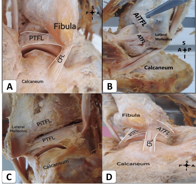
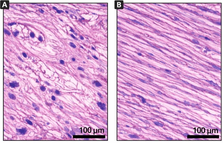
Why instability persists. The cause is multifactorial, and mechanical and functional factors both contribute. Mechanically, the ligament heals incompletely and is left elongated or attenuated. Functionally, mechanoreceptor damage leaves a proprioceptive deficit, the peroneal muscles are weak and neuromuscular control is altered.
The functional deficits. Functional instability shows as:
- Proprioceptive deficit: impaired joint position sense
- Reflex delay: delayed peroneal reaction time
- Muscle weakness: peroneal and inverter weakness
- Postural control deficit: impaired balance
- Somatosensory change: altered afferent input
Classification Systems
Mechanical vs Functional Classification
Mechanical instability is true ligamentous laxity: the anterior drawer or talar tilt is positive, and it needs structural restoration. Functional instability is giving way without laxity, from a proprioceptive deficit and peroneal weakness, and it responds well to rehabilitation. The two can coexist, and most patients with CAI have elements of both, so management has to address both.
- Definition
- True ligamentous laxity
- Examination
- Positive drawer/tilt, stress XR abnormal
- Treatment Focus
- Surgical stabilization
- Definition
- Giving way without laxity
- Examination
- Normal exam, proprioceptive deficit
- Treatment Focus
- Proprioceptive rehabilitation
- Definition
- Both mechanical and functional
- Examination
- Laxity + neuromuscular deficit
- Treatment Focus
- Surgery + comprehensive rehab
Clinical Assessment
History. Establish the index injury and every event since. The symptoms are giving way, swelling, pain and difficulty on uneven ground. Ask about the sport or activity level, with its demands and expectations, and what has already been tried: bracing, rehabilitation or surgery.
Examination. Inspect for swelling and alignment, cavovarus in particular. Palpate the ATFL, CFL, peroneal tendons and sinus tarsi, and record ankle and subtalar range of motion before the stress tests.
- Technique
- Knee flexed 90 degrees, ankle neutral; stabilise tibia, translate talus anteriorly
- Positive Finding
- Greater than 10mm or greater than 3mm vs contralateral
- Structure Tested
- ATFL
- Technique
- Stabilise tibia, invert hindfoot, compare with contralateral
- Positive Finding
- Greater than 10 degrees or greater than 5 degrees asymmetry
- Structure Tested
- CFL (+ ATFL if positive)
- Technique
- Maximal inversion with ankle neutral
- Positive Finding
- Pain, apprehension, increased motion
- Structure Tested
- Lateral ligament complex
- Technique
- Resist eversion, check for subluxation
- Positive Finding
- Weakness, subluxation over malleolus
- Structure Tested
- Peroneal tendons
Absolute values are unreliable because of normal variability, so asymmetry matters more than the absolute measurement. Examine both ankles in the same position and always compare with the contralateral side.
Always assess: subtalar instability (Broden stress), peroneal tendon integrity (subluxation, tears), generalised ligamentous laxity (Beighton score), hindfoot alignment (cavovarus = higher recurrence), and the contralateral ankle for comparison.
Differential Diagnosis of the "Giving-Way" Ankle
Recurrent giving way is not always lateral ligament instability. The patient with normal stress tests may have a problem elsewhere, and missing it is a classic viva trap.
- Distinguishing Features
- Positive drawer/tilt, plantarflexion-inversion mechanism
- Best Test
- Stress radiographs, MRI
- Why It Mimics CAI
- True ATFL/CFL laxity - the index diagnosis
- Distinguishing Features
- Giving way, NORMAL stress tests, peroneal weakness
- Best Test
- Balance/proprioception testing
- Why It Mimics CAI
- Neuromuscular deficit without structural laxity
- Distinguishing Features
- Sinus tarsi pain, hindfoot giving way, Broden views
- Best Test
- Broden stress views, exam under anaesthesia
- Why It Mimics CAI
- Often coexists; tilt may be falsely attributed to CFL
- Distinguishing Features
- Posterolateral pain, snapping, weak eversion
- Best Test
- Dynamic ultrasound, MRI
- Why It Mimics CAI
- Peroneals are dynamic stabilizers - failure feels like instability
- Distinguishing Features
- Deep ankle pain, catching, effusion
- Best Test
- MRI, arthroscopy
- Why It Mimics CAI
- Catching/locking misread as giving way
- Distinguishing Features
- Anterolateral pain on dorsiflexion, no laxity
- Best Test
- MRI, diagnostic arthroscopy
- Why It Mimics CAI
- Pain limits activity, mimics unstable ankle
- Distinguishing Features
- Visible varus, lateral overload, recurrent sprains
- Best Test
- Weight-bearing alignment views, Coleman block
- Why It Mimics CAI
- Structural varus drives recurrent inversion injury
Investigations
Weight-bearing radiographs first. AP, lateral and mortise views, taken weight-bearing because that is essential for assessing alignment. They show malalignment, an osteochondral lesion, talar dome changes and arthritis, and are often normal in isolated ligament injury. A hindfoot alignment view shows cavovarus.
Stress radiographs. Anterior drawer stress views (Telos device) and inversion stress views quantify mechanical instability, read against the same thresholds as the clinical tests and always against the uninjured side. A positive drawer means the ATFL is incompetent; a positive tilt means the CFL is involved as well as the ATFL. Broden stress views assess the subtalar joint.
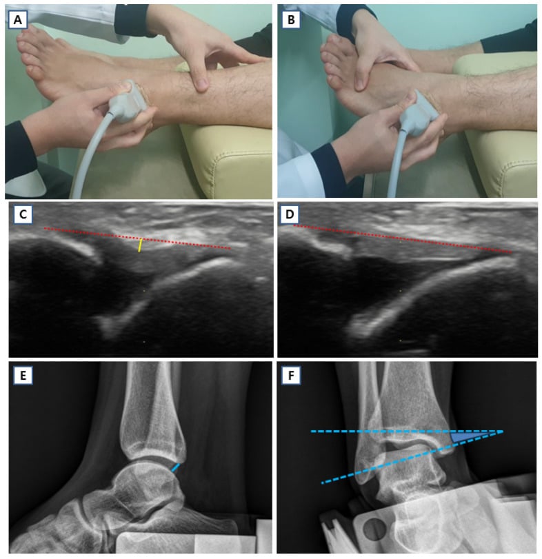
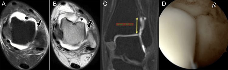
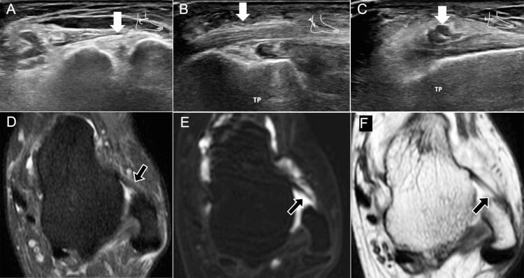
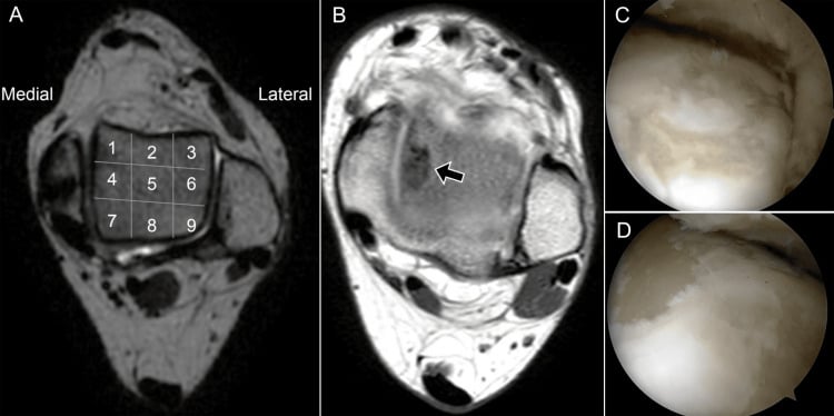
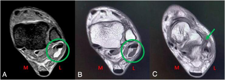
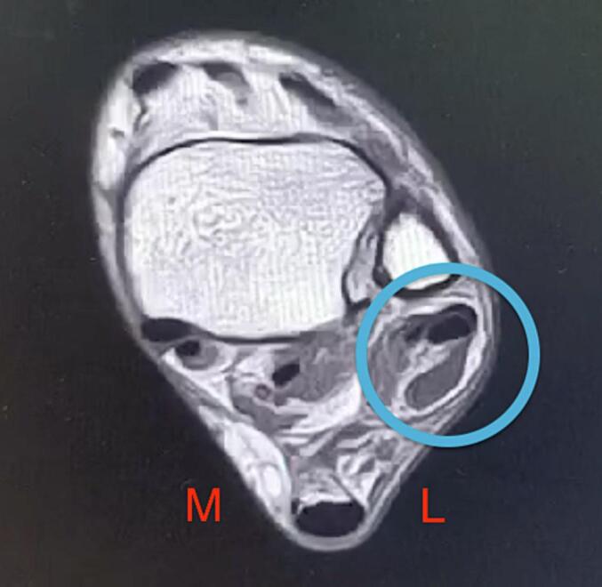
MRI. MRI assesses ligament and tissue quality for surgical planning and finds the associated lesions (osteochondral lesion, peroneal pathology, impingement). Chronic ligament change shows as thickening, discontinuity or scarring. What to report:
- ATFL: thickened, attenuated or absent
- CFL: integrity and scarring
- Osteochondral lesion: location, size and stability
- Peroneal tendons: tears and subluxation
Management Algorithm
Rehabilitation first. Structured, exercise-based rehabilitation comes first, and surgery is reserved for its failure. The guide below matches presentation, examination and imaging to management.
- Examination
- Normal drawer/tilt, weak peroneals
- Imaging
- XR normal, consider MRI
- Management
- 6 months proprioceptive rehab
- Examination
- Positive drawer, minimal tilt
- Imaging
- Stress XR equivocal
- Management
- Trial bracing + rehab first
- Examination
- Positive drawer AND tilt
- Imaging
- Stress XR positive
- Management
- Surgical stabilization
- Examination
- Positive exam, generalized laxity
- Imaging
- MRI shows tissue quality
- Management
- Graft reconstruction
Non-Operative Management Protocol
The goal is to restore proprioception, strength and neuromuscular control.
Rehabilitation Phases
Acute management: RICE for any acute exacerbations. Ankle bracing for activity. Begin ROM exercises. Initiate proprioceptive training (single leg stance, wobble board).
Strengthening and proprioception: Progressive peroneal strengthening. Balance training progression. Sport-specific agility introduction. Continue bracing for activity.
Return to activity: Graduated return to sport. Continue proprioceptive maintenance. Brace use as needed. Monitor for recurrent symptoms.
60-80% success rate with comprehensive rehabilitation. Better outcomes with: functional instability only, good compliance, lower demands, and adequate peroneal strength. Failure indicators: mechanical laxity, cavovarus foot, high-demand athlete.
How long to persist. Give a minimum of 6 months of supervised rehabilitation before considering surgery. The Vuurberg guideline states the standard rather than the clock: surgery is reserved for failure of thorough, comprehensive exercise-based treatment, and it does not define how long that trial must be.
Surgical Technique
Choosing the operation. The modified Brostrom-Gould, an anatomic repair reinforced with the inferior extensor retinaculum (IER), is the first-line operation. Anatomic graft reconstruction, increasingly popular, is for revision, generalised laxity or poor tissue. Non-anatomic tenodesis is largely historical.
Modified Brostrom Procedure (Gold Standard)
Surgical Steps
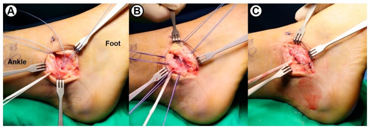
Lateral decubitus or supine with bump. Thigh tourniquet. Mark landmarks: lateral malleolus, ATFL course, peroneal tendons, sinus tarsi.
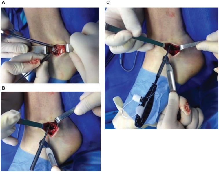
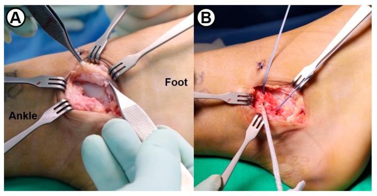
Curvilinear incision anterior and distal to lateral malleolus (following ATFL). Identify and protect superficial peroneal nerve branches. Incise IER (inferior extensor retinaculum).
Identify ATFL (often attenuated, scarred). Assess CFL through same incision or separate distal limb. Evaluate tissue quality for repair vs reconstruction decision.
Incise ATFL capsule longitudinally at its talar attachment. Prepare fibular footprint with decortication. Pass suture anchors (2-3) into anterior fibula. Pants-over-vest imbrication of ATFL with ankle in neutral.
Key step: Advance IER (inferior extensor retinaculum) over ATFL repair and secure to fibula with sutures. This provides critical reinforcement and limits anterior translation.
If CFL involved, extend incision distally. Repair CFL to calcaneus with suture anchor or directly if tissue quality permits. Tension with ankle in neutral to slight eversion.
Tension the repair with the ankle in neutral: over-tightening causes stiffness and under-tightening recurrence. IER augmentation (Gould) is critical for improved outcomes. Consider arthroscopy first to assess and treat an OLT.
After repair the ankle is immobilised in a walking boot for 2-4 weeks, then weight-bearing and rehabilitation progress.
Arthroscopic and Arthroscopic-Assisted Brostrom Repair
The open modified Brostrom-Gould remains the reference standard, but arthroscopic and arthroscopic-assisted ("all-inside") lateral ligament repair has grown rapidly because it treats intra-articular pathology and repairs the ligament in a single setting. Early functional outcomes are equivalent to open repair (Song and Hua meta-analysis); the trade-off is a distinct nerve-injury profile.
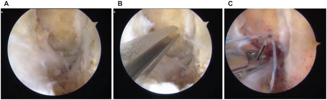
Technique. Diagnostic arthroscopy comes first, through standard anteromedial and anterolateral portals, so that synovitis, loose bodies, anterolateral impingement and any osteochondral lesion are treated in the same sitting. The all-inside repair then follows:
- One or two suture anchors into the anterior fibula at the ATFL footprint, under direct vision
- The sutures capture the distal ATFL remnant and the inferior extensor retinaculum, recreating the Gould reinforcement, and are tied over the retinaculum
- Arthroscopic-assisted variants use a small accessory incision to retrieve and tension the sutures
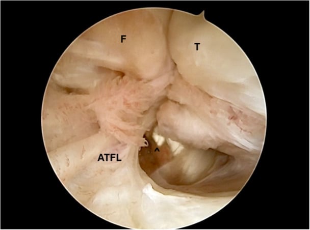
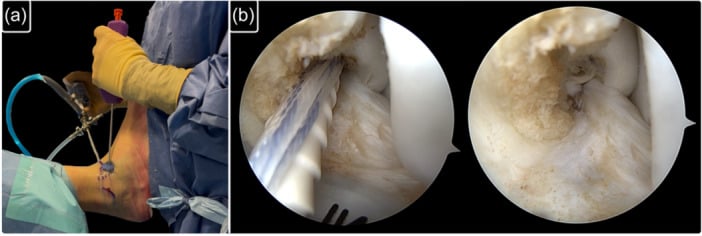
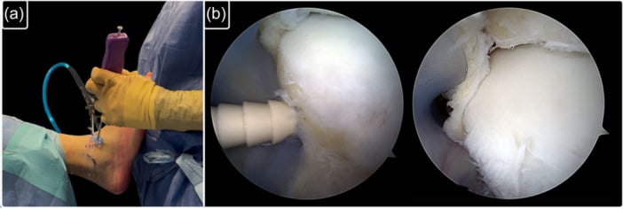
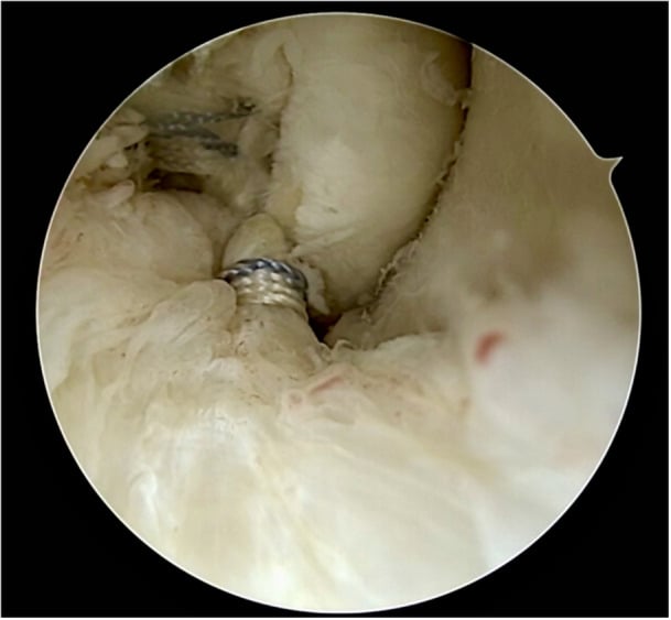
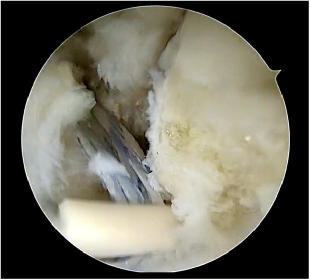
Nerves at risk. The superficial peroneal nerve (intermediate dorsal cutaneous branch) crosses the anterolateral working zone and is the structure most often bruised or caught in a suture. The sural nerve is at risk with more posterior, distal anchor and knot placement near the CFL. Careful portal placement, transillumination and avoiding blind percutaneous suture passage reduce neuritis, the commonest reported complication, but do not eliminate it.
Arthroscopic repair suits primary cases with good-quality remnant tissue and coexisting intra-articular pathology to address. Because it depends on that remnant, poor tissue, generalised hyperlaxity, revision surgery and significant deformity still favour open anatomic repair or graft reconstruction. Long-term durability data for the arthroscopic technique remain immature, so the pragmatic answer is to match technique to tissue quality and surgeon experience.
Complications
- Risk Factors
- Poor tissue quality, cavovarus, non-compliance
- Prevention
- Proper patient selection, address alignment
- Management
- Revision reconstruction with graft
- Risk Factors
- Over-tensioning, prolonged immobilization
- Prevention
- Appropriate tensioning, early ROM
- Management
- Physiotherapy, possible MUA
- Risk Factors
- Surgical approach
- Prevention
- Careful dissection, identify nerve
- Management
- Observation, most resolve
- Risk Factors
- Diabetes, obesity, smoking
- Prevention
- Meticulous technique, optimize health
- Management
- Wound care, possible debridement
- Risk Factors
- Non-anatomic procedures
- Prevention
- Use anatomic techniques
- Management
- PT, accept some limitation
Recurrence runs at 5-15% even with modern techniques, and is higher with a cavovarus foot, generalised ligamentous laxity (use graft augmentation), poor rehabilitation compliance and a return to high-demand sport too early.
The cavovarus foot. Cavovarus alignment places the lateral ligaments under increased stress, and a significant deformity should be addressed surgically. If significant hindfoot varus is present, consider a calcaneal osteotomy (Dwyer) at the time of ligament reconstruction to correct alignment and reduce the risk of recurrence.
Postoperative Care
The protocol runs in four phases, from protection to a criteria-based return to sport.
Rehabilitation Protocol
Immobilization: Below-knee cast or controlled motion boot. Non-weight-bearing initially. Elevate, ice, wound care. Begin toe/knee ROM to prevent stiffness.
Protected mobilization: CAM boot weight-bearing as tolerated. Begin ankle ROM exercises out of boot. Avoid inversion initially. Start isometric strengthening.
Progressive loading: Wean from boot to supportive footwear. Progressive resistance training. Proprioception exercises. Gait training, stationary bike.
Return to activity: Sport-specific drills. Agility and plyometric progression. Criteria-based return to sport. Consider bracing for return.
Return criteria. Return to sport is typically 4-6 months after surgery, once the patient has:
- Greater than 90% strength compared with the contralateral side
- Full range of motion without pain
- Passed functional hop tests
- Tolerated sport-specific drills
Bracing. Consider a brace for the first 6-12 months of sport; it may be used permanently for high-demand activities. Ankle support reduces re-injury risk, and tape is an alternative if the patient prefers it.
Outcomes and Prognosis
- Success Rate
- 85-95%
- Recurrence
- 5-10%
- Return to Sport
- 85-90%
- Success Rate
- 80-90%
- Recurrence
- 5-15%
- Return to Sport
- 80-85%
- Success Rate
- 70-80%
- Recurrence
- 10-20%
- Return to Sport
- 70-80%
- Success Rate
- 70-85%
- Recurrence
- 15-25%
- Return to Sport
- 70-80%
Durability. Brostrom-Gould has long-term outcome data beyond 20 years: at a mean 26 years (Bell), 91% of those who replied rated the ankle good or excellent, though by mailed questionnaire rather than examination. Anatomic reconstruction approaches similar outcomes when properly indicated. The keys to success are appropriate patient selection, treating the associated pathology and comprehensive rehabilitation.
Outcome Measures and Case Identification
Name a validated tool rather than relying on an eyeball assessment. The instruments fall into two groups: case-identification questionnaires, which establish whether a patient genuinely has chronic ankle instability, and outcome scores, which track results and allow studies to be compared.
- Type
- Case identification
- What It Measures
- Severity of functional instability from 9 items, scored 0 to 30 (lower = more unstable)
- Notes
- A score of 27 or below is the commonly cited threshold for instability; validated and assessed per side
- Type
- Case identification
- What It Measures
- History of the first sprain plus current giving-way episodes
- Notes
- Aligns with international consensus (Ankle Consortium) inclusion criteria
- Type
- Outcome
- What It Measures
- Ankle-instability-specific function, 0 to 100
- Notes
- Primary outcome in several instability trials (e.g. the OLT single-stage RCT)
- Type
- Outcome
- What It Measures
- Activities-of-daily-living subscale plus a Sport subscale
- Notes
- Region-specific and responsive to change
- Type
- Outcome
- What It Measures
- Pain, function and alignment, 0 to 100
- Notes
- Widely reported but criticised for limited formal validation
Saying a patient "feels unstable" is not enough. The International Ankle Consortium recommends confirming chronic ankle instability with a validated questionnaire such as the CAIT, together with a documented history of recurrent sprains and giving way persisting for at least 12 months. A defined tool separates true instability from a vague symptom and underpins consistent research and audit.
Guidelines, Registries & Global Practice
Global Epidemiology
Lateral ankle sprain is one of the most common musculoskeletal injuries worldwide, with an estimated incidence around 2-7 per 1000 person-years in the general population and far higher in court and field sports (basketball, volleyball, football/soccer, netball, rugby). Roughly 20-40% of sprains lead to chronic ankle instability. The injury affects all regions and resource settings; the burden is concentrated in young, active populations, and women have a higher rate in indoor court sports.
Side-by-Side Guideline Comparison
- Initial Management
- Functional treatment: brace/tape PLUS supervised exercise; avoid prolonged immobilisation
- Imaging Stance
- Delayed exam at 4-5 days most reliable; imaging for refractory/atypical cases
- Surgical Position
- Reserve surgery for failure of comprehensive exercise-based rehab
- Initial Management
- Functional rehab and neuromuscular training first-line
- Imaging Stance
- Stress radiographs and MRI to confirm mechanical laxity and associated lesions before surgery
- Surgical Position
- Anatomic repair (modified Brostrom-Gould) as standard; reconstruction for revision/poor tissue
- Initial Management
- PRICE/POLICE then exercise rehab; brace for recurrent sprains
- Imaging Stance
- Selective imaging; MRI when associated pathology suspected
- Surgical Position
- Surgery for documented mechanical instability failing rehab
- Initial Management
- Conservative rehabilitation first
- Imaging Stance
- Stress imaging and MRI for surgical planning
- Surgical Position
- Anatomic repair preferred; arthroscopic management of intra-articular lesions encouraged
Across societies the message is consistent: structured exercise-based rehabilitation first, anatomic (Brostrom-type) repair as the surgical default, and reconstruction reserved for revision, hyperlaxity or poor tissue. Differences are mainly in imaging thresholds and how aggressively associated lesions are sought.
- No dedicated international ligament-repair registry exists (unlike arthroplasty); evidence comes from cohort series, RCTs and meta-analyses
- Patient-reported outcome measures used: Karlsson-Peterson, AOFAS, FAAM, CAIT (Cumberland Ankle Instability Tool) for case identification
- Reported recurrence after modern anatomic repair is roughly 5-15%
- High-resource: stress imaging, MRI, arthroscopy, suture-anchor/graft constructs, supervised return-to-sport programmes
- Limited-resource: clinical diagnosis and functional rehab predominate; bracing/taping widely used; non-anatomic tenodesis may still be performed where anchors/grafts are unavailable
- Functional rehabilitation is effective and low-cost - the global first-line everywhere
Examiners worldwide expect: mechanical vs functional instability, anatomy of the lateral ligament complex (ATFL taut in plantarflexion, CFL extra-articular crossing the subtalar joint), stress radiograph thresholds, the Brostrom-Gould technique, and indications for graft reconstruction.
Controversies and Areas of Uncertainty
High-level evidence in chronic ankle instability is limited, and several questions remain genuinely unsettled.
Arthroscopic versus open Brostrom. Early functional outcomes are equivalent, but long-term durability data are immature and nerve-related complications (superficial peroneal, sural) are a concern. Open repair remains the reference standard.
Where repair ends and reconstruction begins. When to abandon anatomic repair for graft reconstruction is not standardised. The proposed triggers are unvalidated: generalised hyperlaxity (a high Beighton score), poor remnant tissue, BMI or high demand, and revision.
Suture-tape (InternalBrace) augmentation. Some advocate primary augmentation of the Brostrom with non-absorbable tape to allow accelerated rehabilitation; the tape may permit earlier loading and protect the repair, but it risks over-constraint, stiffness and a foreign-body construct. The evidence is mostly short-term cohort data without proven outcome benefit over standard Brostrom-Gould, and long-term superiority is unproven.
Concomitant OLT and subtalar instability. RCT data support treating an OLT in a single stage with the stabilisation. Subtalar instability is harder: its diagnosis is unreliable, with no validated stress threshold, and whether it needs specific surgical attention or a combined ATFL/CFL reconstruction remains debated.
When asked "arthroscopic or open?" or "repair or reconstruct?", state that high-quality long-term comparative data are limited, give the pragmatic indication-based answer, and acknowledge that surgeon experience and tissue quality drive the decision. Avoid dogmatic claims.
MCQ Practice Points
Q: Which ligament is the primary restraint against anterior translation of the talus in plantarflexion? A: Anterior talofibular ligament (ATFL) - The ATFL is taut in plantarflexion and provides 100% of resistance to anterior talar translation at 15 degrees plantarflexion. It is the weakest and most commonly injured lateral ligament.
Q: What unique anatomical feature of the CFL makes it important for subtalar stability? A: CFL is extra-articular and crosses the subtalar joint - Unlike the ATFL which is intra-articular, the CFL crosses both the ankle and subtalar joints, contributing to stability of both articulations.
Q: What is considered a positive anterior drawer stress test in chronic ankle instability? A: Greater than 10mm absolute OR greater than 3mm asymmetry compared to contralateral - Side-to-side comparison is more reliable than absolute values due to individual variation in ligamentous laxity.
Q: What is the gold standard first-line surgical procedure for chronic ankle instability? A: Modified Brostrom-Gould - This anatomic repair involves ATFL imbrication to the fibula with suture anchors plus reinforcement with the inferior extensor retinaculum (Gould modification), achieving 85-95% success rates.
Q: What is the purpose of the Gould modification in the Brostrom procedure? A: Reinforcement with inferior extensor retinaculum (IER) - The IER is advanced over the ATFL repair and secured to the fibula, providing additional anterior restraint and improving outcomes compared to ATFL repair alone.
Exam Viva Scenarios
Practise clinical reasoning and management decisions out loud
“A 22-year-old soccer player presents with recurrent ankle sprains over 2 years. He has completed 6 months of physiotherapy with bracing. Examination shows positive anterior drawer (12mm vs 6mm contralateral). He wants to return to competitive sport.”
“A 35-year-old woman had a Brostrom repair 3 years ago that failed after 18 months. She now has persistent instability, giving way episodes, and anterior drawer 14mm (vs 5mm contralateral). MRI shows attenuated ligament remnants.”
“A 28-year-old basketball player has CAI confirmed on stress XR. MRI also shows a 1.2cm medial talar dome osteochondral lesion with overlying cartilage damage. He has failed conservative treatment.”
“A 30-year-old recreational runner has recurrent giving way and ankle sprains. She has completed 3 months of physiotherapy. Examination shows negative anterior drawer and talar tilt. She has significant peroneal weakness and poor single-leg balance.”
Key Anatomy
- ATFL: weakest ligament, intra-articular, taut in PLANTARFLEXION
- CFL: EXTRA-ARTICULAR, crosses subtalar joint, taut in DF
- PTFL: strongest, rarely injured
- Anterior drawer tests ATFL; Talar tilt tests CFL
Instability Types
- MECHANICAL: true laxity (positive stress tests/XR)
- FUNCTIONAL: giving way WITHOUT laxity (proprioceptive deficit)
- Most CAI patients have BOTH components
- Surgery for mechanical; Rehab for functional
Stress Test Thresholds
- Anterior drawer: greater than 10mm OR greater than 3mm asymmetry = positive
- Talar tilt: greater than 10 degrees OR greater than 5 degrees asymmetry = ATFL + CFL
- Always COMPARE to contralateral side
- Asymmetry more reliable than absolute values
Surgical Approach
- Brostrom-Gould = GOLD STANDARD (85-95% success)
- Gould modification = IER reinforcement (critical)
- Graft reconstruction for: revision, hyperlaxity, poor tissue
- Non-anatomic tenodesis largely HISTORICAL
Exam Pearls
- 6 months conservative before surgery
- ATFL provides 100% resistance to anterior translation at 15 degrees PF
- Always assess for OLT (15-25%), peroneals, subtalar instability
- Cavovarus = higher recurrence - consider calcaneal osteotomy
Evidence Base
DiGiovanni BF et al. Associated injuries found in chronic lateral ankle instability
- In 61 patients undergoing lateral ligament reconstruction, NO ankle had isolated ligament injury. Most common associated lesions: peroneal tenosynovitis 77%, anterolateral impingement 67%, attenuated peroneal retinaculum 54%, ankle synovitis 49%, loose body 26%, peroneus brevis tear 25%, talar osteochondral lesion 23%.
Bell SJ et al. Twenty-six-year results after the Brostrom procedure
- Thirty-one male midshipmen (32 ankles) at the US Naval Academy underwent the Brostrom procedure; 22 of the 31 were reached at a mean 26.3 years. Mean ankle function 91 of 100 and 91 per cent rated the ankle good or excellent - figures that belong to the 22 who replied, not to the original 32 ankles.
Krips R et al. Long-term outcome of anatomical reconstruction versus tenodesis
- Multicentre comparison (25 anatomical reconstruction vs 29 tenodesis), mean 12.3-year follow-up. Tenodesis had more positive anterior drawer, greater talar tilt and translation on stress radiographs, more medial degenerative change, and fewer excellent results. Tenodesis does NOT restore normal ligament anatomy.
Vuurberg G et al. Diagnosis, treatment and prevention of ankle sprains: update of an evidence-based clinical guideline
- Functional treatment (tape/brace plus a supervised exercise programme) is favoured over immobilisation for acute lateral ligament rupture. Surgery is reserved for cases failing comprehensive exercise-based treatment. Bracing reduces recurrent sprains. NSAIDs may impair healing.
Song YJ, Hua YH. Arthroscopic vs open repair of chronic ankle instability: systematic review and meta-analysis
- 4 comparative studies (1 RCT), 207 patients. No statistically significant difference in functional outcome scores between arthroscopic and open Brostrom repair at early term; both gave favourable results, with one study showing faster return to daily activity after arthroscopy.
Wei Y et al. Single-stage versus staged treatment of OLT with chronic lateral ankle instability: RCT
- RCT of 103 patients with both CLAI and osteochondral lesion of the talus. Concurrent (single-stage) microfracture plus modified Brostrom-Gould was non-inferior to staged surgery at 24 months, and gave better Karlsson-Peterson and AOFAS scores at short-term follow-up.