Fleck Sign | Modified Thompson | Olecranon Avulsion | Active Extension Loss
- Most ruptures are AVULSIONS off the olecranon - the tendon-bone junction (enthesis) is the weak point, not the tendon midsubstance
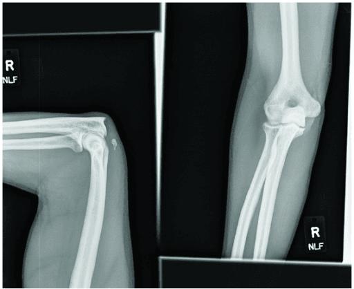
- Fleck sign on a lateral elbow radiograph (a small avulsed bony flake from the olecranon) is the pathognomonic plain-film clue - always get an X-ray
- Test active extension against gravity: ask the patient to extend the elbow with the arm overhead/horizontal. A modified Thompson-type squeeze of the triceps belly should produce passive extension if the tendon is intact
- Complete tears in active patients are repaired surgically; partial tears (less than 50%) with preserved antigravity extension can be treated non-operatively
- Risk factors: anabolic steroids, local or systemic corticosteroid, chronic kidney disease, hyperparathyroidism, weightlifting and olecranon bursitis injection
- “Antigravity extension can be preserved despite a complete tear because the anconeus and intact lateral tendinous expansion compensate - so always palpate for a gap AND get an X-ray
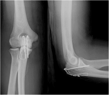
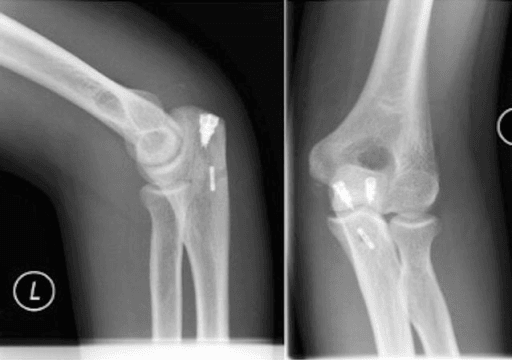
- “Suture anchor repair has lower re-rupture and complication rates than transosseous bone-tunnel repair in recent systematic reviews
- “A partial tear that fails a non-operative trial, or any tear in a high-demand athlete, should be repaired
- “Delayed/chronic tears retract and scar - they may need V-Y turndown or tendon (e.g. Achilles) allograft augmentation
Triceps Tendon Rupture
Antigravity elbow extension may be partly preserved even with a complete tear (anconeus and the intact lateral tendinous expansion compensate). A patient who can "just about" straighten the arm can still have a complete rupture - palpate for a gap and image.
The fleck sign (avulsed olecranon flake) is pathognomonic and easy to miss. A normal X-ray does not exclude a tear - proceed to MRI or ultrasound if clinical suspicion persists.
SATRepair Construct Choices
Hook:SAT down on the footprint: suture anchors, anatomic, then transosseous as the fallback.
Overview & Epidemiology
Triceps tendon rupture is the rarest of the major tendon ruptures, accounting for roughly 1-2% of all tendon injuries. According to PubMed, it occurs most often in men aged 40-60, classically during weightlifting (forced eccentric contraction at the end of a bench-press lockout) or after a fall on an outstretched hand that suddenly flexes a contracting elbow.
Key facts students should know:
- The injury is almost always an avulsion at the tendon-bone junction (enthesis) off the olecranon, not a midsubstance tear, because the enthesis is the mechanically weakest link.
- A large insurance-database cohort identified male sex, age over 60, ischaemic heart disease, rheumatoid arthritis and chronic kidney disease as risk factors for complications, and prior anabolic steroid use as the strongest predictor of needing revision surgery.
- Up to a third of cases have an associated injury (radial head fracture, olecranon bursitis, or wrist/forearm injury from the same fall) - examine the whole limb.
- Because it is so rare, the diagnosis is frequently missed at first presentation, leading to chronic, retracted tears that are harder to repair.
Pathophysiology & Anatomy
- The triceps brachii has three heads (long, lateral, medial) that converge into a common tendon inserting on the posterior olecranon. It is the primary elbow extensor.
- The distal tendon is bilaminar: a thick superficial layer (from the long and lateral heads) inserts more proximally, and a deep layer (mainly the medial head) inserts more distally and broadly. MRI tear-classification systems are built on this two-layer structure.
- A lateral tendinous expansion blends with the forearm fascia and anconeus. When intact, it allows weak residual extension even after a complete central tear - the source of the "antigravity trap".
- Eccentric overload: a sudden extension force against an actively contracting (decelerating) triceps - the lockout phase of a heavy bench press is the textbook example.
- The avulsion propagates through the enthesis, sometimes lifting a thin cortical flake of olecranon (the fleck sign).
- Pre-existing tendinopathy weakens the insertion. Anabolic steroids, fluoroquinolones, repeated local steroid injection for olecranon bursitis, and metabolic bone disease (renal failure, hyperparathyroidism) all degrade collagen and predispose to rupture at lower loads.
Classification
Clinical Presentation
- Sudden pain and a "pop" at the back of the elbow during a lift or after a fall.
- Weakness extending the elbow, or difficulty pushing up from a chair / doing press-ups.
- Ask specifically about steroid use, renal disease and previous olecranon injections.
- Inspection: Posterior elbow swelling, ecchymosis, sometimes a visible defect.
- Palpation: A palpable gap just proximal to the olecranon is the most useful sign.
- Active extension against gravity: Position the shoulder so the elbow must extend against gravity (arm overhead or horizontal). Inability, or marked weakness, suggests a complete tear - but remember weak residual extension does not exclude it.
- Modified Thompson test (squeeze test): With the arm supported and elbow flexed, squeeze the triceps muscle belly. In an intact tendon the elbow passively extends; absence of extension indicates a complete rupture.
Investigations
- Radiographs first (AP + lateral): Look for the fleck sign - an avulsed flake of olecranon cortex, pathognomonic of a triceps avulsion. Also excludes associated radial head/olecranon fractures.
- Ultrasound: Cheap, dynamic, operator-dependent; good for confirming a tear and assessing gap, useful in resource-limited settings.
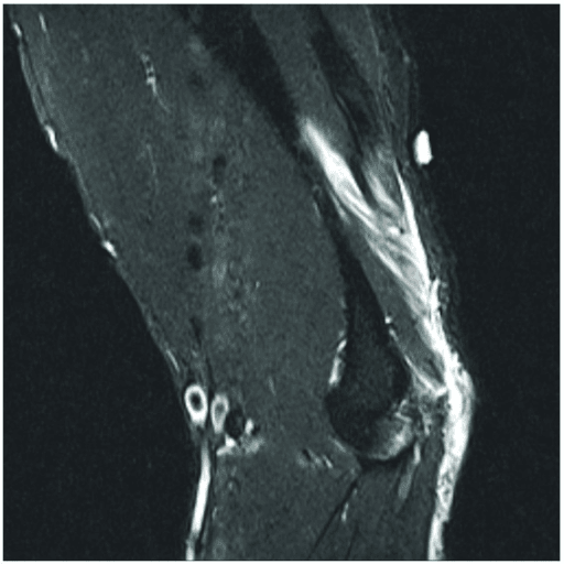
- MRI: Best for equivocal cases, partial tears and surgical planning - quantifies the percentage of tendon torn, which layers are involved, gap/retraction, and muscle quality in chronic tears. Sagittal fat-saturated sequences show fluid at the footprint.
A confident clinical diagnosis (gap + antigravity loss + fleck sign) needs no advanced imaging before surgery, but MRI is invaluable when the examination is equivocal or a partial tear is suspected.
GAPDiagnosis Triad
Hook:Mind the GAP: gap on palpation, antigravity loss, plain-film fleck.
Reading the MRI: The Layered (Bilaminar) Tear Pattern
The anatomy section notes the distal triceps is bilaminar, and the classification refers to an "MRI layered classification" — this is the framework MRI actually uses to grade the tear and plan the repair.
- The two layers. A thick superficial layer (long and lateral heads) inserts more proximally on the olecranon; a deep layer (mainly the medial head) inserts more distally and broadly. Sagittal fat-saturated MRI shows each layer separately.
- The commonest real-world pattern. The typical injury is a superficial-layer full-thickness tear with the deep layer still partly attached — which is exactly why a patient can retain weak extension and why the tear is easily under-called: the intact deep layer (plus the anconeus and lateral expansion) masks the superficial avulsion.
- Why it changes the operation. MRI quantifies (1) which layers are torn, (2) the percentage of tendon width involved, and (3) the retraction/gap. A superficial tear with a substantial intact deep layer may suit a single-row/limited repair, whereas a full bilaminar avulsion needs a footprint-restoring double-row/suture-bridge construct.
- Practical rule: when the extension weakness is worse than the tear "looks", suspect an occult superficial-layer avulsion sitting over a still-attached deep layer — image both layers before committing to partial versus complete. Systematic elbow imaging is developed in the imaging-elbow-systematic topic.
Q: Why can MRI under-call a distal triceps tear, and how does the bilaminar anatomy guide repair? A: The commonest pattern is a superficial-layer full-thickness tear with the deep (medial-head) layer still attached, so residual deep fibres (plus anconeus/lateral expansion) preserve weak extension and the tear looks partial. Read both layers, the percentage torn and the retraction: an intact deep layer may allow a single-row repair, a full bilaminar avulsion needs a footprint-restoring double-row construct.
Management
Treatment hinges on completeness of the tear and patient demand. Use the tabs below.
- Partial tears (less than 50%) with preserved antigravity extension.
- Low-demand or high-surgical-risk patients who decline or cannot tolerate surgery.
- Splint/brace at 30-40 degrees of flexion for ~4 weeks, then progressive range of motion.
- Avoid resisted extension for 6 weeks; graded strengthening thereafter.
- Monitor closely - a partial tear that progresses, stays painful, or fails to regain strength should be converted to surgical repair.
Reconstructing the Chronic, Retracted Triceps
The classification and management repeatedly flag that a chronic tear (beyond about 6 weeks) retracts and scars so that primary footprint repair becomes impossible, requiring "V-Y turndown or Achilles allograft" — here are those reconstruction options in full.
- Why primary repair fails late. By around 6 weeks the muscle-tendon unit shortens and adheres, so dragging it back to the olecranon either fails under tension or blocks flexion. First assess muscle quality on MRI — a fatty, atrophic triceps predicts a poorer result whatever is done.
- V-Y triceps turndown (lengthening). A proximally-based inverted-V (tongue) of the scarred triceps aponeurosis is turned distally to bridge the gap and reattached to the olecranon, lengthening the musculotendinous unit so native tissue reaches bone; the donor defect is closed side-to-side. It uses the patient's own tissue with no graft.
- Tendon allograft augmentation. An Achilles tendon allograft — its calcaneal bone block fixed to the olecranon and the tendinous fan woven into the triceps — is the workhorse for a large irreparable gap; semitendinosus/hamstring or fascia lata grafts are alternatives.
- Anconeus rotation flap. The anconeus can be rotated to reconstruct a smaller distal-triceps defect.
- Counselling. Reconstruction outcomes are less reliable than acute repair (weaker extension, some extension lag, stiffness) — which is exactly why the topic stresses diagnosing and repairing within ~3 weeks. The underlying tendon-healing biology is developed in the tendon-healing topic.
Q: A triceps rupture presents at 8 weeks with a retracted, scarred tendon — how does management differ from an acute tear? A: Direct footprint repair is usually impossible, so reconstruct: a V-Y triceps turndown lengthens the patient's own aponeurosis to reach bone, or an Achilles tendon allograft (calcaneal block to olecranon) bridges a large gap; an anconeus flap suits smaller defects. Check muscle quality on MRI and counsel that outcomes are poorer than acute repair.
STEROIDRisk Factors for Triceps Rupture
Hook:A patient on STEROIDs with a weak elbow extensor should make you think triceps rupture.
Complications & Prognosis
- Approx. rate
- 2-7%
- Notes
- Higher with transosseous tunnels and in anabolic steroid users; early non-compliance a key cause
- Approx. rate
- Up to ~10%
- Notes
- Nerve runs medially near the field; usually transient, occasionally needs release/transposition
- Approx. rate
- Common (~9 degrees mean)
- Notes
- More frequent after suture-anchor-only repair in one large cohort; usually minor
- Approx. rate
- Low (a few %)
- Notes
- Higher in dialysis/transplant and diabetic patients
- Approx. rate
- ~3-15%
- Notes
- Overall surgery has a relatively high reoperation rate regardless of technique
Prognosis: Across systematic reviews, surgical repair gives excellent patient-reported outcomes (mean Mayo Elbow Performance Score ~92, high satisfaction), with about 92% returning to pre-injury function and essentially all patients regaining at least grade 4/5 extension strength. Outcomes are best for acute repairs; chronic, retracted tears requiring grafting do less well.
Key Exam Points
Because the anconeus and the intact lateral tendinous expansion of the triceps provide weak residual extension. This is the "antigravity trap" - never exclude a complete tear on partial extension alone. Palpate for a gap and obtain a radiograph (fleck sign) or MRI.
The fleck sign - a small avulsed flake of cortical bone pulled off the posterior olecranon - best seen on the lateral radiograph. It confirms an avulsion-type rupture at the enthesis.
A suture-anchor (double-row/footprint-restoring) repair is reasonable: systematic reviews show lower complication and re-rupture rates than transosseous tunnels, and biomechanically it restores far more of the native footprint with higher load to failure. Transosseous tunnels remain an acceptable, familiar alternative.
Acute repair (less than 3 weeks) allows direct anatomic reattachment. In chronic tears the tendon retracts and scars, so plan for V-Y triceps turndown or Achilles tendon allograft augmentation, and counsel the patient that outcomes are less reliable than acute repair.
Guidelines, Registries & Global Practice
There is no large randomised trial or dedicated society guideline for this rare injury, so practice is guided by systematic reviews and expert consensus. Practice is broadly consistent worldwide.
- Consensus position
- Surgical repair, ideally within ~3 weeks
- Source/evidence
- Systematic reviews (Tran 2022; Alnaji 2021)
- Consensus position
- Non-operative trial; repair if it fails or progresses
- Source/evidence
- Narrative reviews (Keener & Sethi, Hand Clin 2015)
- Consensus position
- Footprint-restoring suture anchors trending over transosseous tunnels
- Source/evidence
- Lower complication/retear rates (Tran 2022; biomechanics Scheiderer 2018)
- Consensus position
- Counsel renal, rheumatoid, cardiac and steroid users on higher complication/revision risk
- Source/evidence
- Large database cohort (Lee 2022)
Registry note: Unlike arthroplasty, there are no national joint-registry data for triceps tendon repair; the strongest population-level evidence comes from large insurance-claims database cohorts (e.g. Lee 2022), which inform risk-factor counselling rather than implant choice.
Exam Viva Scenarios
Practise clinical reasoning and management decisions out loud
“A 46-year-old male recreational weightlifter presents 4 days after feeling a sudden pop at the back of his right (dominant) elbow during the lockout phase of a heavy bench press. He has posterior elbow swelling and bruising and complains of weakness straightening the arm. On examination there is a tender boggy swelling and a subtle palpable defect just above the olecranon. When you ask him to extend the elbow with the shoulder flexed to 90 degrees, he can produce weak extension. A lateral radiograph shows a small flake of bone lifted off the olecranon. He mentions he occasionally uses anabolic steroids. How do you assess, investigate and manage him, and how would you counsel him about his steroid use?”
“A 39-year-old manual labourer fell onto an outstretched hand and has posterior elbow pain and mild weakness extending the elbow against resistance. There is tenderness over the olecranon insertion but you cannot feel a definite gap, and he can extend the elbow against gravity, though it is painful and slightly weak. The plain radiograph is normal with no fleck sign. How do you work out whether this is a partial or complete tear, and how does that change your management?”
Must-Know Facts
- Rarest major tendon rupture (1-2% of tendon injuries); men aged 40-60
- Almost always an AVULSION off the olecranon (enthesis is the weak link)
- Mechanism: eccentric load (bench-press lockout) or fall on outstretched hand
- Risk: anabolic steroids, corticosteroid, renal failure, hyperparathyroidism, olecranon bursitis injection
Diagnosis
- GAP: palpable defect proximal to olecranon
- Antigravity extension loss (but weak residual extension does NOT exclude complete tear)
- Modified Thompson (squeeze) test: no passive extension = complete tear
- Fleck sign on lateral X-ray = pathognomonic; MRI for equivocal/partial tears
Management
- Partial (less than 50%) with preserved extension: non-operative brace trial + close follow-up
- Complete / high-grade partial / athlete: surgical repair, ideally within 3 weeks
- Suture anchors (footprint-restoring): lower complication and re-rupture rates than transosseous tunnels
- Chronic retracted tear: V-Y turndown or Achilles allograft augmentation
Outcomes & Complications
- ~92% return to pre-injury function; mean Mayo Elbow score ~92; high satisfaction
- Re-rupture 2-7% (higher with transosseous and in steroid users)
- Ulnar neuropathy up to ~10% (usually transient); minor extension loss common
- Counsel renal/rheumatoid/cardiac patients and steroid users on higher complication/revision risk
Evidence Base
The evidence below was retrieved from PubMed for this topic. Triceps rupture is rare, so the literature is dominated by systematic reviews of observational series, large database cohorts, and biomechanical/cadaveric studies rather than randomised trials.
Surgical Repair Outcomes - Systematic Review (suture anchor vs transosseous)
- Systematic review of 16 studies of distal triceps repair; mean DASH 4, mean Mayo Elbow Performance Score 92, mean isokinetic strength 87% of normal
- 95% of patients were satisfied and 92% returned to pre-injury function; 100% regained at least grade 4/5 gross strength
- Overall complication rate 15%, with retears ~5%
- Transosseous repair had a higher complication rate (18% vs 8%) and higher retear rate (7% vs 2%) than suture anchor repair
Surgical Management - Systematic Review of Techniques
- 19 studies, 560 patients (565 tendons), 78.6% male, mean age 46 years, mean follow-up 32 months
- Overall complication rate 14.9%; by technique: direct repair 29.2%, transosseous 15.2%, suture anchor 7.7%
- Re-rupture: transosseous 4.3%, suture anchor 2.1%, direct repair 0%
- Significant postoperative improvement in pain, strength and range of motion across all techniques
Footprint Restoration & Repair Strength - Biomechanics
- 24 cadaveric elbows: transosseous cruciate vs knotless suture-bridge vs V-shaped unicortical button repair
- Transosseous repair restored only 37% of the native footprint vs ~85-89% for the suture-bridge and V-shaped button techniques
- Mean peak load to failure: V-shaped button 732 N, suture-bridge 505 N, transosseous cruciate 281 N
- Footprint-restoring constructs also showed less gap formation than transosseous repair
Primary Repair in a General Population - Multicentre Series
- 28 primary triceps reinsertions (23 acute, 5 chronic), mean age 45 years, mean follow-up 47.5 months
- Mean Mayo Elbow Performance Score 94, mean QuickDASH 10; satisfactory results in 93%
- Strength 5/5 in 18 and 4/5 in 10 tendons; only one re-rupture (a patient with chronic renal failure)
- Both transosseous and suture-anchor reinsertion gave satisfactory results in acute and chronic tears
Trends, Outcomes & Risk Factors - Large Database Cohort
- 8143 distal triceps repairs from an insurance claims database; most common in men aged 40-59
- 90-day complication rate 5.8%; 1-year revision rate 2.6%
- Risk factors for 90-day complications: male sex, age over 60, ischaemic heart disease, rheumatoid arthritis, chronic kidney disease
- Prior anabolic steroid use significantly increased the risk of 1-year revision surgery