Angular Deviation in Coronal Plane
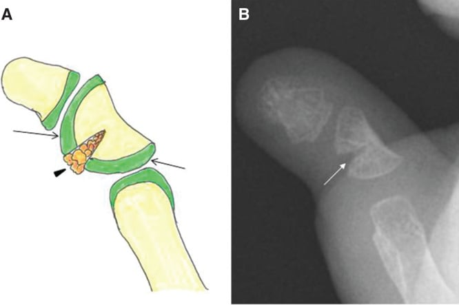
- Delta phalanx: Triangular bone with C-shaped bracket epiphysis that spans two sides causing angular growth.
- Most Common: Small finger (radial deviation) - middle phalanx.
- Down syndrome: Over 50% have clinodactyly.
- Surgical indications: progression, digital overlap or functional impairment at ANY angle; over 30 degrees is the conventional prompt, but 15-30 degrees is inside the published osteotomy indication (PMID 19362790).
- Procedures: Opening wedge osteotomy (most common) or physiolysis for younger children.
- “Delta phalanx = C-shaped bracket epiphysis
- “30 degrees is a prompt, not a gate - osteotomy is published for 15-30 too
- “Small finger most commonly affected
- “Down syndrome strong association
- “Opening wedge needs bone graft
Overview and Epidemiology
Clinodactyly is angular deviation of a digit in the coronal (radioulnar) plane. The word is Greek, klinein (to bend) and daktylos (finger), and the deviation it names is most often a little finger bent radially. Camptodactyly is the other bent little finger of the congenital hand clinic, but that is a flexion deformity in the sagittal plane. Kirner deformity is the eponym for a volar-radial curvature of the little finger distal phalanx.
How common. Between 1 and 10% of the population, the figure depending on the angle at which a bent finger is called abnormal. The sexes are affected equally, 60-70% of cases are bilateral, and inheritance is often autosomal dominant with variable penetrance. The strongest association is with Down syndrome, in which over 50% of children are affected, though most clinodactyly is isolated and non-syndromic.
- Incidence
- Over 50%
- Other Hand Features
- Single palmar crease, short 5th finger
- Incidence
- Common
- Other Hand Features
- Complex syndactyly, broad thumb
- Incidence
- Common
- Other Hand Features
- Small hands, asymmetry
- Incidence
- Variable
- Other Hand Features
- Symbrachydactyly, absent pectoralis
- Incidence
- Most common
- Other Hand Features
- No other anomalies
Embryology. The digits form between weeks 4 and 8 of gestation and the phalanges chondrify in weeks 5-6. A normal physis forms as a straight transverse plate; abnormal chondrification produces a delta phalanx, in which a bracket epiphysis develops in place of the linear physis.
Where its siblings are. Clinodactyly is one of the digit-shape anomalies in the congenital hand overview, and the one it is most often confused with is camptodactyly - bent in a different plane, flexion rather than angulation, and a different operation entirely. Syndactyly matters here specifically because its presence pushes the choice away from physiolysis. The association everyone tests is with Down syndrome.
Pathoanatomy and Biomechanics
The normal phalanx. A tubular bone with parallel sides and a single straight transverse physis at one end. Growth is equal on the radial and ulnar sides, so the bone lengthens without turning and the digital cascade stays straight in the coronal plane.
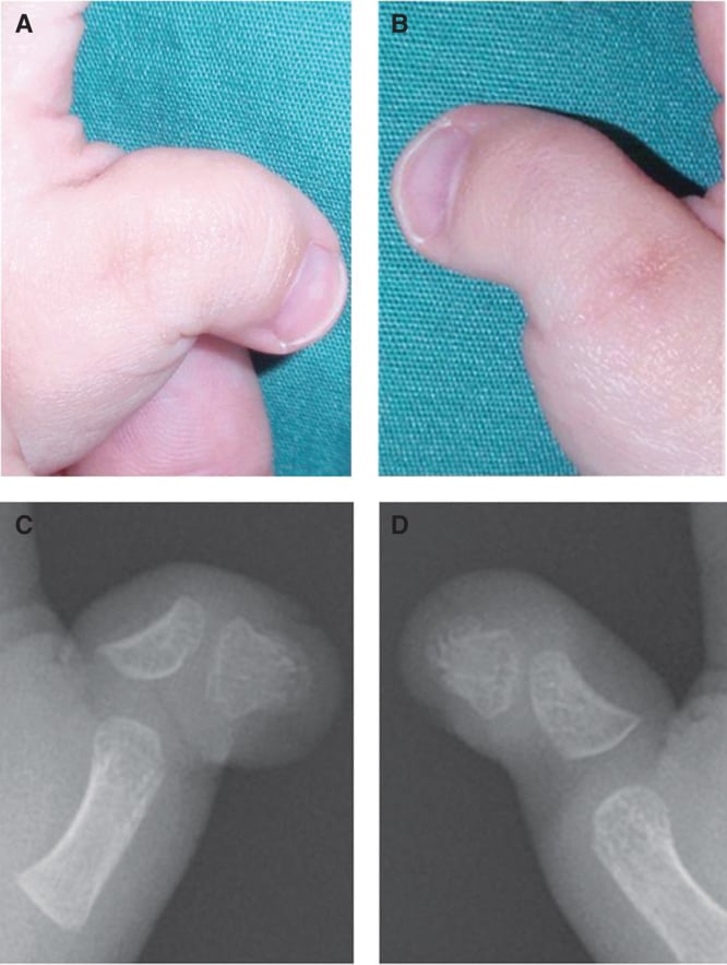
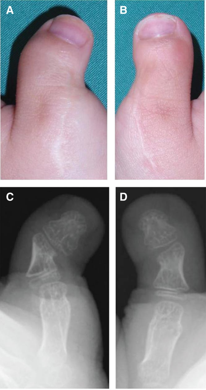
The delta phalanx. The bone is triangular or wedge-shaped rather than rectangular, and its epiphysis is a C-shaped bracket that curves round to span two sides of the bone instead of one. The bracketed side, usually the radial, grows less than the free side, so the bone lengthens unequally and the digit deviates a little further with every increment of skeletal growth.
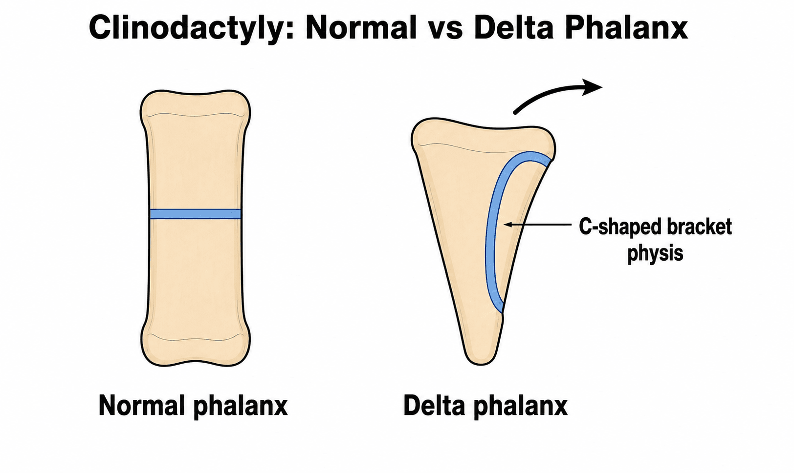
Biomechanics. The Hueter-Volkmann principle applies: compression inhibits physeal growth and tension stimulates it. The bracket tethers one side while the other continues to grow, so the deformity worsens through growth spurts and stabilises only at skeletal maturity. Remodelling potential in a coronal-plane deformity is limited.
Physiological or pathological. Most of the common 1-10% population little-finger clinodactyly is physiological: a mild, non-progressive, often familial (autosomal dominant) deviation from a slightly trapezoidal middle phalanx without a true longitudinal epiphyseal bracket, needing only observation and reassurance. The minority that is progressive and surgically relevant has a genuine delta phalanx, a longitudinal epiphyseal bracket that tethers one side and worsens with growth.
Confirm a true bracket epiphysis on the AP radiograph before labelling a clinodactyly "delta phalanx" or recommending surgery. A mild, non-progressive deviation is not a delta phalanx.
Where. The little finger is the commonest site, at the middle phalanx with radial deviation, often bilateral, and it is the pattern associated with Down syndrome. The index finger is second. The thumb is rare and, when involved, often syndromic; involvement of several digits also points to a syndrome.
The thumb. A delta phalanx in the thumb, often producing a triphalangeal thumb with thumb clinodactyly, is far more likely to be syndromic than little-finger clinodactyly and should trigger a targeted screen. The associations to know:
- Holt-Oram syndrome - cardiac septal defects
- Fanconi anaemia
- Thrombocytopenia-absent-radius (TAR) syndrome
- Blackfan-Diamond anaemia
- Rubinstein-Taybi syndrome - broad thumbs
A thumb, which is to say radial-ray, anomaly therefore warrants a cardiac and haematological work-up (echocardiography, full blood count and film, and chromosomal-breakage testing where Fanconi is suspected), not just a hand assessment.
Clinical Assessment
History. The questions that decide between observation and surgery:
- When the deformity was first noticed
- Whether it is progressing or stable
- Family history, looking for the autosomal dominant pattern
- Functional limitation: overlapping, grip problems
- Syndromic features: developmental delay, cardiac problems
- What the child and parents make of the appearance
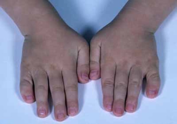
Inspection. Record the angle of deviation in degrees, the digit or digits involved and the direction, radial or ulnar, whether the other hand is affected, and any accompanying syndactyly or polydactyly. Look for a rotational component, the alignment of the nail and the pattern of the skin creases. Then look at the child: the flat facial profile and single palmar crease of Down syndrome, the craniosynostosis and mitten hand of Apert, the growth retardation and asymmetry of Russell-Silver.
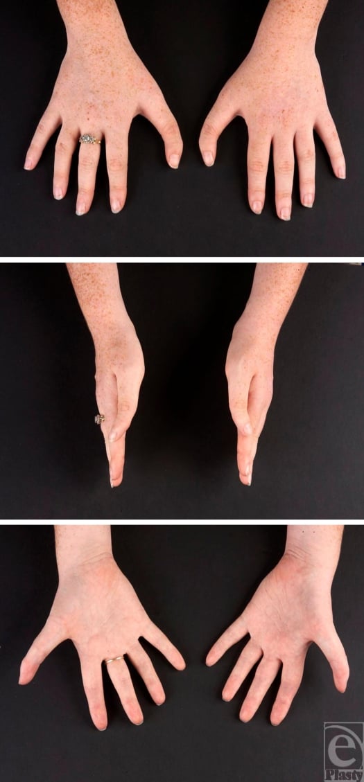
Palpation. Palpate the apex of the deformity and judge the size and quality of the bone, any abnormal prominence and the stability of the joints. Then the soft tissues: skin quality and elasticity, the absence of contracture, and the neurovascular status. This is what separates a bony deformity from a soft-tissue one.
Function. Measure active and passive motion at the PIP and DIP joints against the other side and document any stiffness. Grip strength is usually normal; check pinch, fine motor tasks such as writing and keyboard use, whether the finger overlaps its neighbour, and what the patient reports. Most clinodactyly is a cosmetic concern with minimal functional impairment: overlapping digits and trouble with keyboard or fine motor work belong to severe deformity, and grip interference is rare. QuickDASH, satisfaction with appearance and a functional limitations questionnaire serve as outcome measures.
Measurement. Clinically, a goniometer on the extended digit gives the angle between it and the longitudinal axis, in degrees, compared with the other hand when the deformity is bilateral. Radiographic measurement is more accurate: on an AP film with the hand flat, measure the angle between the longitudinal axes of the proximal and distal phalanges, and compare serial films for progression. The angle is then graded into the severity bands used in Management.
Differential diagnosis. The other bent, short or stiff digits, and how each declares itself:
- Plane of Deformity
- Coronal (radioulnar)
- Key Distinguishing Feature
- Delta phalanx with C-shaped bracket epiphysis; little finger radial deviation
- Plane of Deformity
- Sagittal (flexion)
- Key Distinguishing Feature
- Fixed flexion at PIPJ, usually little finger; no bony wedge phalanx
- Plane of Deformity
- Volar-radial
- Key Distinguishing Feature
- Curvature of the little finger DISTAL phalanx; bilateral, adolescent females
- Plane of Deformity
- Coronal with shortening
- Key Distinguishing Feature
- GDF5-related shortening of index/middle phalanges with clinodactyly component
- Plane of Deformity
- No angulation (stiffness)
- Key Distinguishing Feature
- Congenital interphalangeal joint fusion; absent skin creases, no motion
- Plane of Deformity
- Coronal or sagittal
- Key Distinguishing Feature
- History of injury; normal physeal morphology, no bracket epiphysis
Investigations
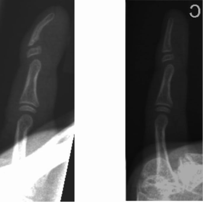
Radiographs. Plain films make the diagnosis, and the AP is the one that matters:
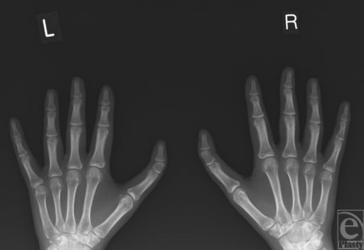
- AP hand - essential; shows the angular deformity and the delta phalanx
- Lateral hand - supplementary; it may miss a deformity that lies in the coronal plane, and a delta phalanx may look relatively normal on it
- Oblique - sometimes helpful for showing the bracket epiphysis
What to read off the film.
- The delta phalanx: a triangular or wedge-shaped outline with a C-shaped or bracket epiphysis, pathognomonic and best seen on the AP; the adjacent bones are usually of normal morphology
- The angle: measured as described under Clinical Assessment, documented, and followed on serial films for progression
- The physis: open or closing, which affects the surgical options; the bracket configuration; any physeal asymmetry
- The adjacent structures: joint congruity, collateral ligament stress and any rotational component
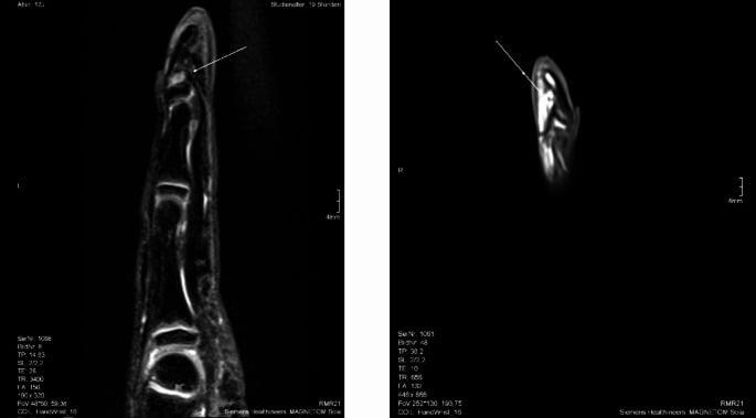
Further imaging. Advanced imaging is rarely needed; plain radiographs are sufficient in most cases. CT is reserved for complex deformity or pre-operative planning, and MRI is not routinely indicated.
Beyond the hand. If there are syndromic features: karyotype for Down syndrome, echocardiography for the congenital heart disease that accompanies the syndromes, a global developmental assessment, and genetic counselling for familial cases. Isolated clinodactyly needs none of this. The radiographs confirm the diagnosis and the child is monitored for progression.
Management Algorithm
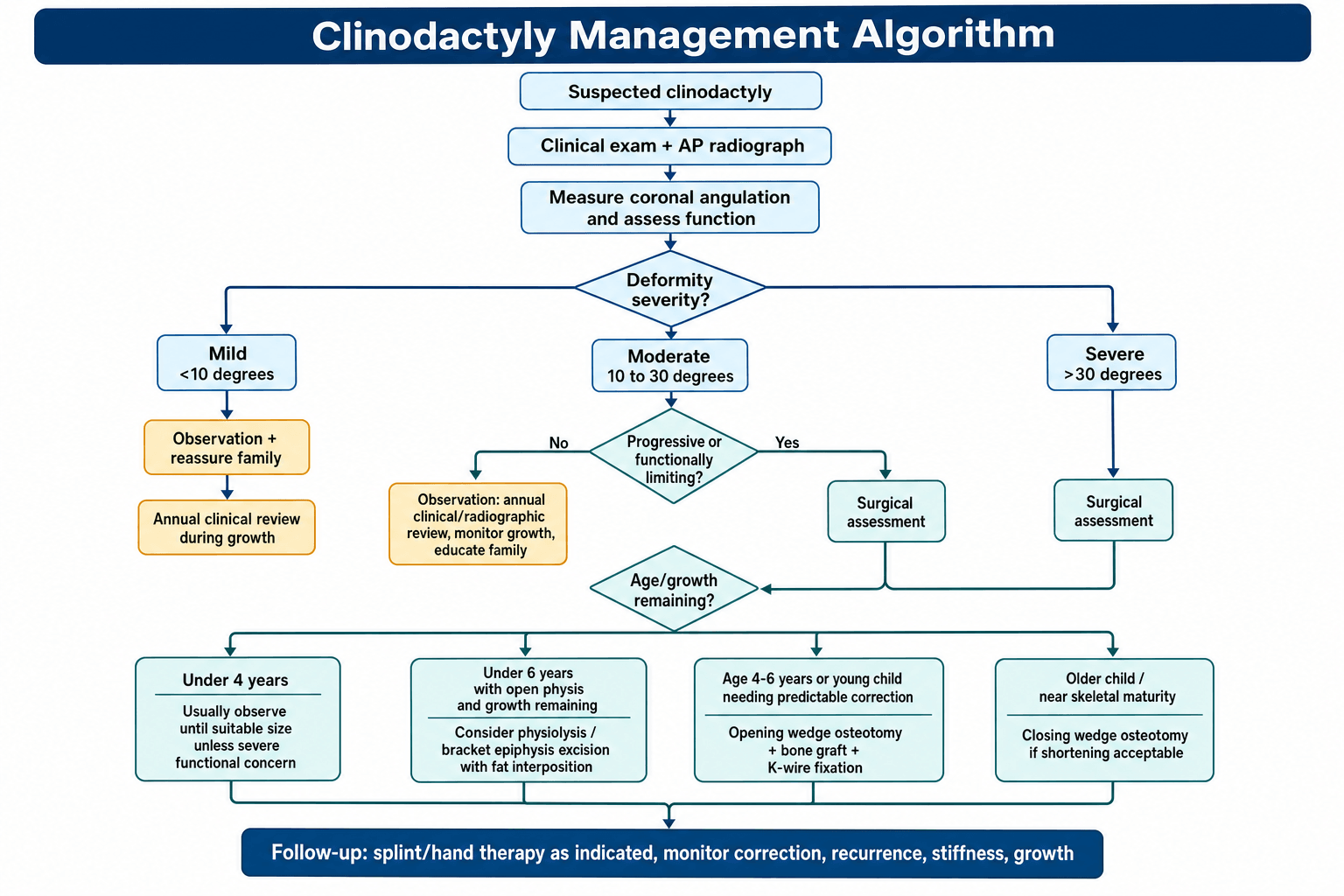
The decision. The angle, whether it is progressing, what it costs the hand and how much the appearance troubles the child and family decide it together. The angle is graded on the AP radiograph:
- Mild, under 10 degrees - observation
- Moderate, 10-30 degrees - observation unless progressive or causing functional problems
- Severe, over 30 degrees - surgery usually indicated
Progression on serial radiographs lowers the threshold for surgery; a stable deformity can be observed. Overlapping digits, grip interference, fine motor limitation and cosmetic concern from the child or parents are weighed alongside the angle.
Thirty degrees is a prompt, not a gate. "Over 30 degrees" is the conventional teaching and worth quoting, but this page's own citations do not support it as a go/no-go line. In 25 fingers treated by closing wedge osteotomy the authors explicitly recommended the procedure for moderate (15 to 30 degrees) as well as severe (over 30 degrees) deformity (PMID 19362790), so a 25-degree finger that is progressing, overlapping or bothering the child is not below a threshold; it is inside the published indication. Below about 30 degrees most clinodactyly is a cosmetic concern with minimal functional impact and observation is reasonable, but progression, digital overlap and functional impairment convert observation into an operation at any angle.
Observation. The child with a deviation under 30 degrees that is stable, without functional impairment and of little cosmetic concern, or whose family declines surgery, is reviewed clinically and radiographically once a year through growth, with any functional change documented and the surgical indications reconsidered at each visit. Tell the family what to expect: most deformities are stable and some progress with growth, the functional impact is usually minimal, surgery remains available if progression or concern develops, and the outcome for mild deformity is good with or without it. Documented progression, a new functional limitation, increasing cosmetic concern or the patient's own request bring the decision back to the table.
Surgery. The indications are the four already named: a severe angle, progression with growth, functional impairment (overlap, grip) and significant cosmetic concern from the patient or family. The harder question is which operation.
Which operation: the physis is the variable. What the evidence stratifies is the choice of procedure rather than the indication. A physiolysis works with the remaining growth, so it has a window in which it can be done and a ceiling on what it can correct; an osteotomy corrects what is already there and can be done later, but does not redirect growth. Age and the presence of syndactyly therefore drive the choice at least as much as the angle does.
- Where the evidence puts it
- Most effective under 55 degrees and in isolated clinodactyly, not with syndactyly (PMID 30762436)
- Result
- 43.0 to 23.9 degrees (46.2% correction)
- Where the evidence puts it
- 27 fingers, minimum 6-year follow-up (PMID 26972556)
- Result
- 38 to 8 degrees (79%); residual under 10 degrees in 15 of 27
- Where the evidence puts it
- 25 fingers, all over 25 degrees (PMID 19362790)
- Result
- 33 to 9 degrees; DIP arc essentially preserved, 84 to 81 degrees
- Where the evidence puts it
- 13 digits, mean 36 degrees (PMID 25754787)
- Result
- Mean correction 32 degrees, maintained in 12 of 13; pre-operative pain resolved in all 6 who had it
- For whom, and what is done
- Young children, under 6 years, with an open physis, substantial remaining growth and a progressive deformity, where an osteotomy is to be avoided. The bracket epiphysis is excised and a fat graft interposed
- Trade-off
- Gradual correction with no bone graft; depends on remaining growth and is unpredictable
- For whom, and what is done
- The most common procedure, at ages 4-6: an open physis with limited remaining growth, or a family wanting immediate correction. Osteotomy at the apex, the wedge opened, bone graft, K-wire fixation
- Trade-off
- Immediate, predictable, single-stage correction; needs a bone graft and later wire removal
- For whom, and what is done
- Older children near skeletal maturity with a closed or closing physis, when a graft is to be avoided and slight shortening is acceptable. A wedge is removed, the gap closed and fixed with K-wires
- Trade-off
- No graft and stable fixation; shortens the digit
Age. Under 4 years the structures are small and the surgery technically difficult, so these children are usually observed unless the deformity is severe. 4-6 years is the optimal window when surgery is indicated: the bone is of adequate size and there is growth left for remodelling. After 10 the operation is still possible, but with less remodelling to come a closing wedge may be the better choice, and at skeletal maturity the closing wedge is preferred.
Surgical Technique
Planning. Measure the deformity on the AP radiograph, calculate the wedge angle needed and template the correction. Decide the graft source: iliac crest or olecranon autograft, or allograft.
Steps.
- Exposure. Dorsal longitudinal incision over the affected phalanx; identify and protect the radial and ulnar digital nerves; incise the periosteum longitudinally and elevate it from the bone.
- Osteotomy. At the apex of the deformity, usually mid-shaft, with a small sagittal saw or osteotome: a transverse cut perpendicular to the long axis, kept incomplete so that a hinge remains on the concave (shorter) side.
- Opening. Open the cut gently on the long, convex side with a small lamina spreader or osteotomes to the templated angle, checking alignment clinically and on fluoroscopy.
- Graft. Insert the graft into the wedge defect and make sure it sits stably; it holds the correction and is what unites.
- Fixation. Smooth K-wires, usually two, in a crossed configuration for rotational stability, passed across the osteotomy and left out through the skin for later removal. Absorbable pins are an alternative in young children.
- Closure. Periosteum over bone and graft, then subcutaneous and skin layers, and a dorsal splint.
Pearls. Stay dorsal to keep clear of the neurovascular bundles. Use enough graft to fill the defect. Slight overcorrection, 5-10 degrees, is acceptable.
The cut is incomplete on purpose. The hinge left on the concave (shorter) side gives the construct its stability and prevents overcorrection while the wedge is opened in a controlled way on the convex side. Cut through it and the correction floats.
Complications
On the table.
- Neurovascular injury - the digital nerves are at risk; stay dorsal, identify and protect them
- A complete osteotomy - the hinge is lost and the construct is unstable
- Undercorrection - may need revision
- Overcorrection - usually minor, and remodels
- Graft dislodgement - make sure the graft sits stably
Early, under 6 weeks. Wound infection occurs in 1-2%, alongside dehiscence over the K-wires and pin-site infection, and is treated with local wound care and antibiotics. Loss of fixation, wire migration or breakage with loss of correction, is rare with proper technique and may need revision fixation. Digital artery injury with compromised perfusion of the digit is rare but serious: recognise it immediately and explore urgently if suspected.
Recurrence. 10-20% after physiolysis, higher than after osteotomy, from incomplete excision of the physis or recurrence through growth. The choice is between observation and revision osteotomy.
Nonunion. Under 5% with adequate bone graft. Inadequate graft and excessive motion are the risk factors; revision grafting if symptomatic.
Residual angulation. Undercorrection is the commonest problem, at 10-15%; overcorrection is less common and usually minor. Revise a severe residual deformity and observe a mild one.
Stiffness. Some PIP joint stiffness in 10-20%, usually a mild loss of 10-20 degrees. Early range of motion once the wires are out prevents it, hand therapy treats it, and manipulation is rarely needed. The cited opening-wedge series recorded its stiffness at the DIP joint instead, in three of 13 digits (Piper 2015), and counselled families about that joint specifically.
Growth disturbance. Physeal damage is rare with careful technique. Shortening of 2-3 mm is expected after a closing wedge and has minimal functional impact; asymmetric growth arrest can produce angular deformity.
Postoperative Care and Rehabilitation
The first four weeks. A dorsal blocking splint including the MCP, PIP and DIP joints of the digit, buddy tape to the neighbour for stability, and the hand elevated for the first 48-72 hours. The K-wires protrude through the skin: clean the pin sites with saline daily, watch for signs of infection and keep the splint clean and dry. Pain is usually minimal after the first 2-3 days and paracetamol or ibuprofen suffices, with ice for swelling.
Four to six weeks. A radiograph at 4 weeks checks healing, maintenance of alignment and incorporation of the graft. The wires come out at 4-6 weeks once union is evident, in clinic and usually without anaesthesia in a cooperative child, and part-time splinting continues afterwards.
Six weeks to three months. Wean the splint gradually after the wires are removed, with night splinting for a further 2-4 weeks, and begin gentle active motion, PIP and DIP joints especially, buddy-taped during activity; hand therapy if stiffness is significant. Progressive strengthening starts at 8-10 weeks and unrestricted, age-appropriate activity at 12 weeks.
Follow-up.
- 3 months - clinical and radiographic assessment
- 6 months - final alignment and function
- Annually - through skeletal maturity, for recurrence
- Skeletal maturity - final assessment; discharge if stable
Outcomes and Prognosis
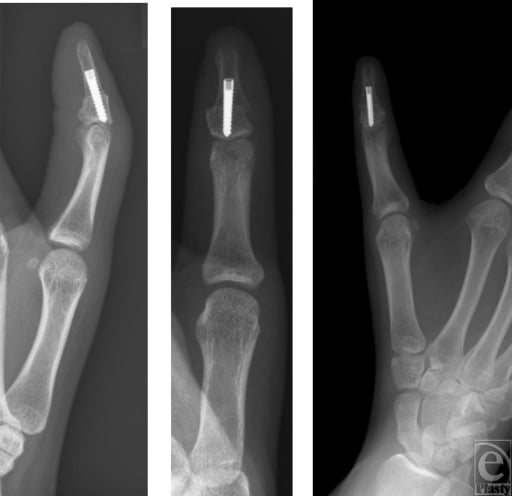
Opening wedge osteotomy. Excellent angular correction in over 90%, recurrence under 5% with adequate technique, minimal effect on function with good range of motion, and high satisfaction from patients and parents.
Physiolysis. Correction is variable: 60-80% achieve a satisfactory result, it accrues over 2-3 years, recurrence is higher than after osteotomy (see Complications), and 15-20% require a later osteotomy. The cited cohorts are kinder than those figures: in 27 fingers with a minimum six-year follow-up after early bracket resection and fat interposition, none needed a later closing wedge (Medina 2016), and in the comparative series the reoperations fell on the osteotomy side (Gillis 2020).
Observation. Deformity under 30 degrees is usually stable with good function; 10-20% progress during growth spurts. The functional impact is minimal in most cases and satisfaction with observation is good for mild deformity.
- Good Outcome
- Moderate deformity (30-45 degrees)
- Poorer Outcome
- Severe deformity (over 60 degrees)
- Good Outcome
- 4-6 years
- Poorer Outcome
- Very young (under 3) or late (over 10)
- Good Outcome
- Opening wedge osteotomy
- Poorer Outcome
- Physiolysis (unpredictable)
- Good Outcome
- Autograft iliac crest
- Poorer Outcome
- No graft or inadequate graft
- Good Outcome
- Isolated clinodactyly
- Poorer Outcome
- Syndromic (may have other issues)
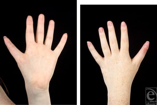
Function. Range of motion is usually normal or near it, within 10 degrees of the other side; grip strength is unaffected by surgery and fine motor function returns to baseline. The cosmetic improvement is significant and the scar usually inconspicuous, and over 85% are satisfied or very satisfied.
Long term. The prognosis for isolated clinodactyly treated surgically is excellent: low recurrence with the opening wedge technique, normal hand function, no increased risk of arthritis from the surgery, and improved confidence with correction.
Guidelines, Registries & Global Practice
Global Epidemiology
Clinodactyly is one of the most common congenital digital differences worldwide, though reported figures vary widely with diagnostic threshold (most series quote a 1-10% population frequency for mild deviation). Population-based registry data place congenital upper limb anomalies as individually rare but collectively a meaningful paediatric workload: in the Stockholm County cohort, 562 affected children were identified among 261,914 live births, with "deformations" (the Oberg-Manske-Tonkin subgroup that contains clinodactyly) accounting for 124 of 577 anomalies (Ekblom et al, J Hand Surg Am 2014).
- Reported value
- 1-10% (threshold-dependent)
- Source
- Clinical series consensus
- Reported value
- Majority of children affected
- Source
- Trisomy 21 phenotype literature
- Reported value
- Little (5th) finger, radial deviation, middle phalanx
- Source
- Operative series (Piper 2015; Ali 2009)
- Reported value
- 562 children / 261,914 births (Stockholm)
- Source
- Ekblom 2014 (PMID 24480684)
Classification & Guidance Framework
There is no single high-level society guideline (e.g. AAOS clinical practice guideline) dedicated to clinodactyly; management rests on classification frameworks and Level III-IV operative evidence. The table summarises the principal frameworks referenced internationally.
- Region
- International (IFSSH-endorsed)
- Role in clinodactyly
- Places clinodactyly within the malformation/deformation axis; current standard for congenital hand differences
- Evidence basis
- Registry-validated (Ekblom 2014, Level III)
- Region
- International
- Role in clinodactyly
- Earlier descriptive classification, now largely superseded by OMT
- Evidence basis
- Descriptive consensus
- Region
- UK
- Role in clinodactyly
- Paediatric congenital hand pathways; refer functionally/cosmetically significant cases to specialist congenital hand units
- Evidence basis
- Expert/pathway guidance
- Region
- USA
- Role in clinodactyly
- Surgeon education and operative principles for delta phalanx correction
- Evidence basis
- Expert/Level IV
- Region
- Europe
- Role in clinodactyly
- Continuing-education curricula and consensus on paediatric hand reconstruction
- Evidence basis
- Expert/consensus
Registry & Practice Variation
Dedicated arthroplasty-style registries do not exist for clinodactyly; the relevant evidence is from national congenital-anomaly and hand-surgery registries (e.g. Swedish registry data above) plus single-centre operative cohorts. Practice variation centres on procedure choice rather than indication threshold:
- Procedure selection varies by age and growth remaining: Vickers physiolysis (epiphyseal bracket resection with fat-graft interposition) is favoured in skeletally immature children, while opening or closing wedge osteotomy predominates nearer skeletal maturity. A comparative cohort found physiolysis achieved similar correction to osteotomy with fewer reoperations for deformity under 55 degrees (Gillis et al, Hand 2020).
- Surgical threshold is broadly consistent internationally: observation for mild/stable deformity, intervention for deformity greater than ~30 degrees, documented progression, or functional impairment (digital overlap, grip interference).
- Multidisciplinary syndromic screening is standard worldwide where clinodactyly is associated with Down syndrome or other syndromes, with genetic and developmental follow-up integrated into care.
Long-term follow-up through skeletal maturity to detect recurrence is standard practice across health systems, and telehealth has broadened access for serial radiographic and clinical monitoring of geographically remote families.
Viva Scenarios
Practise clinical reasoning and management decisions out loud
“A 5-year-old child with Down syndrome presents with bilateral small finger radial deviation. Radiographs show delta phalanx of middle phalanx with 35 degrees angulation. Parents are concerned about appearance. What is your management?”
“A 3-year-old child presents with small finger clinodactyly. Current deformity is 20 degrees on radiograph, but parent brings old X-ray from age 1 showing only 10 degrees. The deformity is clearly progressive. What is your recommendation?”
“You are performing opening wedge osteotomy for 40 degrees clinodactyly in a 5-year-old. After making the osteotomy, you open the wedge but the correction seems unstable. What are the potential issues and how do you manage?”
DEFINITION & PATHOANATOMY
- Angular deviation in CORONAL plane
- Delta phalanx = triangular bone
- C-shaped BRACKET epiphysis (2 sides)
- Normal physis = straight (1 side only)
- Asymmetric growth = progressive deformity
EPIDEMIOLOGY
- Incidence: 1-10% general population
- Down syndrome: Over 50%
- Small finger most common (radial deviation)
- Middle phalanx typical location
- Often bilateral (60-70%)
SURGICAL INDICATIONS
- Over 30 degrees angulation
- Progressive deformity (documented on X-rays)
- Functional impairment (overlap, grip)
- Significant cosmetic concern
- Age 4-6 years optimal for surgery
SURGICAL OPTIONS
- Opening wedge: Age 4-6, needs bone graft
- Physiolysis: Age under 6, unpredictable
- Closing wedge: Older child, no graft
- Bone graft: Iliac crest or olecranon
- Fixation: Crossed K-wires x 4-6 weeks
OPENING WEDGE TECHNIQUE
- Osteotomy at apex of deformity
- INCOMPLETE cut - hinge on concave side
- Open wedge on convex (long) side
- Bone graft to fill defect
- Crossed K-wires for stability
COMPLICATIONS
- Recurrence: 10-20% (physiolysis higher)
- Undercorrection: Most common issue
- Stiffness: 10-20% (usually mild)
- Nonunion: Under 5% with graft
- Neurovascular injury: Rare with technique
Evidence Base
Piper, Goldfarb & Wall - Opening Wedge Osteotomy for Little Finger Clinodactyly
- 13 digits in 9 children, all with more than 20 degrees pre-operative clinical angulation (mean 36 degrees)
- Significant improvement in clinical and radiographic angle (mean correction 32 degrees), maintained in 12 of 13 digits
- Pre-operative pain in 6 patients resolved in all post-operatively
- Three digits developed distal interphalangeal joint stiffness; one recurrence
Gillis et al - Vickers Physiolysis versus Osteotomy for Clinodactyly
- Comparative cohort of 30 digits (Vickers physiolysis) versus 11 digits (osteotomy)
- Physiolysis corrected angulation 43.0 to 23.9 degrees (46.2%); osteotomy 39.2 to 22.4 degrees (55.3%)
- More reoperations occurred in the osteotomy group; physiolysis had minimal complications
- Vickers physiolysis most effective for deformity of less than 55 degrees and isolated (non-syndactyly) clinodactyly
Medina, Lorea, Elliot & Foucher - Early Physiolysis: 6-Year Results
- 27 fingers in 22 patients treated by central epiphyseal (bracket) resection with fat-graft interposition, minimum 6-year follow-up
- Mean angle corrected from 38 degrees pre-operatively to 8 degrees at final follow-up (mean correction 79%)
- Residual deformity of less than 10 degrees - effectively full correction - in 15 of 27 fingers
- No patient required a later closing wedge osteotomy for insufficient correction
Ali, Jackson & Rayan - Closing Wedge Osteotomy of the Middle Phalanx
- 25 fingers in 17 patients, all with more than 25 degrees of angulation, treated by closing wedge osteotomy and K-wire fixation
- Clinical deformity corrected from a mean of 33 degrees to 9 degrees; radiographic from 29 degrees to 5 degrees
- Distal interphalangeal joint arc of motion essentially preserved (84 to 81 degrees)
- High parental satisfaction; recommended for moderate (15 to 30 degrees) and severe (over 30 degrees) deformity
Ekblom, Laurell & Arner - Epidemiology of Congenital Upper Limb Anomalies (Stockholm Registry)
- Population-based cohort of 562 children with congenital upper limb anomalies among 261,914 live births in Stockholm County (1997-2007)
- All anomalies could be organised within the Oberg, Manske and Tonkin (OMT) classification, now the international standard
- Deformations (which include clinodactyly) comprised 124 of 577 anomalies
- Registry data confirm congenital hand differences are individually rare but collectively a meaningful paediatric workload