Irreducible Dorsal Dislocation | Kaplan Lesion | Volar Plate Interposition
- Index MCP is most common site for complex dorsal dislocation
- Volar plate and sesamoids interpose between metacarpal head and proximal phalanx base
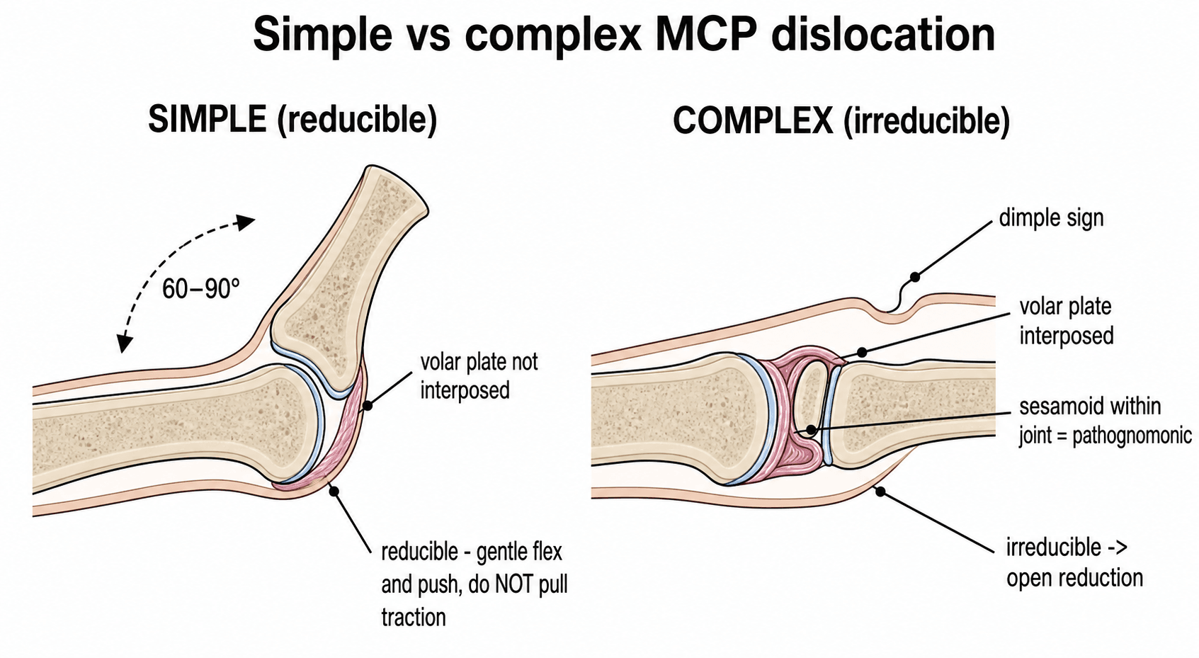
- Skin dimple at proximal palmar crease is pathognomonic for complex injury
- Traction alone tightens the noose around the metacarpal neck and fails
- The interposed plate sits DORSAL to the metacarpal head - so the dorsal approach, not the volar, looks directly at the block
- “Always examine for skin dimple before attempting reduction
- “Attempt closed reduction only once under adequate anaesthesia
- “Dorsal approach splits the volar plate LONGITUDINALLY, after which the joint reduces spontaneously (Becton)
- “Neither approach is proven superior: dorsal favours motion, volar favours grip - and volar carries the nerve risk
- “Post-reduction stability determines early motion versus immobilisation
Overview and Epidemiology
A complex dorsal metacarpophalangeal dislocation is a true orthopaedic emergency in the hand. The index finger is involved in over 50 percent of cases (53.8% in the largest surgical series, PMID 41906838). Misdiagnosis as a simple dislocation leads to repeated failed reduction attempts, cartilage damage, and ultimately an open reduction that could have been planned from the outset; recognising the pathognomonic skin dimple allows immediate preparation for surgery and avoids iatrogenic injury from forceful traction.
Where it sits. The wider set of digital injuries is covered in finger dislocations; the systematic films that accompany any of them are in imaging of the wrist and hand, and the sensory mapping that localises the nerve at risk is in hand examination and clinical localisation.
Mechanism. A hyperextension force at the MCP joint:
- Fall on the outstretched hand with the MCP hyperextended
- Direct blow - dorsal force driving the proximal phalanx into hyperextension
- Sports injury - ball sports, gymnastics, martial arts
- High-energy trauma - road traffic accidents, with associated fractures
Why it matters. Volar plate interposition makes the joint irreducible without surgery. The radial digital nerve is stretched over the metacarpal head, prolonged dislocation abrades the articular cartilage, and delayed treatment leads to permanent loss of motion and pain.
Pathophysiology
The joint. The metacarpal head is cam-shaped, with a wider dorsal articular surface. Dorsally the sagittal bands hold the extensor tendon centred over the head, the proper and accessory collateral ligaments flank it, and volar to the joint lies the volar plate, tethered to its neighbours by the deep transverse metacarpal ligament, with flexor digitorum profundus and superficialis passing beneath it through the A1 pulley. In a complex dislocation the metacarpal head is trapped among these structures.
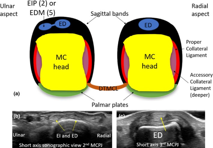
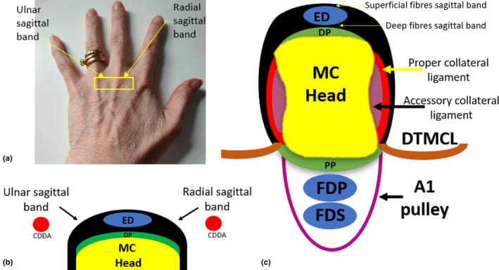
The volar plate. A thick fibrocartilaginous structure on the palmar surface of the joint, containing the sesamoids in the index and middle fingers, with two very different attachments. Distally, onto the base of the proximal phalanx, it is firm, fibrocartilaginous and strong; proximally, onto the metacarpal neck, it is thin, membranous and weak, and this is the critical zone. The asymmetry is the whole pathology: it is what lets a hyperextension force produce an irreducible joint. The same volar-plate mechanics govern the interphalangeal injuries in finger dislocations.
What happens in hyperextension. The base of the proximal phalanx rides over the metacarpal head and the plate avulses or ruptures at its weak proximal origin while staying firmly attached to the phalanx. As the phalanx displaces dorsally the freed plate is carried with it and flips to lie dorsal to the metacarpal head, buttonholed between the head and the phalangeal base, with the sesamoids adding a bony wedge to the block. This is the Kaplan lesion, the classic complex dislocation: the metacarpal head buttonholed through the volar structures, with the volar plate, sesamoids and flexor sheath as the irreducible block. It is the anatomical basis of the simple-versus-complex distinction.
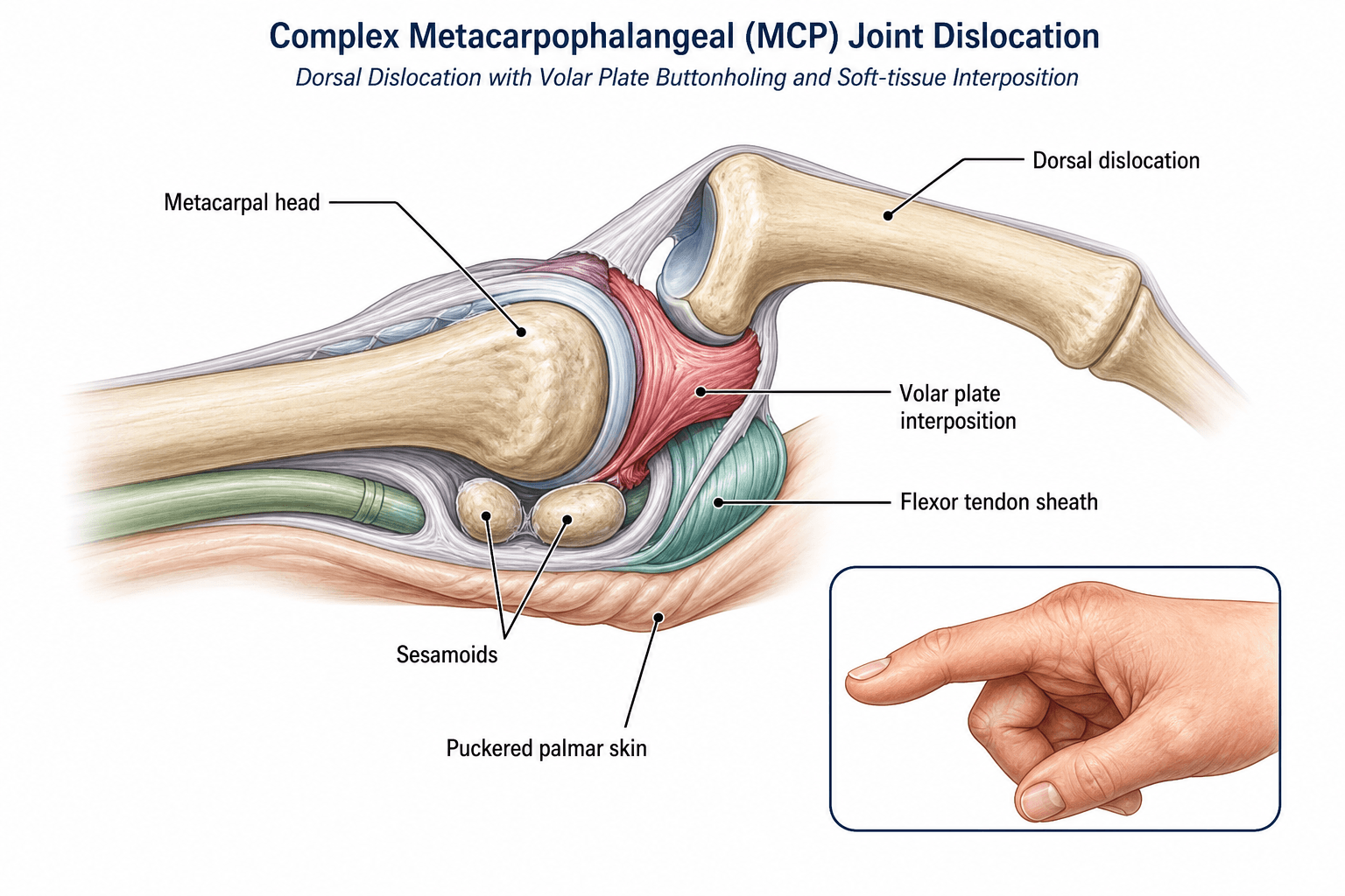
The noose. The deep transverse metacarpal ligament tethers the plate to its neighbours, which is why it cannot simply slide back once it has flipped, and with the lumbrical on the radial side and the flexor tendons on the ulnar side it completes a constricting noose around the metacarpal neck. Traction increases the tension on these structures, so the proximal phalanx stays locked in hyperextension however hard it is pulled. Forceful or repeated attempts add cartilage abrasion and soft-tissue swelling, converting a surgical case into a more difficult one.
- Role in Block
- Primary interposed structure
- Anatomic Location
- Folds between metacarpal head and phalanx base
- Release Method
- Longitudinal split - from dorsal it comes into full view (Becton)
- Role in Block
- Bony component of volar plate
- Anatomic Location
- Embedded in volar plate substance
- Release Method
- Divided with volar plate or excised if fractured
- Role in Block
- Form constricting noose
- Anatomic Location
- Ulnar and radial to metacarpal neck
- Release Method
- A1 pulley release if required
- Role in Block
- Radial side interposition
- Anatomic Location
- Between metacarpal head and volar plate
- Release Method
- Gentle retraction or partial release
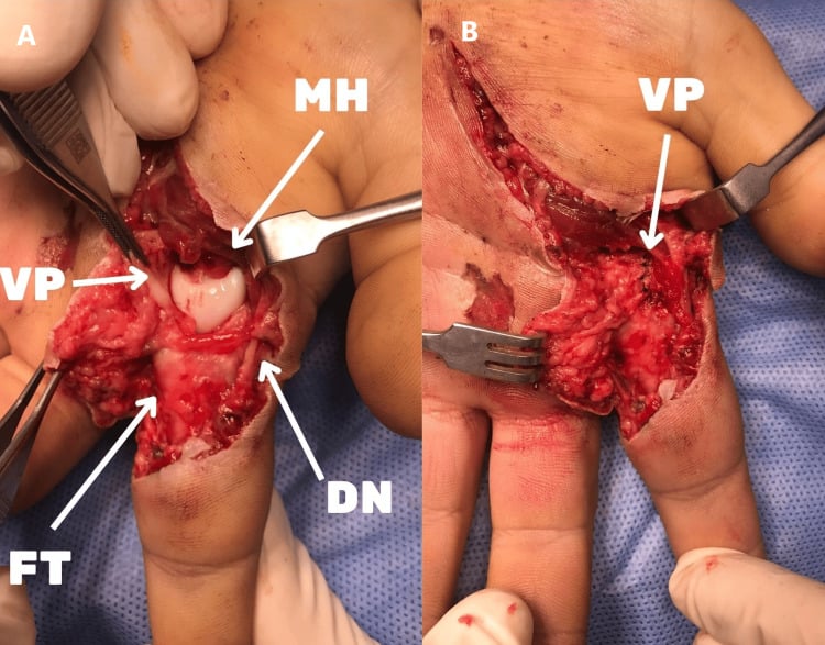
The structures at risk. The radial digital nerve is stretched over the prominent metacarpal head in index finger dislocations; the ulnar digital nerve is less commonly at risk. Both must be identified and protected during any volar approach. The radial collateral ligament is frequently torn and should be repaired after reduction. The destroyed-joint endpoint of a missed injury is the subject of MCP joint arthritis.
Classification and Types
By digit. The index finger predominance relates to its position at the radial border and the presence of a radial sesamoid that contributes to the bony block.
- Frequency
- Most common (over 50 percent)
- Anatomic Features
- Radial sesamoid present, narrow web space
- Surgical Notes
- Protect the radial digital nerve, especially if approaching from volar
- Frequency
- Second most common
- Anatomic Features
- Two sesamoids, wider head
- Surgical Notes
- Similar volar plate interposition pattern
- Frequency
- Less common
- Anatomic Features
- No radial sesamoid in little finger
- Surgical Notes
- May reduce more easily, check for associated injury
By direction. Dorsal is the pattern this topic is about. Volar dislocations are uncommon and usually require open reduction because the extensor mechanism becomes interposed.
- Direction
- Proximal phalanx dorsal and hyperextended
- Block to Reduction
- Volar plate, sesamoids, lumbricals
- Approach Preference
- Dorsal (full view, longitudinal split) or volar (Kaplan)
- Direction
- Proximal phalanx volar to metacarpal
- Block to Reduction
- Extensor hood or sagittal band
- Approach Preference
- Dorsal approach usually required
- Direction
- Sideways displacement
- Block to Reduction
- Collateral ligament interposition
- Approach Preference
- Dorsal or lateral approach
Clinical Assessment
History. The answers that change management:
- Mechanism - a hyperextension injury, typically a fall onto the outstretched hand
- Timing - acute presentation, or a delayed and missed injury
- Previous attempts - how many closed reductions, and how forceful
- Associated injuries - other hand trauma, open wounds, nerve symptoms
Examination. The MCP sits hyperextended, the finger slightly extended and ulnarly deviated, with the metacarpal head prominent and tender in the palm, so the deformity looks far less dramatic than the radiograph. Test the radial and ulnar digital nerves and capillary refill before anything is manipulated. A single gentle reduction attempt, under digital block or sedation, is part of the assessment; the technique has its own section below.
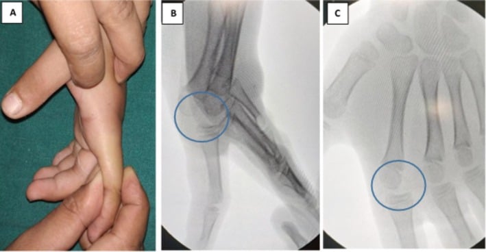
The skin dimple. Examine the palm with the MCP in maximal hyperextension. A transverse or oblique dimple at the proximal palmar crease, directly over the metacarpal head, means the volar plate has buttonholed and is interposed: the dimple is pathognomonic of a complex, irreducible dislocation. Its absence does not completely exclude complexity, but makes a simple dislocation more likely. Document it before any reduction attempt, because its presence changes management from attempted closed reduction to planned open reduction.
Differential. The main differential for a locked digit without trauma is trigger finger.
- Posture
- MCP hyperextension, PIP flexed
- Discriminating Finding
- Skin dimple, irreducible on traction
- Key Test / Imaging
- Clinical diagnosis, X-ray confirms direction
- Posture
- MCP hyperextension, reducible
- Discriminating Finding
- No skin dimple, reduces with traction-flexion
- Key Test / Imaging
- Post-reduction X-ray, assess stability
- Posture
- MCP flexed, proximal phalanx volar
- Discriminating Finding
- Extensor interposition, rare
- Key Test / Imaging
- Dorsal approach usually required
- Posture
- PIP flexed, MCP neutral
- Discriminating Finding
- History of triggering, no trauma
- Key Test / Imaging
- A1 pulley injection trial
Associated injuries. Metacarpal head fracture, proximal phalanx base fracture, collateral ligament avulsion and sesamoid fracture all change the post-reduction protocol and may require internal fixation. The metacarpal-head osteochondral fracture is the one that should influence the choice of approach, for the reason given under the dorsal approach below.
Investigations
Radiographs. PA, true lateral and oblique of the affected ray, before and after any reduction attempt. The lateral confirms dorsal versus volar displacement and guides approach planning; overlap on the AP alone can hide the dislocation. Look for:
- Direction of dislocation
- Associated fractures of the metacarpal head or proximal phalanx base
- Sesamoid displacement - a sesamoid sitting within the joint space is pathognomonic
- Joint incongruity
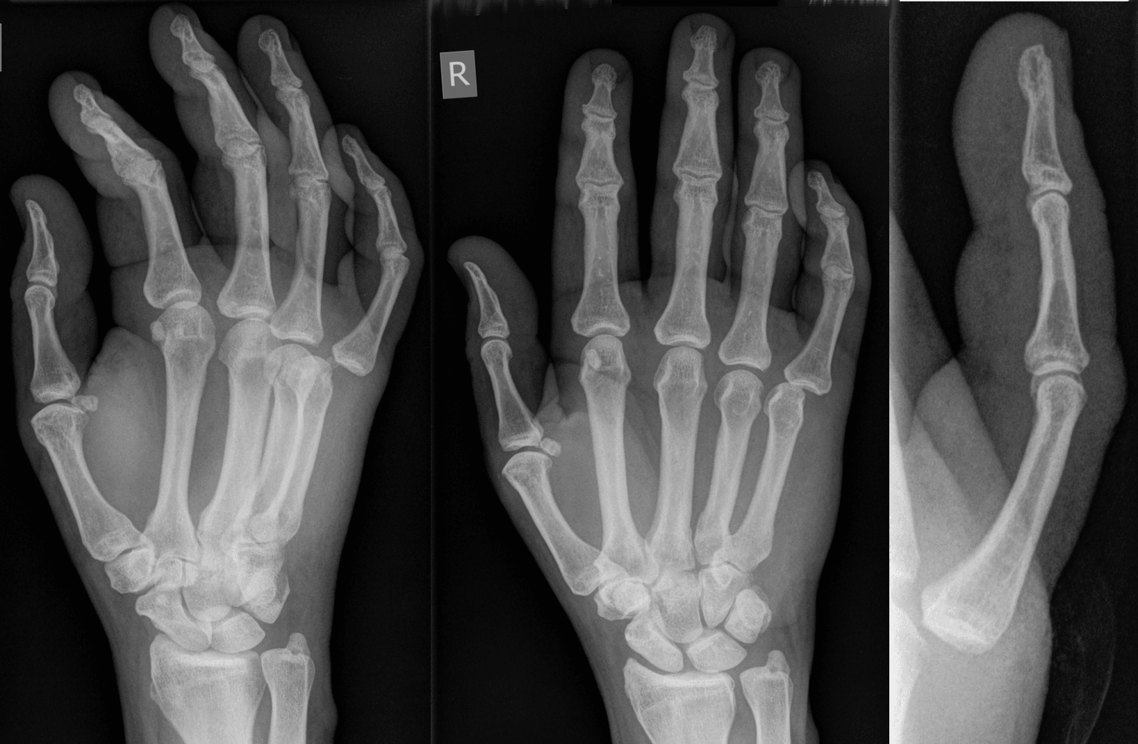
CT when an intra-articular fracture or a comminuted metacarpal head is suspected, or to assess sesamoid integrity. Fragments involving greater than 20 percent of the articular surface or with greater than 2 mm of step-off warrant consideration of ORIF, and the scan helps decide between volar plate repair and excision of comminuted fragments.
MRI for the chronic or missed case: a delayed presentation with fibrosis, where cartilage status and volar plate integrity decide between joint salvage and arthroplasty. It shows interposed volar plate scarring, chondral loss and possible osteochondral defects.
The decision is clinical. Radiographs confirm the diagnosis and exclude fracture, but open reduction is decided on the skin dimple and a failed closed attempt. Do not delay surgical exploration waiting for advanced imaging in an acute irreducible dislocation.
Closed Reduction Technique: One Gentle Attempt, Push Not Pull
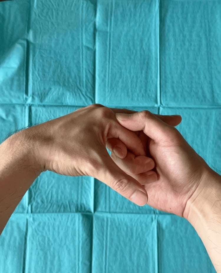
A single gentle closed attempt is appropriate for an acute dorsal MCP dislocation before complexity is confirmed: it is curative for a simple dislocation or subluxation, which has an excellent prognosis, and does no harm when done correctly. If a skin dimple is present, or the one attempt fails, proceed to open reduction and do not repeat it. The technique matters because the common instinct, longitudinal traction, is exactly what locks a complex injury.
- Anaesthesia and relaxation. Wrist block or sedation, never an awake, guarding hand. Flex the wrist and the IP joints to slacken the long flexors.
- Direction of force. Accentuate the hyperextension first, then push the base of the proximal phalanx distally and volarly so that it rolls over the metacarpal head into flexion, keeping the base in contact with the head throughout. Do not pull along the axis of the finger: traction tightens the volar plate, flexor and lumbrical noose and can convert a reducible subluxation into an entrapped complex dislocation.
- One attempt. Then reassess. Repeated forceful attempts abrade cartilage and worsen the interposition.
- After reduction. Confirm concentric reduction on radiographs, test stability and apply an extension-block splint, for 3 weeks in a simple dislocation. Nobody is discharged without a post-reduction film and a stability check.
Irreducible without a dimple. A dislocation that will not reduce but has no skin dimple may have the radial collateral ligament interposed; it is approached dorsally, and stability is confirmed after reduction as for any other.
Management Algorithm
Timing and planning. Open reduction within 24 hours if possible, before swelling peaks, aiming for a concentric reduction with minimal articular damage and stable motion afterwards. Consent covers stiffness, nerve injury, the possible need for collateral repair and infection. Equipment: hand table, fluoroscopy, fine rongeurs, 3-0 or 4-0 nonabsorbable suture.
Choosing the approach. Neither is proven superior, but know the anatomical argument, because it runs the opposite way to what most candidates say. The interposed plate lies dorsal to the metacarpal head, so it is the dorsal approach that looks straight at it. Becton's two stated objections to the volar (Kaplan) approach are that it gives a limited view of the plate and that the digital nerves are easily damaged during the exposure; the comparative series bears the second out (see Complications). In that same series, the only head-to-head comparison, the dorsal approach favoured motion and the volar approach favoured grip.
Dorsal approach (Becton).
- Direct dorsal longitudinal incision through skin and extensor tendon over the MCP joint
- The entrapped plate, trapped over the dorsal aspect of the head, comes into full view; this is the anatomical reason Becton proposed the approach
- Split the plate longitudinally; the dislocation reduces spontaneously as the flexor tendons and lumbrical slip past the metacarpal head, with no forceful manipulation
- Reduce and fix a metacarpal-head osteochondral fracture, which frequently accompanies this injury, through the same exposure
- Repair the extensor tendon split and any sagittal band rent to restore stability
Volar approach (Kaplan).
- Zigzag or Brunner incision centred over the MCP joint, extended proximally and distally as needed
- Identify and protect both digital neurovascular bundles before dividing any volar structure: the radial digital nerve is stretched over the metacarpal head, can be mistaken for a fibrous band, and is the structure at risk in this approach
- Divide the A1 pulley if needed and identify the interposed plate and sesamoids
- Divide the plate longitudinally, retract the lumbricals, and reduce the phalanx by flexing it and pushing the base over the head
- Once the plate has been incised and extracted from the joint, repair it above the metacarpal head
The trade-off is a limited view of the plate and exposed nerves against direct access to the sesamoids, lumbricals and collateral ligaments, and the better grip strength in the one comparative series.
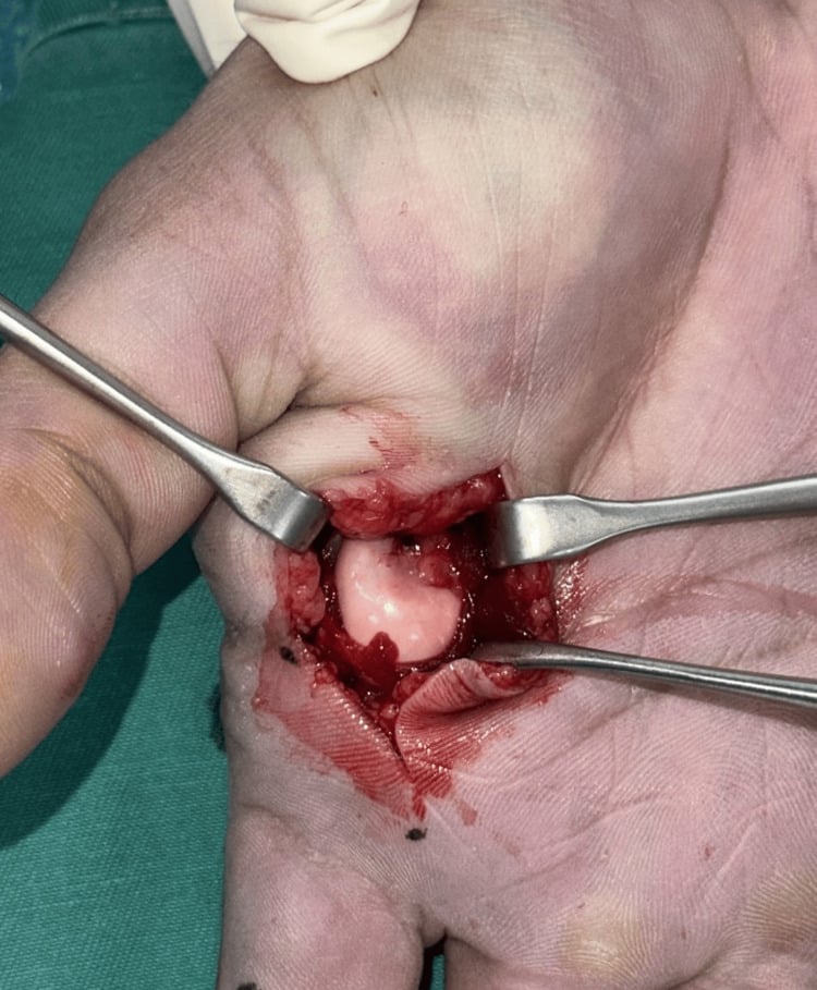
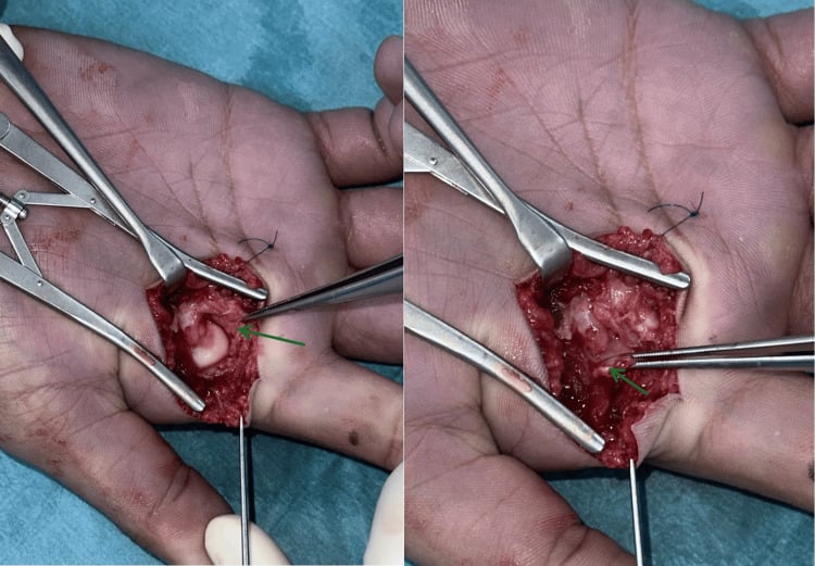
Before closure. Test passive MCP extension under fluoroscopy. If the joint tends to redislocate in extension, repair the radial collateral ligament and consider temporary K-wire stabilisation in 30 degrees of flexion for 3 weeks. Document stability before closure; it decides the splint position.
Rehabilitation.
- Extension block splint at 20-30 degrees of flexion for 2-3 weeks
- Protected active range of motion from week 2-3 under therapist supervision
- Grip strengthening from week 6, return to sport at 10-12 weeks
Missed or chronic (greater than 3 weeks). The goal is to restore motion and stability while addressing fibrosis and cartilage damage, and the extensor hood may also contribute to the block in a chronic or missed case. Through a volar approach the additional steps are:
- Extensive release of volar plate scar and contracted collateral ligaments
- Possible excision of fractured or arthritic sesamoids
- Check the metacarpal head for osteochondral defects requiring microfracture or grafting
- Consider a temporary spanning external fixator if stiffness is severe
Chronic cases often require more aggressive soft-tissue release and have higher rates of residual stiffness.
Salvage when the joint is destroyed. Severe cartilage loss ends any attempt at joint preservation. Silicone arthroplasty suits the low-demand, older patient and arthrodesis the young labourer with high demands; vascularised joint transfer is rare, for the young patient with a single digit involved.
- Indication
- Low demand, age greater than 60, destroyed cartilage
- Outcome
- Pain relief, 40-60 degrees arc
- Complications
- Implant fracture, silicone synovitis
- Indication
- Young labourer, single digit, high demand
- Outcome
- Stable pinch, pain-free
- Complications
- Loss of motion, adjacent joint stress
- Indication
- Young patient, multiple failed procedures
- Outcome
- Potential motion restoration
- Complications
- Donor morbidity, technical complexity
Complications
Read the percentages as conventional teaching ranges, not measured rates. No cohort of this injury is large enough to support precise complication rates: the published literature is case reports, technique descriptions and small series, the largest comparative series has 13 patients, and no registry collects MCP dislocations. The figures in the table are the ranges conventionally quoted in hand-surgery teaching. Quote them as orders of magnitude with the uncertainty attached; "stiffness is the commonest problem and is the main thing we are trying to prevent with early motion" is both truer and more useful than a spuriously precise percentage.
- Conventionally quoted range
- 30-50 percent of open reductions
- Risk Factors
- Delayed presentation, repeated reduction attempts
- Management
- Early protected motion, aggressive hand therapy
- Conventionally quoted range
- 5-10 percent (mostly neuropraxia)
- Risk Factors
- Volar approach, stretched nerve over metacarpal head
- Management
- Meticulous identification, microsurgical repair if transected
- Conventionally quoted range
- Less than 10 percent with proper repair
- Risk Factors
- Unrepaired collateral ligament, inadequate splinting
- Management
- Collateral repair, extension block splint, possible temporary pinning
- Conventionally quoted range
- Less than 5 percent
- Risk Factors
- Open injury, delayed surgery, diabetes
- Management
- Prophylactic antibiotics, meticulous wound care
- Conventionally quoted range
- Common after greater than 6 weeks delay
- Risk Factors
- Prolonged dislocation, cartilage abrasion
- Management
- Arthroplasty or arthrodesis if symptomatic
Digital nerve injury is approach-specific, and that is the single most defensible reason to consider the dorsal approach: it is the disadvantage Becton named in 1975, and the only nerve complications in the 2026 comparative series were in volar cases, transient hypoaesthesia in two of three volar patients (PMID 41906838), the one nerve figure on this page with a real denominator. A missed nerve transection causes permanent sensory loss and is a common source of litigation; see digital nerve compression for the sensory territory involved.
Outcomes and Prognosis
The trend. Earlier reduction does better, consistently across every published series, and that is the point the examiner wants. The percentages in the table are conventional teaching estimates rather than measured figures, since no cohort of this injury is large enough to generate them, and "functional ROM" is not a defined endpoint; quote the trend and the mechanism rather than the numbers if pressed on where they come from.
- Treatment
- Open reduction, collateral repair if needed
- Expected Outcome
- 80-90 percent achieve functional ROM
- Long-term Function
- Excellent return to work and sport
- Treatment
- Open reduction, more extensive release
- Expected Outcome
- 60-75 percent functional ROM
- Long-term Function
- Good for daily activities, some stiffness
- Treatment
- Open reduction or salvage procedure
- Expected Outcome
- 40-60 percent useful arc, variable pain relief
- Long-term Function
- Functional improvement but rarely normal motion
Prognostic factors. The best results follow acute presentation, a single gentle reduction attempt, concentric reduction, a repaired collateral ligament and a compliant early-motion protocol. Diagnosis delayed beyond 3 weeks, multiple failed closed attempts, chondral damage on the metacarpal head and non-compliance with therapy predict poorly: outcomes decline significantly after the 3-week window, with higher rates of stiffness and post-traumatic arthritis, and the conventional figure in the complications table has arthritis common once the delay exceeds 6 weeks.
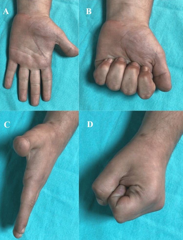
Guidelines, Registries & Global Practice
- Index finger MCP is the most common site worldwide for complex dorsal dislocation
- Hyperextension falls are the dominant mechanism across all regions and age groups
- Sports and occupational injuries account for the majority of presentations
- Missed diagnosis remains a problem in emergency settings where skin dimple is not routinely sought
- High-resource centres: early MRI, hand therapist involvement, fluoroscopic stability assessment
- Limited-resource settings: clinical diagnosis, plain radiographs only, volar approach with loupe magnification
- Universal principle: prompt recognition of the skin dimple and avoidance of repeated closed attempts determines outcome more than technology
- Surgery: concentrated in specialist hand units globally, with similar techniques reported from Europe, North America, Asia and Australia
- Diagnosis emphasis
- Skin dimple recognition, single closed attempt only
- Acute treatment
- Open reduction within 24 hours, nerve protection
- Surgical approach
- No approach mandated; choose on the obstructing anatomy
- Diagnosis emphasis
- Clinical diagnosis, radiographs to exclude fracture
- Acute treatment
- Urgent referral to hand surgery, avoid repeated manipulation
- Surgical approach
- Volar or dorsal according to surgeon preference
- Diagnosis emphasis
- High index of suspicion in hyperextension injuries
- Acute treatment
- Open reduction, collateral ligament assessment
- Surgical approach
- Both approaches accepted, stability testing essential
- Diagnosis emphasis
- Assess for associated metacarpal head fracture
- Acute treatment
- Anatomic reduction, protect soft tissues
- Surgical approach
- Approach dictated by fracture pattern and surgeon experience
There is no dedicated international registry for MCP dislocations, and no society guideline mandates a surgical approach — the row above should be read as the emphasis each body places on diagnosis and timing, not as a quotable position on volar versus dorsal. Evidence is derived from small case series, technique descriptions and anatomic studies; the largest comparative series has 13 patients. The consistent message is that recognition of the skin dimple changes management from closed to open reduction, that nerve protection during the volar approach is mandatory, and that outcomes are time-dependent with best results when reduction occurs within 24-48 hours. Where the literature is this thin, the examiner is testing whether you know how thin it is — say so, and reason from the anatomy of the block rather than citing a preference as though it were established.
Record in every dorsal MCP dislocation:
- Presence or absence of skin dimple at proximal palmar crease
- Number and result of closed reduction attempts
- Neurovascular status before and after any manipulation
- Intraoperative stability assessment and structures repaired
- Post-reduction splint position and rehabilitation plan
A missed complex dislocation leading to permanent stiffness or nerve injury is a recurring source of complaints worldwide. Always document the skin dimple examination and the rationale for proceeding to open reduction.
Controversies & Areas of Uncertainty
The one head-to-head comparison is a 13-patient retrospective series, so treat the direction of travel as a hint, not a verdict (PMID 41906838). The dorsal approach was used in 76.9% and gave greater MCP flexion (85.8° vs 78.4°), a smaller extension lag (−1.8° vs −4.2°), lower disability (QuickDASH 4.0 vs 8.8) and earlier return to sport (11.5 vs 14.4 weeks). Volar reconstruction gave the better grip strength (107.5% vs 90.9% of the other hand). None of these differences reached statistical significance — with 13 patients that is expected — though effect sizes were large (d ≥ 0.8). The one asymmetry that is not a wash: the only reported nerve complication was transient post-operative hypoesthesia, in two cases, and both were volar — two of the three volar patients in the series — which matches Becton's fifty-year-old objection. Surgeon experience still reasonably drives the choice; what is no longer defensible is claiming the volar approach gives the better view of the plate.
Some authors advocate routine repair of the radial collateral ligament after reduction, while others repair only when residual instability is demonstrated on stress testing. No comparative studies exist. Most surgeons repair obvious tears and test stability intraoperatively before deciding.
Extension block splinting at 20-30 degrees for 2-3 weeks is widely practised, but the precise angle and duration lack high-quality evidence. Early protected motion is favoured to minimise stiffness, yet too much motion risks recurrent subluxation. Therapist-led protocols vary between centres.
Temporary K-wire stabilisation across the reduced MCP joint is used by some when stability is marginal. Others avoid pinning to allow early motion. No randomised data exist. Decision is individualised based on intraoperative stability and patient compliance.
MCQ Practice Points
Q: Which structure is the primary block to reduction in a complex dorsal MCP dislocation? A: The volar plate with embedded sesamoids. In hyperextension the volar plate folds into the joint space between the metacarpal head and the base of the proximal phalanx. The sesamoids act as a bony wedge. The flexor tendons, lumbricals and deep transverse metacarpal ligament form a secondary constricting noose around the metacarpal neck.
Q: What is the pathognomonic clinical finding in a complex MCP dislocation? A: A skin dimple at the proximal palmar crease. This transverse or oblique dimple directly overlies the metacarpal head and indicates that the volar plate has buttonholed through the joint capsule. Its presence confirms that closed reduction will fail and open reduction is required.
Q: Why does longitudinal traction fail to reduce a complex MCP dislocation? A: Traction tightens the noose. The volar plate, flexor tendons and lumbricals already encircle the metacarpal neck. Longitudinal force increases tension on these structures, locking the proximal phalanx in hyperextension rather than disengaging the interposed tissue. A single gentle attempt is acceptable, but repeated traction causes further damage.
Q: What are the advantages of the volar versus dorsal approach for complex MCP dislocation? A: This is the question most candidates answer backwards. The interposed volar plate is trapped dorsal to the metacarpal head, so the dorsal approach is the one that brings the main blocking structure into full view — Becton's explicit objection to the volar (Kaplan) approach was that it gives a limited view of the plate and that the digital nerves are easily damaged during the exposure. From dorsal, the plate is split longitudinally and the joint reduces spontaneously; the approach also permits fixation of the metacarpal-head osteochondral fracture that often accompanies this injury. The volar approach still has a place: it gives direct access to the sesamoids, lumbricals and collateral ligaments, and in the only head-to-head series it produced the better grip strength. Neither is proven superior — say that, then justify whichever you choose on the obstructing anatomy in front of you.
Q: What is the recommended post-reduction protocol after open reduction of a complex MCP dislocation? A: Extension block splint at 20-30 degrees flexion for 2-3 weeks, followed by protected active motion under hand therapy supervision. Early motion prevents stiffness while the block splint maintains reduction. Strengthening begins at 6 weeks with return to full activity at 10-12 weeks. Stability must be confirmed intraoperatively before deciding on splint position.
Q: Which nerve is most at risk during the volar approach to an index finger MCP dislocation? A: The radial digital nerve. It is stretched over the prominent metacarpal head and can be mistaken for a fibrous band. Careful identification and protection of both digital nerves before dividing any volar structures is mandatory. Nerve injury causes permanent sensory loss on the radial border of the index finger.
Exam Viva Scenarios
Practise clinical reasoning and management decisions out loud
“A 32-year-old mechanic presents 6 hours after a fall onto his outstretched right hand. His index finger MCP is locked in 60 degrees of hyperextension. There is a clear skin dimple at the proximal palmar crease. Gentle traction under digital block fails to reduce the joint. What is your diagnosis and management plan?”
“A 45-year-old woman presents 5 weeks after a fall. She was told her index finger was 'sprained' and was treated with buddy strapping. She now has a stiff, painful MCP joint with 40 degrees hyperextension deformity, numbness on the radial side of the index finger, and a fixed skin dimple. Radiographs show early joint space narrowing. How would you manage this?”
Key Anatomy
- Volar plate with sesamoids is primary block to reduction
- Flexor tendons, lumbricals and deep transverse ligament form constricting noose
- Index finger most commonly affected due to radial sesamoid and border position
- Radial digital nerve stretched over metacarpal head and at risk in volar approach
Diagnosis
- Skin dimple at proximal palmar crease is pathognomonic for complex injury
- Irreducible on gentle traction confirms volar plate interposition
- X-ray: PA, lateral and oblique to confirm direction and exclude fracture
- High index of suspicion in any hyperextension MCP injury
Treatment Algorithm
- Simple dislocation: single closed reduction attempt, extension block splint 3 weeks
- Complex (skin dimple or failed reduction): open reduction, volar or dorsal approach
- Acute (under 24 hours): best outcomes with prompt open reduction
- Chronic (over 3 weeks): consider salvage arthroplasty if cartilage destroyed
Surgical Pearls
- Dorsal approach (Becton): plate lies DORSAL to the head so this gives the FULL view; split it LONGITUDINALLY and it reduces spontaneously; less nerve risk; allows MC-head fracture fixation
- Volar approach (Kaplan): direct access to sesamoids and collaterals, better grip in the one head-to-head series - but limited view of the plate and the digital nerves are at risk
- Neither approach is proven superior (best comparison n=13, no significant differences)
- After reduction: test stability, repair collateral ligament if torn
- Splint: extension block 20-30 degrees flexion for 2-3 weeks then early motion
Complications
- Residual stiffness: 30-50 percent, mitigated by early protected motion
- Digital nerve injury: 5-10 percent, mostly neuropraxia, prevent by identification
- Recurrent instability: less than 10 percent with proper collateral repair
- Post-traumatic arthritis: common after delayed presentation greater than 3 weeks
Evidence Base and Key Trials
Closed dislocation of the metacarpophalangeal joint of the index finger
- Classic early description of dorsal metacarpophalangeal dislocation of the index finger, distinguishing the simple (reducible) from the complex (irreducible) injury
- The complex/irreducible dislocation is characterised by interposition of the volar plate, which blocks closed reduction
- A puckered skin dimple in the proximal palm is a clinical sign of the complex injury
- Open reduction is required once a dislocation is irreducible by closed means
A simplified technique for treating the complex dislocation of the index MCP joint
- Described a direct dorsal longitudinal approach (through skin and extensor tendon) for the complex dislocation of the index MCP joint, as an alternative to Kaplan's volar approach
- States two explicit disadvantages of the volar (Kaplan) approach: the digital nerves are easily damaged during exposure, and there is a LIMITED VIEW of the entrapped volar plate because the plate lies dorsal to the metacarpal head
- The interposed fibrocartilaginous volar plate is brought into full view and split longitudinally, after which the dislocation reduces SPONTANEOUSLY as the flexor tendons and lumbrical slip past the metacarpal head
- Stated advantages over the volar approach: full exposure of the volar plate (the main structure blocking reduction), digital nerves less apt to be damaged, and accurate reduction and fixation of the frequently-associated metacarpal-head osteochondral fracture
Irreducible metacarpophalangeal joint dislocations: Clinical characteristics, surgical approaches, and outcomes
- Retrospective series of 13 surgically-treated irreducible MCP dislocations (mean age 29.2 years, range 7 to 78); the index finger was most often involved (53.8%) and dorsal dislocations predominated (76.9%)
- A dorsal approach was used in 76.9% and gave greater MCP flexion (85.8 vs 78.4 degrees), a smaller extension lag (-1.8 vs -4.2 degrees) and lower disability (QuickDASH 4.0 vs 8.8)
- Volar reconstruction gave superior grip strength at 107.5 percent of the contralateral hand versus 90.9 percent - exceeding baseline
- Return to sport was earlier after the dorsal approach (11.5 vs 14.4 weeks)
- No difference reached statistical significance, but effect sizes were large (d of 0.8 or more); transient post-operative hypoesthesia occurred in two cases, both volar
Kaplan's Lesion: A Case Report On The Complex Dislocation Of The Index Finger In A Weightlifter
- Case report of complex index finger MCP dislocation (Kaplan lesion) in a weightlifter successfully managed with open reduction