Rhabdomyolysis | Hyperkalemia | Myoglobinuria | Acute Kidney Injury
- Crush syndrome = systemic manifestation after release; crush injury = localized damage
- IV fluids MUST start BEFORE extrication - prevents reperfusion cardiac arrest
- Lethal triad: Hyperkalemia (cardiac arrest), metabolic acidosis, hypocalcemia
- Target urine output 200-300mL/hour to flush myoglobin and prevent AKI
- Fasciotomy threshold: delta pressure under 30mmHg SUSTAINED beyond 2 hours - an absolute pressure over 30mmHg alone is NOT an indication
- “Pre-hospital IV fluids before release distinguishes survivors from non-survivors
- “Hyperkalemia kills in minutes - treat before other priorities if K+ greater than 6.5
- “Dark tea-colored urine = myoglobinuria until proven otherwise
- “Hypocalcemia from calcium sequestration in damaged muscle - do NOT aggressively replace
Overview & Epidemiology
Two diagnoses, one injury. A crush injury is the direct local tissue damage caused by sustained mechanical compression of a body part. Crush syndrome (traumatic rhabdomyolysis) is the systemic sequel: the rhabdomyolysis, electrolyte disturbance and acute kidney injury (AKI) that follow compression and, critically, the moment of reperfusion when the limb is released. The condition was first characterised by Bywaters and Beall during the London Blitz of 1941, who linked limb crush to dark urine and fatal renal failure.
Who gets it. Earthquakes and building collapse are the leading cause of mass-casualty crush. It also follows industrial and motor-vehicle entrapment, and prolonged immobilisation: the "long-lie" after collapse, overdose, or being found down. After major earthquakes a significant minority of rescued survivors develop crush-related AKI, and the proportion requiring dialysis depends heavily on rescue speed and access to renal services.
What raises the risk. Risk rises sharply with:
- Entrapment beyond roughly 4-6 hours
- Large muscle-mass involvement: thigh, trunk, bilateral limbs
- Delayed or absent pre-extrication resuscitation
Pathophysiology
Crush syndrome is the systemic expression of traumatic rhabdomyolysis. That page covers the non-traumatic causes and the biochemistry in more depth; what follows is specific to the compressed and then reperfused limb.
Compression. Direct pressure plus arterial occlusion produces myocyte ischaemia. ATP depletion disables the Na+/K+ and Ca2+ pumps, so sodium, water and calcium flood into the cells while potassium and myoglobin leak out. The venous outflow is occluded too, so while the limb is still compressed the muscle is relatively protected from acute systemic toxicity: the toxins are trapped.
Release. On extrication, restored blood flow flushes the accumulated potassium, myoglobin, phosphate, urate, lactate and creatine kinase into the systemic circulation. This sudden potassium and acid load can precipitate ventricular fibrillation within minutes, the classic "smiling death" or extrication death.
Fluid sequestration. Reperfused muscle sequesters massive volumes of fluid, litres per limb, causing profound hypovolaemia, hypotension and worsening renal hypoperfusion. Compartment pressures rise, perpetuating the ischaemia.
The kidney. AKI results from three converging mechanisms:
- Renal vasoconstriction: hypovolaemia plus scavenging of nitric oxide by myoglobin
- Intratubular cast formation: myoglobin precipitating with Tamm-Horsfall protein in acidic urine
- Direct myoglobin/ferrihaemate tubular toxicity with oxidative injury
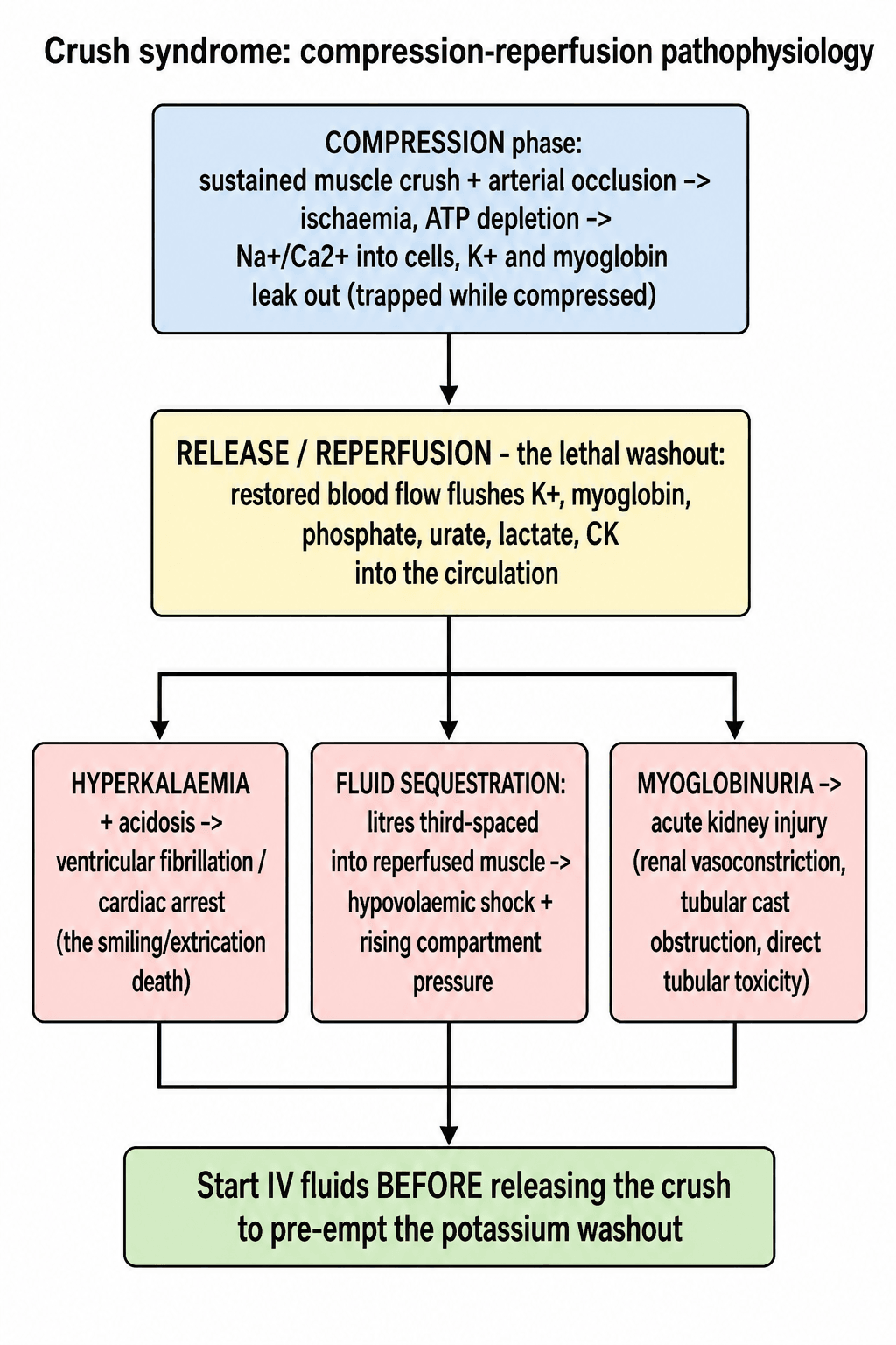
The single most examined concept: the trapped patient is often haemodynamically deceptive, with a normal blood pressure because the compressed limb is not yet reperfused, then deteriorates or arrests on release.
Do not be reassured; the metabolic load is building and will be released the moment compression ends. Intravenous fluids must be running before the crushing force is removed, to dilute the potassium load and pre-empt the reperfusion washout.
Clinical Presentation
In the limb. A swollen, tense and often pulseless limb, with pain out of proportion and pain on passive stretch, sensory loss, paraesthesia and motor weakness, and skin that shows necrosis, blistering or fixed compression marks.
Systemically. After release the patient becomes hypovolaemic and shocked as fluid third-spaces into the muscle, passes dark tea- or cola-coloured urine from myoglobinuria, and is at risk of cardiac arrhythmia from hyperkalaemia. Oliguria progresses to anuria, and nausea, confusion and the features of metabolic acidosis complete the picture.
Laboratory investigations. The findings to expect, and what each one is for:
- Expected Finding
- Markedly elevated; greater than 5,000 U/L
- Significance
- In 2,083 trauma ICU admissions, CK over 5,000 U/L carried 19% renal failure versus 8% below it; over 15,000-40,000 U/L predicts dialysis. CK stratifies risk - it does not itself trigger treatment
- Expected Finding
- Hyperkalaemia, often rapid
- Significance
- Immediate cardiac life-threat. The rate of rise matters more than any single value, and it is fastest in the hours around extrication - so the sampling interval must be short enough to catch a rise between checks, not fixed by routine
- Expected Finding
- Rising (AKI)
- Significance
- Track renal trajectory and dialysis need
- Expected Finding
- Hypocalcaemia early
- Significance
- Sequestered in muscle; do NOT over-correct
- Expected Finding
- Hyperphosphataemia, hyperuricaemia
- Significance
- Released from damaged muscle
- Expected Finding
- Metabolic acidosis, high lactate
- Significance
- Guides bicarbonate and resuscitation
- Expected Finding
- Dipstick strongly positive for blood, few or no red cells on microscopy
- Significance
- The bedside signature of myoglobinuria: the dipstick detects myoglobin, and negative microscopy means myoglobinuria
- Expected Finding
- May show DIC
- Significance
- Poor prognostic marker
CK, not myoglobin. CK is the more reliable quantitative marker because serum myoglobin clears rapidly.
The ECG. Potassium writes its progress on the trace, and the sequence is worth knowing in order:
- Early: tall, tented T waves and a short QT
- Moderate: P-wave flattening and loss of P waves, PR prolongation, progressive QRS widening
- Severe: the broad QRS merges with the T wave into a sine-wave pattern, heralding ventricular fibrillation or asystole
ECG changes can lag or precede a given potassium value. If a crush patient has any hyperkalaemic ECG change, give calcium for membrane stabilisation immediately while awaiting confirmatory bloods.
Differential Diagnosis
- Distinguishing Features
- Prolonged compression, rising CK, hyperkalaemia, myoglobinuria, AKI after release
- Key Discriminator
- History of entrapment plus systemic rhabdomyolysis
- Distinguishing Features
- Tense compartment, pain on passive stretch, usually after fracture/reperfusion
- Key Discriminator
- Local limb-threat without the systemic metabolic load (they overlap)
- Distinguishing Features
- Pulseless, cold, pale limb; hard signs of vascular injury
- Key Discriminator
- Vascular imaging and absent distal flow rather than diffuse muscle necrosis
- Distinguishing Features
- Statins, exertion, seizures, NMS, toxins; high CK without crush history
- Key Discriminator
- No mechanical entrapment
- Distinguishing Features
- Dipstick blood positive, plasma pink, low haptoglobin
- Key Discriminator
- Plasma colour and haemolysis markers, not muscle injury
- Distinguishing Features
- Fever, vasodilatation, infection source
- Key Discriminator
- Sepsis markers and absence of massive CK rise
Management
Priorities. Never extricate a long-entrapped limb without intravenous (or intraosseous) fluids running and a plan for hyperkalaemia, and treat hyperkalaemia and hypovolaemia before any orthopaedic intervention. In order:
- Pre-extrication fluids, which prevent cardiac arrest at release
- Treat hyperkalaemia, the immediate life threat
- Massive intravenous fluid resuscitation, which prevents AKI
- Continuous cardiac monitoring
- Consider dialysis early if oliguria persists
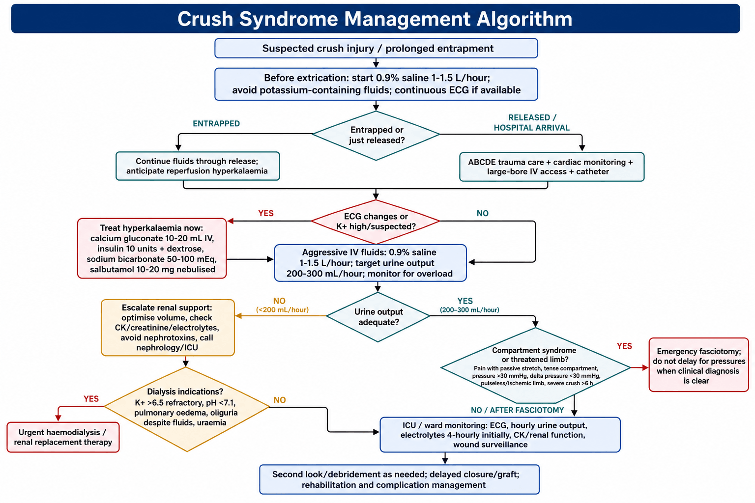
Before release. 1-1.5 L/hour of 0.9% saline, started before extrication and continued through it. If extrication is delayed, the patient may need 1 L every 30 minutes.
Why saline. Normal saline is preferred because it contains no potassium; Hartmann's and Ringer's lactate do, and lactated solutions should be avoided in crush syndrome for that reason.
In hospital. 1-1.5 L/hour continues through the first six hours, and the patient may require 10-12 L in the first 24 hours; a central line and an arterial line are recommended. High-volume fluids may continue for 48-72 hours, adjusted to urine output, electrolytes and clinical status, watching for fluid overload.
The target is the urine, not the litres. Aim for 3 mL/kg/hour, which is about 200-300 mL/hour in a 70-100 kg adult. The two figures coincide only at that weight; below it they diverge, and the weight-based one is the one to follow. The hourly rates are adult figures too: 1-1.5 L/hour is not weight-adjusted, and it is unsafe transposed to a small adult or a child without scaling.
Every figure above - 1-1.5 L/hour, 1 L per 30 minutes during prolonged extrication, 10-12 L in the first 24 hours - is a renoprotective regimen in a patient whose kidneys are responding. The same volume given to a patient who is not passing urine is a route to fatal pulmonary oedema, not renal protection.
Once a urinary catheter is in and resuscitation is under way, if urine output does not establish despite adequate volume, the answer is renal replacement therapy, not more fluid. The dialysis indications on this page - oliguria under 0.5 mL/kg/hour despite fluids, and pulmonary oedema - exist precisely because the high-volume protocol has to be abandoned in the patient it is not working for.
Controversies: Fluid Choice and Drug Therapy
Isotonic saline is favoured to avoid potassium, but large-volume saline risks hyperchloraemic acidosis; some advocate balanced (low-potassium) crystalloid once hyperkalaemia is controlled. Note the scope of the evidence: the commonly cited 1-1.5 L/h, titrated to urine output, is consensus-based, not trial-proven - which is exactly why it must be titrated rather than run as a fixed prescription.
Bicarbonate to raise urine pH above 6.5 is widely taught, but Brown et al. and others show no clear added benefit over volume alone; alkalinisation risks worsening hypocalcaemia and metabolic alkalosis.
Once routine, now largely abandoned - no proven benefit and potential harm (osmotic nephrosis, volume shifts) in hypovolaemic patients. Reserve, if ever, for well-hydrated patients with persistent oliguria.
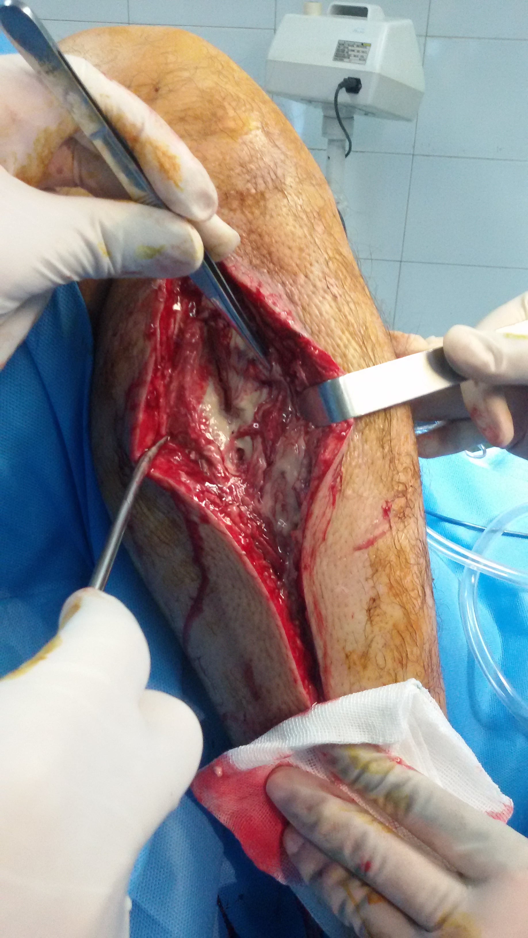
Routine prophylactic fasciotomy in crush limbs is debated: it can prevent ischaemic necrosis but converts a closed injury into an open wound with infection and bleeding risk, especially in disaster settings with limited sterility. Decompress on clear indication; individualise the prophylactic decision.
Surgical Management
The threshold. Crush injuries carry a high risk of compartment syndrome. Fasciotomy is indicated when the delta pressure (diastolic blood pressure minus compartment pressure) is under 30 mmHg sustained for more than 2 hours, or when the clinical picture of pain on passive stretch and a tense compartment is clear; do not delay for pressure measurements if the clinical diagnosis is evident. The duration is part of the threshold, not an optional refinement, and severe crush with swelling keeps the threshold low on high clinical suspicion.
What is not an indication. An absolute compartment pressure over 30 mmHg on its own has been superseded: it is common and usually transient after injury, and decompressing on that reading alone would have sent 43 per cent of continuously monitored tibial fractures to an unnecessary fasciotomy in the study that defined the delta-P threshold. Prolonged ischaemia over 6 hours is not an indication by itself either; it raises suspicion, and viability is assessed first.
The reflex from general trauma teaching is that prolonged ischaemia plus anticipated swelling equals a prophylactic fasciotomy. In crush syndrome specifically, disaster-nephrology guidance (the Renal Disaster Relief Task Force / Sever) cautions against routine or prophylactic fasciotomy, and the reason is not squeamishness about the operation:
- Cutting down on muscle that is already devitalised converts a closed injury into an open one and creates a portal for infection and sepsis, a leading cause of late death after crush.
- Sepsis, not renal failure alone, drove the late deaths in the Marmara earthquake experience. A fasciotomy that cannot save dead muscle can still supply the infection that kills the patient - and in a disaster setting the wound care that might mitigate that is exactly what is unavailable.
The reconciled position, and the one to give: fasciotomy is for a tense compartment in a still-viable limb, on unequivocal clinical signs or the sustained delta-pressure threshold above. Measured pressures therefore matter more here than elsewhere; see compartment syndrome for the measurement technique and the delta-pressure evidence.
Do not reflexively decompress a limb whose muscle is already dead: that limb may be better served by resuscitation, demarcation or amputation, and mangled extremity and limb salvage decision-making covers the limb that is past decompressing.
Complications
- Incidence
- 50% (30-50% need dialysis)
- Timing
- 24-72 hours
- Management
- Fluids, dialysis if refractory
- Incidence
- 30-40%
- Timing
- Minutes to hours (at extrication)
- Management
- Calcium, insulin, dialysis
- Incidence
- 20-30%
- Timing
- Hours to days
- Management
- Emergency fasciotomy
- Incidence
- 15-20%
- Timing
- 24-48 hours
- Management
- Treat underlying cause, FFP, platelets
- Incidence
- 10-20%
- Timing
- Days to weeks
- Management
- Debridement, antibiotics
- Incidence
- 10-15%
- Timing
- 24-72 hours
- Management
- Ventilatory support, lung protective strategy
- Incidence
- 10-20%
- Timing
- Days
- Management
- ICU support, treat underlying cause
- Incidence
- 10-15%
- Timing
- Days to weeks
- Management
- For non-viable limb, uncontrolled infection
- Incidence
- 10-20%
- Timing
- Variable
- Management
- Prevention through early aggressive treatment
Who does badly. Early aggressive management significantly improves survival; the poor prognostic factors are:
- Entrapment greater than 6 hours
- Trunk or bilateral limb involvement
- A very high CK: a figure of 75,000 U/L is conventionally quoted, but we could not trace it to a primary source and it should be treated as a rule of thumb rather than a validated cut-off
- Delayed fluid resuscitation
- DIC
- Multi-organ failure
Do not prognosticate from the renal stage. When modified RIFLE criteria were applied to 416 Marmara crush victims, a worse category predicted more medical complications, more dialysis sessions and a lower discharge GFR, but did not discriminate survival at all. Renal severity tells you what resources the patient will consume; it does not tell you whether they will live.
What kills these patients is extra-renal: the Marmara series attributed death to sepsis, thrombocytopenia, DIC, ARDS and thoraco-abdominal trauma rather than to renal failure itself, and in a mass-casualty triage that matters, because the sickest kidney is not the same patient as the sickest victim.
Guidelines, Registries & Global Practice
Global Epidemiology
Crush syndrome arises in three broad contexts worldwide: natural disasters (earthquakes are the dominant cause of mass-casualty crush, e.g. Marmara 1999, Kashmir 2005, Haiti 2010, Turkey-Syria 2023), industrial and motor-vehicle entrapment (mining, construction, building collapse, prolonged extrication after road traffic collisions), and prolonged immobilisation (collapse with "long-lie", drug overdose, the elderly found-down). After a major earthquake, crush-related AKI develops in a substantial minority of rescued survivors; the proportion needing dialysis varies enormously with rescue speed and access to renal services - illustrated by the Marmara series (74.6% of renal victims dialysed) versus the rural Kashmir earthquake, where far fewer reached dialysis at all.
Side-by-Side Guidance
- Emphasis
- Disaster nephrology
- Key Position
- Pre-extrication and early high-volume isotonic fluids; early dialysis mobilisation; avoid potassium-containing fluids
- Emphasis
- Renal protection
- Key Position
- Volume expansion as primary renoprotection; routine bicarbonate/mannitol not supported by strong evidence
- Emphasis
- Limb & compartment
- Key Position
- Low threshold for fasciotomy; serial compartment assessment; damage-control surgery
- Emphasis
- Pre-hospital & field
- Key Position
- IV/IO access and fluids before release; cardiac monitoring; treat hyperkalaemia empirically in the field
- Emphasis
- Cardiac arrest
- Key Position
- Hyperkalaemia is a reversible cause of arrest; give calcium early, consider dialysis
High- vs Limited-Resource Practice Variation
In well-resourced settings, rapid extrication, point-of-care potassium, continuous renal replacement therapy and intensive care allow aggressive support and comparatively low renal-cause mortality. In limited-resource or austere disaster settings, delayed rescue, scarce dialysis and limited transport drive higher rates of hyperkalaemic death and amputation; field priorities shift toward early fluids, empirical hyperkalaemia treatment, and triage of who can be transferred for renal replacement. International coordination (e.g. deploying mobile dialysis capacity after earthquakes) materially changes outcomes.
Controversies and Areas of Uncertainty
Tourniquet at extrication. A proximal tourniquet applied before releasing a long-entrapped, clearly non-salvageable (mangled) limb can blunt the lethal reperfusion washout of potassium and myoglobin, and is part of field-amputation decision-making in mass-casualty settings. It is selective and debated, not routine, and must not delay fluid resuscitation; for a salvageable limb the priority remains pre-extrication fluids rather than ischaemic tourniquet time.
See tourniquet use for safe pressures and duration, and damage control orthopaedics for how these decisions sit within the wider resuscitation of a polytrauma patient.
Amputation or limb salvage. For a non-viable, heavily contaminated or overwhelmingly toxic limb, early amputation can be life-saving by removing the source of ongoing potassium and myoglobin load and infection, and is sometimes the correct disaster decision over repeated debridement of dead muscle. The threshold is a clinical judgement balancing systemic toxicity against limb salvage.
Exam Viva Scenarios
Practise clinical reasoning and management decisions out loud
“You are called to a building collapse where a 35-year-old construction worker has been trapped under concrete debris for 5 hours. His right leg is crushed. Rescue teams are preparing to extricate him. What is your management?”
“A 28-year-old woman was trapped in a car accident for 3 hours with her legs crushed. She was extricated by paramedics and appeared stable initially. Ten minutes after extrication, she develops VF arrest. What is the likely cause and how would you manage this?”
“A 40-year-old man is admitted following a mining accident. His right thigh was crushed for 4 hours. He is hypotensive, has dark urine, and his thigh is massively swollen and tense. Potassium is 6.8, CK is 85,000, creatinine is rising. His leg is pulseless. How would you manage him?”
Definitions
- Crush injury = localized tissue damage from compression
- Crush syndrome = SYSTEMIC manifestation after release (rhabdomyolysis, AKI, hyperkalemia)
- Develops after greater than 4-6 hours of compression
- Reperfusion injury = metabolic derangement at moment of release
Lethal Triad
- Hyperkalemia - causes cardiac arrest (K+ greater than 6.5 is dangerous)
- Metabolic acidosis - lactic + phosphoric acid
- Hypocalcemia - sequestered in muscle (do NOT aggressively replace)
Pre-Extrication Protocol
- IV access BEFORE release - never extricate without IV
- Normal saline 1-1.5L/hour (NOT Hartmann's - contains K+)
- Cardiac monitoring if available
- Intraosseous access if IV impossible
Fluid Targets
- Urine output 200-300mL/hour (3mL/kg/hr)
- May need 10-12L in first 24 hours
- Alkalinize urine to pH greater than 6.5 with bicarbonate
- Avoid nephrotoxins (NSAIDs, aminoglycosides, contrast)
Hyperkalemia Treatment
- Calcium gluconate 10% 10-20mL IV (membrane stabilization)
- Bicarbonate 50-100mEq IV (K+ shift)
- Insulin 10U + Dextrose 50mL 50% (K+ shift)
- Salbutamol 10-20mg nebulized (K+ shift)
- Kayexalate/Dialysis (K+ removal)
Fasciotomy Indications
- Absolute compartment pressure greater than 30mmHg is NOT an indication on its own
- Delta pressure (DBP - CP) less than 30mmHg
- Clinical: pain on passive stretch + tense compartment
- Prolonged ischemia greater than 6 hours raises suspicion - not an indication by itself
Dialysis Indications
- Refractory hyperkalemia (K+ greater than 6.5 despite treatment)
- Severe acidosis (pH less than 7.1)
- Fluid overload/pulmonary edema
- Oliguria (less than 0.5mL/kg/hr despite fluids)
Key Numbers
- CK greater than 5,000 U/L = high risk AKI
- CK greater than 15,000-20,000 U/L = almost certain dialysis
- Entrapment greater than 4-6 hours = high risk crush syndrome
- 20% overall mortality; 50% of AKI need dialysis
Evidence Base
Management of Crush-Related Injuries After Disasters (Landmark Review)
- Definitive review of disaster-related crush injury drawing on the Renal Disaster Relief Task Force experience
- Establishes pre-extrication and early intravenous fluid loading as the central preventive intervention against crush-induced AKI
- Frames hyperkalaemia, hypovolaemia and myoglobinuric AKI as the principal early killers
- Calls for early mobilisation of dialysis and intensive-care resources in mass-casualty settings
Marmara Earthquake - Largest Crush Syndrome Renal Series
- 639 patients with crush-related acute renal failure across 35 hospitals after the 1999 Marmara earthquake
- 477 patients (74.6%) received one or more dialysis treatments
- Overall mortality 15.2% (17.2% in dialysed vs 9.3% in non-dialysed patients)
- Death was driven by sepsis, thrombocytopenia, DIC, ARDS and thoraco-abdominal trauma rather than renal failure alone