Denis Three-Column Classification of Spinal Fractures
If the middle column is disrupted, the injury is mechanically unstable — regardless of what the radiograph looks like at first glance. Burst, flexion-distraction, and fracture-dislocation all cross the middle column. A wedge compression that leaves the middle column intact is a different problem from a burst that violates it, even when the wedge is more impressive on the lateral view.
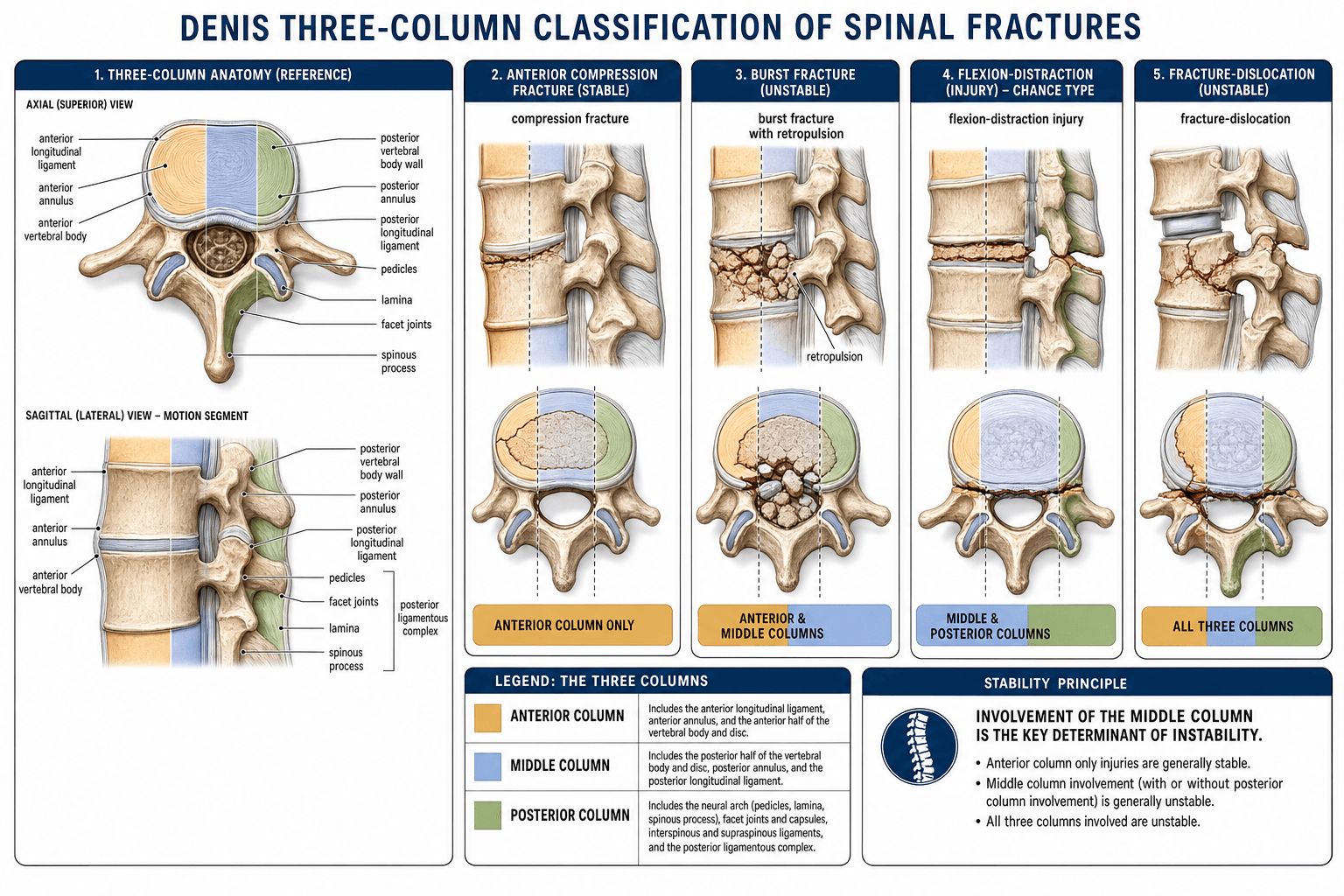
The Three Columns
The spine is conceptualised as three longitudinal load-bearing columns. The division runs through the vertebral body, disc, and soft tissues, not through the pedicles.
- Bony structures
- Anterior two-thirds of vertebral body and disc
- Soft-tissue structures
- Anterior longitudinal ligament, anterior annulus
- Mechanical role
- Resists compression; bears the majority of axial load
- Bony structures
- Posterior one-third of vertebral body and disc (including the posterior wall of the body)
- Soft-tissue structures
- Posterior annulus, posterior longitudinal ligament
- Mechanical role
- Keeps the vertebral ring intact — the key to stability
- Bony structures
- Pedicles, facets, laminae, spinous and transverse processes
- Soft-tissue structures
- Supraspinous, interspinous, ligamentum flavum, facet capsules
- Mechanical role
- Resists tension; tether for flexion and rotation
Anterior · Middle · PosteriorThe three columns
Hook:The dividing line between anterior and middle runs through the vertebral body — the middle column is the back third (the posterior wall is its key structure).
The middle column runs from the posterior longitudinal ligament forward through the posterior third of the vertebral body and disc. It is not a bony column in the sense of a separate bone — it is a functional column whose integrity prevents the vertebral ring from collapsing into a wedge. On a sagittal CT or MRI, look at the posterior wall of the vertebral body: if it is retropulsed, the middle column is disrupted.
Stability and the Middle Column
- Columns involved
- Anterior only
- Middle column
- Intact
- Mechanical stability
- Usually stable if less than 50 percent height loss and no posterior ligamentous complex injury
- Columns involved
- Anterior and middle
- Middle column
- Disrupted (posterior wall fragments retropulsed)
- Mechanical stability
- Unstable — risk of kyphosis and neural compromise
- Columns involved
- Middle and posterior (tension failure)
- Middle column
- Disrupted
- Mechanical stability
- Unstable — often missed if purely ligamentous
- Columns involved
- All three (translation/rotation)
- Middle column
- Disrupted
- Mechanical stability
- Grossly unstable — almost always neurological injury
Middle intact → stable · Middle broken → unstableThe middle-column rule
Hook:Don't be fooled by an impressive-looking wedge with an intact posterior wall — that is a compression, not a burst.
Do not confuse Denis stability with clinical decision-making. Denis is an anatomical and biomechanical descriptor. A stable Denis pattern (anterior compression) can still need surgery in a young patient with marked kyphosis, and an unstable pattern may be managed conservatively in a frail patient with no neurology. Use Denis to describe the injury, then add morphology, PLC integrity, and neurology to decide what to do.
The four patterns above are Denis's major injuries; the original paper also defined two things the topic should not omit:
- Minor injuries — isolated fractures of the transverse process, articular (facet) process, pars interarticularis, or spinous process. These are generally stable because they spare the weight-bearing three-column ring — but a transverse-process fracture can flag the energy of the injury and associated damage (for example an L5 transverse-process fracture with a pelvic-ring injury, or lumbar plexus involvement), so look beyond the fracture itself.
- Three degrees of instability — Denis graded instability rather than treating it as simply present/absent: First-degree = mechanical instability (a tendency to late kyphosis/deformity — e.g. severe compression and seat-belt injuries); Second-degree = neurological instability (a risk of late neurological deterioration — e.g. burst fractures); Third-degree = both mechanical and neurological instability (fracture-dislocations, and burst fractures with a neurological deficit).
The Four Denis Fracture Patterns
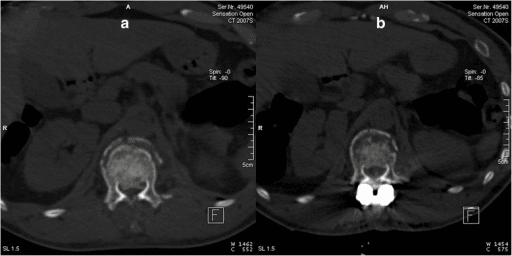
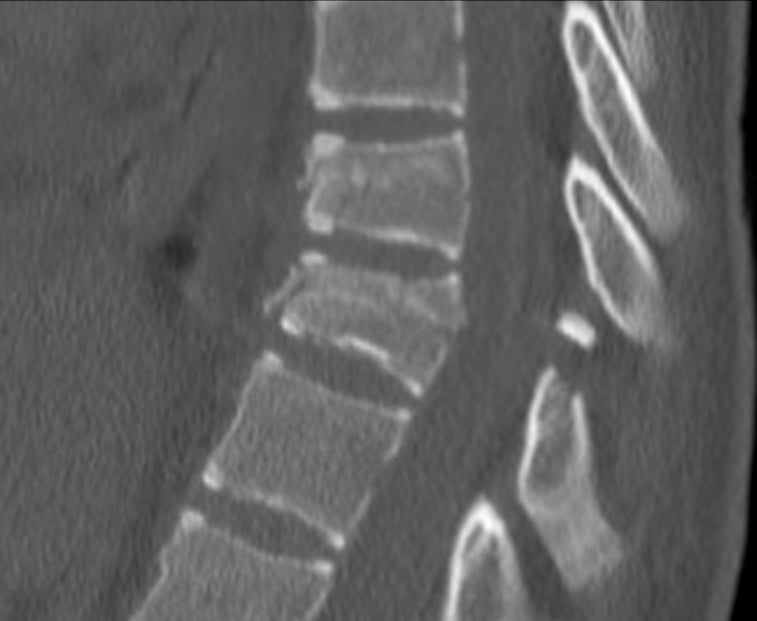
Compression · Burst · Distraction · DislocationThe four fracture patterns
Hook:Climb the severity ladder: one column (compression) → two columns crossing the middle (burst, distraction) → all three (fracture-dislocation).
The key distinction between compression and burst is the posterior wall of the vertebral body. Both can produce anterior wedging; only a burst has a retropulsed posterior wall fragment. If the posterior cortex of the body is intact on CT and MRI, the injury is a compression, not a burst.
- Mechanism
- Pure flexion / axial load on the anterior column
- Imaging clue
- Anterior wedge; posterior wall intact; no retropulsion
- Typical behaviour
- Stable if less than half the body height is lost and the PLC is intact
- Mechanism
- Pure axial load — nucleus pulposus is driven through the endplate into the vertebral body
- Imaging clue
- Posterior wall fragment retropulsed into the canal; widened pedicles on AP; loss of body height
- Typical behaviour
- Unstable; neurological deficit common when canal compromise is significant
- Mechanism
- Flexion over a fixed axis (lap-belt, fall)
- Imaging clue
- Horizontal split through the spinous process, lamina, pedicles, and posterior body, OR pure ligamentous failure posteriorly and through the disc
- Typical behaviour
- Unstable; the spine hinges open posteriorly; classic ‘empty’ or ‘opened’ posterior elements on CT
- Mechanism
- High-energy flexion-rotation, shear, or compression-flexion
- Imaging clue
- Translation or rotation of one vertebra on the next; lateral view shows offset
- Typical behaviour
- Grossly unstable; complete spinal cord injury in over half of cases
Classification in Practice — Using Denis at the Bedside
- Identify the level — thoracolumbar junction (T11–L2) is the commonest site for burst, Chance, and fracture-dislocation injuries because it is the transition from stiff thoracic to mobile lumbar spine.
- Look at the lateral X-ray first — wedge or burst? Is the posterior wall broken? Is there translation?
- CT to confirm the posterior wall of the vertebral body and to assess pedicle widening, fragment size, and canal compromise. MRI to assess the posterior ligamentous complex, the disc, and cord/canal signal.
- Apply the three-column rule — does the middle column cross the fracture line? If yes, the injury is unstable. If no, consider it a one-column problem until proven otherwise.
- Document the pattern as compression, burst, flexion-distraction, or fracture-dislocation, and add morphology, PLC, and neurology to feed into modern classification systems (TLICS / AO Spine Thoracolumbar Injury Classification System).
Limitations and Modern Context
- Denis is purely morphological. It does not score neurological status, posterior ligamentous complex integrity, or patient factors; modern classifications (TLICS, AO Spine) add these.
- It is poorly reproducible for subtle injuries. Inter-observer agreement for distinguishing mild burst from compression and for distinguishing bony from ligamentous Chance-type injuries is modest; CT and MRI are essential.
- The middle column is a functional, not an anatomical, structure. Different authors have placed the dividing line between columns at slightly different points in the posterior wall of the body. The pragmatic rule — is the posterior wall of the body intact? — is more reproducible than reciting textbook lines.
- Burst and flexion-distraction can coexist (a ‘burst-variant Chance’ or ‘distraction-burst’). Do not force a complex injury into a single Denis box.
- The AO Spine Thoracolumbar Injury Classification System (2013, revised 2020) is now the dominant global teaching tool for thoracolumbar trauma. Denis is still the framework most trainees use to think about columns and stability, and examiners expect you to be able to apply it.
The topic repeatedly says to "score with TLICS or AO Spine" — here are the actual systems the examiner expects you to apply:
- TLICS (Thoracolumbar Injury Classification and Severity Score) sums three categories: morphology (compression 1, burst 2, translation/rotation 3, distraction 4 — take the highest), posterior ligamentous complex (intact 0, indeterminate 2, disrupted 3), and neurology (intact 0, nerve root 2, complete cord/conus 2, incomplete cord/conus 3, cauda equina 3). Total 3 or less → non-operative; 4 → surgeon's discretion; 5 or more → operative.
- AO Spine Thoracolumbar — morphology Type A (compression; A0–A4, with A3/A4 the burst patterns), Type B (tension-band failure; B1 bony Chance, B2 posterior ligamentous, B3 hyperextension) and Type C (translation/displacement) — plus a neurological grade (N0–N4, NX) and case modifiers (M1 indeterminate PLC, M2 patient comorbidity). The AO Spine severity (TL AOSIS) score weights these to guide treatment: Type C and most B are operative, A0–A2 usually non-operative, and A3/A4 (burst) the debated middle ground.
- The mapping — Denis compression/burst ≈ AO Type A, flexion-distraction ≈ Type B, fracture-dislocation ≈ Type C — so you describe columns with Denis and score/communicate with TLICS or AO Spine.
Guidelines, Registries and Global Practice
- No single international society publishes a guideline dedicated to the Denis classification itself. Denis is a teaching and descriptive tool, and modern guidelines use it as background context rather than as a treatment-decision rule.
- AO Spine (international) — the AO Spine Thoracolumbar Injury Classification System (Vaccaro et al., 2013; revised 2020) is the most widely taught system globally and is mapped onto Denis patterns (A = compression/burst, B = flexion-distraction, C = translation/rotation). The AO Foundation provides free online teaching modules that explicitly cross-reference the Denis three-column concept.
- Orthopaedic Trauma Association (OTA / AAOS, US) — the OTA Open Fracture Classification and the AAOS Appropriate Use Criteria for thoracolumbar injuries treat Denis as foundational knowledge; treatment decisions incorporate AO Spine morphology, neurology, and PLC status.
- British Orthopaedic Association (BOA) and AO Spine Europe — teaching materials for the FRCS(Orth) exam still teach Denis as the first language for describing columns and stability, before introducing AO Spine and TLICS.
- AO Trauma and SICOT (international) — global fellowship teaching (including in low- and middle-income settings) relies on the three-column model because it is intuitive and requires only a plain lateral radiograph and a CT to apply in a basic form.
- No procedure-specific registry scores Denis directly; AO Spine’s international multicentre outcome studies use AO Spine type as the morphological variable. Denis is, however, the most common ‘first pass’ descriptor in registry entries and operative notes worldwide.
- Practice variation — the same Denis pattern (for example, an intact-neurology L1 burst) is operated on far more frequently in some regions than others. Modern international comparative-effectiveness work (AO Spine studies) has shown broadly similar long-term patient-reported outcomes between operative and nonoperative treatment in selected neurologically intact bursts, supporting a more conservative global trend.
Viva practice
Exam Viva
Practise clinical reasoning and management decisions out loud
“A 28-year-old man falls from a height and lands on his feet. He has back pain but no neurological deficit. A lateral radiograph shows a wedge of L1 with loss of about 40 percent of anterior height. CT shows a retropulsed posterior wall fragment with 30 percent canal compromise. Using the Denis classification, how would you describe this injury, and is it stable?”
“A 35-year-old front-seat passenger is involved in a high-speed lap-belt motor vehicle collision. He is tender over the upper lumbar spine with a palpable gap between the spinous processes. He has no neurological deficit. Plain films show an ‘empty’ appearance of the posterior elements at L2 with anterior wedging. Describe the injury using Denis, explain why it is unstable, and outline the importance of MRI.”
Exam cheat sheet
The three columns
- Anterior: anterior two-thirds of the body and disc, ALL, anterior annulus
- Middle: posterior one-third of the body and disc, posterior annulus, PLL
- Posterior: pedicles, facets, laminae, spinous process, posterior ligamentous complex
The stability rule
- Middle column intact = potentially stable (one-column injury)
- Middle column disrupted = mechanically unstable (two- or three-column injury)
- Always confirm the posterior wall of the vertebral body on CT before deciding stability
The four Denis patterns
- Compression: anterior column only (wedge; posterior wall intact) — usually stable
- Burst: anterior and middle columns (retropulsed posterior wall) — unstable
- Flexion-distraction (Chance): middle and posterior columns in tension — unstable
- Fracture-dislocation: all three columns with translation/rotation — grossly unstable
Denis in modern practice
- Use Denis to think about columns and stability; pair with AO Spine or TLICS to score the injury
- MRI is mandatory to assess the posterior ligamentous complex, disc, and neurology
- Stability is descriptive, not a treatment rule — intact neurology, alignment, and patient factors all matter
Evidence
The three column spine and its significance in the classification of acute thoracolumbar spinal injuries
- Three-column model derived from a retrospective study of 412 thoracolumbar injuries; introduced the middle (osteoligamentous) column — posterior wall of the body, PLL and posterior annulus.
- Established that the mode of middle-column failure correlates with both fracture type and neurological injury, and defines mechanical stability.
- Defined four major patterns: compression, burst, seat-belt-type (flexion-distraction), and fracture-dislocation.
The value of computed tomography in thoracolumbar fractures: an analysis of one hundred consecutive cases and a new classification
- 100 consecutive potentially-unstable injuries studied with multiplanar CT, classified by the MODE of middle-column failure: axial compression (73), axial distraction (15), and translation (12).
- Translational injuries were the most unstable, with the most severe neural deficits (and a contraindication to routine Harrington distraction).
- CT was more sensitive than other modalities for posterior-element disruption in burst fractures — corroborating the middle-column concept independently of Denis.
Acute thoracolumbar burst fractures in the absence of neurologic deficit: a comparison between operative and nonoperative treatment
- Retrospective comparison of operative versus nonoperative treatment in 52 neurologically intact thoracolumbar burst fractures (of 104).
- All surgically treated patients without unrelated disability returned to full-time work; 25% of the nonoperative group could not, and 17% of the nonoperative group developed neurological problems.
- The authors concluded that prophylactic stabilisation and fusion had significant advantages over conservative management.
According to PubMed, the three-column concept (412 injuries) is from Denis 1983 (Spine 1983;8(8):817-31; DOI 10.1097/00007632-198311000-00003), with independent CT corroboration and the three failure modes from McAfee et al. 1983 (PMID 6833320). Denis's own 1984 series (PMID 6478691) favoured operative treatment of intact-neurology bursts (17% of nonoperative developed neurological problems) — a position later tempered by randomised data. The AO/Magerl A/B/C language is summarised by Gertzbein 1994 (DOI 10.1097/00007632-199403000-00022) and formalised, with substantial reliability (kappa 0.72), by the AO Spine system of Vaccaro et al. 2013 (DOI 10.1097/BRS.0b013e3182a8a381).