Internal Disc Disruption | Degenerative Disc Disease | Mechanical Back Pain
- Discogenic pain = internal disc disruption with intact annulus but painful fissures
- Diagnosis requires concordant provocation discography - controversial gold standard
- 80% improve with 6-12 months conservative care - surgery last resort only
- Fusion indicated for single-level disease with concordant pain reproduction
- Disc replacement for preserved disc height, no facet arthropathy, under 60 years
- “Key distinction: discogenic pain (internal) vs herniation (external compression)
- “Modic changes on MRI correlate with discogenic pain (Type 1 = inflammation)
- “High-Intensity Zone (HIZ) = annular tear on MRI - 80% sensitivity for discogenic pain
- “MRC trial and Brox RCT: fusion no clearly better than intensive rehabilitation; NICE advises against fusion for low back pain outside a trial
Overview and Epidemiology
Discogenic back pain is internal disc disruption (IDD): a degenerative condition in which the nucleus pulposus loses its hydration and the annulus fibrosus develops painful fissures, without the frank herniation that compresses neural tissue. It is a mechanical and chemical pain syndrome arising from disrupted disc architecture, and it presents as axial low back pain from an annulus that is damaged but intact.
The key distinction. Discogenic pain is internal; a herniation is external compression.
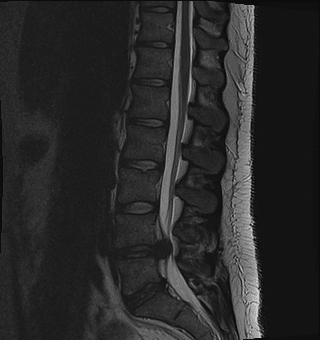
How the pain arises. The nucleus loses proteoglycan, dehydrates, and the disc loses height. Annular tears propagate from the nucleus outward until they reach the pain-sensitive outer annular fibres, and inflammatory cytokines leak into the tears and sensitise nociceptors. The anatomy and chemistry behind this are in the next section.
The degenerative cascade. Four stages, each with the age band attached to it.
Degenerative Progression
The nucleus pulposus loses proteoglycan and water. Disc height is maintained but the internal architecture is disrupted, and annular tears begin centrally.
Annular fissures propagate radially outward and reach the pain-sensitive outer one-third of the annulus, where they become visible on MRI as the high-intensity zone (HIZ). Inflammatory mediators sensitise the nociceptors there.
Disc height is lost and the endplates respond with Modic changes. Facet loading increases, biomechanical instability develops and the segment may become unstable.
The disc collapses and osteophytes form. The segment stabilises paradoxically (auto-fusion), and pain may improve as motion decreases: the disc "burns out".
Natural history. Variable. Approximately 80% of patients improve with conservative care over 6-12 months, and the disc may burn out with progressive collapse and fibrosis, reducing pain. Younger patients with single-level disease and Modic Type 1 changes have a worse prognosis.
Pathophysiology and Anatomy
The disc. Three components.
Nucleus pulposus. A central gelatinous core, 80% water in youth, held by a proteoglycan matrix (aggrecan) that retains the water and given structure by type II collagen. It is aneural and avascular; its nutrients arrive by diffusion from the endplates.
Annulus fibrosus. Concentric lamellae of type I collagen, 15-25 layers with the fibre orientation alternating at 30 degrees between layers, anchored to the vertebral endplates by Sharpey fibres. Only the outer one-third is innervated.
Cartilaginous endplates. The interface between vertebral body and disc: the nutrient diffusion pathway for the avascular disc, and the surface that distributes load across the vertebral body. Endplate injury causes Modic changes and disc degeneration.
Innervation, the key to discogenic pain. The outer third of the annulus is supplied by:
- Sinuvertebral nerve (recurrent branch of the ventral ramus) - posterior and posterolateral annulus
- Grey rami communicantes - anterolateral annulus
- Dorsal rami - posterior annulus
The inner two-thirds is normally aneural. In discogenic pain, nerve growth factor (NGF) drives aberrant nerve ingrowth into the inner annulus along radial tears, creating pain-sensitive tissue in a zone that should have none.
The chemistry of the painful disc. Inflammatory cytokines (IL-1, IL-6, TNF-alpha and PGE2) leak into the fissures and sensitise nociceptors. Matrix metalloproteinases degrade collagen and proteoglycans, NGF drives the nerve ingrowth above, and substance P, a nociceptive neurotransmitter, is found in painful discs.
The mechanics of the failing disc. Annular incompetence distributes stress abnormally across the disc, and endplate fracture produces the Modic Type 1 change. The table sets the rest of the sequence against its clinical effect.
- Normal Disc
- 80% water content, high proteoglycan
- Degenerated Disc
- Reduced to 70% water, proteoglycan loss
- Clinical Effect
- Loss of shock absorption, increased annular stress
- Normal Disc
- Normal pressure distribution
- Degenerated Disc
- Altered load transfer, peak pressures
- Clinical Effect
- Annular tears propagate, endplate injury
- Normal Disc
- Normal height maintains lordosis
- Degenerated Disc
- Collapse causes kyphosis
- Clinical Effect
- Facet overload, foraminal stenosis, sagittal imbalance
- Normal Disc
- Controlled motion within physiologic range
- Degenerated Disc
- Abnormal translation/rotation or stiffening
- Clinical Effect
- Instability (early) or autofusion (late)
Load. In flexion, sitting raises intradiscal pressure by 200-300% compared with standing. That single figure explains why discogenic pain is characteristically worse with sitting and flexion activities.
Classification Systems
Three systems describe three tissues. Pfirrmann grades the disc itself on T2 MRI, Modic describes the marrow response in the adjacent endplates, and the Dallas discogram grades the annular tear by how far contrast escapes the nucleus.
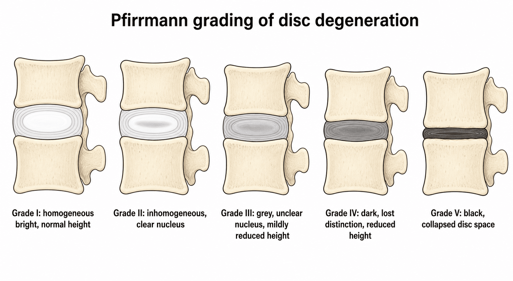
Pfirrmann MRI Classification
A five-grade system based on T2 signal intensity, disc structure and disc height, and the most widely used MRI grading of disc degeneration.
- Structure
- Homogeneous, bright white
- Distinction
- Clear nucleus-annulus distinction
- Height
- Normal
- Signal (T2)
- Hyperintense (bright)
- Structure
- Inhomogeneous with horizontal band
- Distinction
- Clear distinction maintained
- Height
- Normal
- Signal (T2)
- Hyperintense with central horizontal band
- Structure
- Inhomogeneous, grey
- Distinction
- Unclear distinction
- Height
- Normal to decreased
- Signal (T2)
- Intermediate (grey)
- Structure
- Inhomogeneous, dark grey
- Distinction
- Lost distinction
- Height
- Decreased
- Signal (T2)
- Intermediate to hypointense (dark grey)
- Structure
- Inhomogeneous, black
- Distinction
- Lost distinction
- Height
- Collapsed
- Signal (T2)
- Hypointense (black)
Grade 3-5 correlates with symptomatic disc degeneration, but Grade 3-4 changes are common in asymptomatic individuals over 40. Grade 4-5 with Modic Type 1 changes and HIZ has the highest specificity for a discogenic pain source.
Clinical Presentation and Assessment
History. Axial, mechanical low back pain centred on the low back (L4-5 or L5-S1), worse with prolonged sitting, bending and lifting, and better standing, walking or lying supine. Sitting hurts because intradiscal pressure is highest there, and flexion because it increases annular stress. The pain may refer to the buttocks or posterior thighs but does not radiate below the knee and is not dermatomal, which is what rules out radiculopathy. Morning stiffness improves with movement. There is no nocturnal pain (unlike tumour) and there are no constitutional symptoms (unlike infection).
Examination. The neurological examination is normal and the straight leg raise negative, which distinguishes this from a herniation. Flexion is painful and range of motion is limited by pain; provocative testing makes discogenic pain worse in flexion. Facet loading (extension and rotation) is negative, which rules out a facet source.
A diagnosis of exclusion. Discogenic pain is confirmed only after the other sources have been ruled out:
- Facet arthropathy - pain worse with extension, positive facet loading tests
- Sacroiliac joint - provocative SI tests (FABER, Gaenslen)
- Myofascial pain - trigger points, muscle spasm
- Spondylolisthesis - palpable step-off, instability on dynamic radiographs
- Red flags - infection, tumour, fracture, cauda equina syndrome
- Pain pattern
- Central axial, may refer to buttock/posterior thigh, not below knee
- Aggravating / relieving
- Worse with sitting and flexion; better standing/walking/lying
- Key positive finding
- Concordant pain on provocation; HIZ, Modic 1, Pfirrmann 3-5 at one level
- Discriminator from discogenic
- The diagnosis of exclusion - confirmed only after facet, SIJ, hip and radicular sources are ruled out
- Pain pattern
- Axial, paraspinal, may refer to buttock/thigh
- Aggravating / relieving
- Worse with extension and rotation; better with flexion
- Key positive finding
- Positive facet loading; relief with medial branch block
- Discriminator from discogenic
- Extension-worse (discogenic is flexion-worse)
- Pain pattern
- Below L5, over the joint, groin/buttock
- Aggravating / relieving
- Worse with transitional movements and single-leg loading
- Key positive finding
- Cluster of provocative tests (FABER, thigh thrust, Gaenslen, compression)
- Discriminator from discogenic
- Pain below L5 and positive SIJ cluster
- Pain pattern
- Dermatomal leg pain often exceeding back pain
- Aggravating / relieving
- Worse with sitting/Valsalva; positive tension signs
- Key positive finding
- Positive SLR/femoral stretch, dermatomal deficit, nerve-root compression on MRI
- Discriminator from discogenic
- Neurological signs and leg-dominant pain
- Pain pattern
- Insidious, age under 45, alternating buttock pain
- Aggravating / relieving
- Worse with rest and at night; better with exercise; prolonged morning stiffness
- Key positive finding
- Raised CRP, HLA-B27, sacroiliitis on MRI/X-ray
- Discriminator from discogenic
- Inflammatory rhythm (rest pain, night pain, AM stiffness)
- Pain pattern
- Constant, progressive, may be night pain
- Aggravating / relieving
- Often unrelated to posture
- Key positive finding
- Red flags - fever, weight loss, malignancy history, focal tenderness, neurology
- Discriminator from discogenic
- Red flags and constitutional symptoms - exclude first
In 92 consecutive patients with chronic low back pain who underwent CT discography, not one conventional clinical test could distinguish those who met the criteria for internal disc disruption from those who did not. That is a more useful finding than the prevalence figure from the same study, and it explains the shape of this diagnosis: there is no Lachman for the disc. Examination excludes the alternatives - radiculopathy, facet, sacroiliac joint, hip - and confirms nothing.
What examination should therefore add is an assessment of the factors that actually predict outcome. Psychological distress and occupational disability predicted a painful discogram better than the disc did, and they predict the response to every intervention offered here. Screen for them explicitly rather than treating them as a footnote:
- Yellow flags: belief that pain means damage, fear-avoidance and activity withdrawal, catastrophising, low mood, poor sleep, expectation that only a procedure can help, dissatisfaction at work, and an active compensation or litigation claim.
- Function, not just pain score: what they have stopped doing, and what returning to it would take. A pain score falls with a drug; function is what surgery is being asked to restore.
Waddell's signs - what they are and what they are not. The eight signs in five groups - superficial or non-anatomical tenderness, pain on axial loading or simulated rotation, distraction (a straight-leg raise that differs supine versus seated), regional weakness or sensory loss that follows no neurological pattern, and overreaction - were described to identify patients whose presentation is amplified by distress. Three or more groups is the conventional threshold.
They are not a test of malingering, and using them that way is both wrong and harmful. They do not detect deception, they correlate with distress rather than dishonesty, and they can be positive in people with entirely genuine and severe pathology. Their legitimate use is as a prompt: a patient with several is unlikely to do well from an operation aimed at a structural target, and needs the psychological and functional side of the problem addressed first. In a viva, offer them with that qualification attached - quoting them as a lie detector is a reliable way to lose the mark.
Investigations
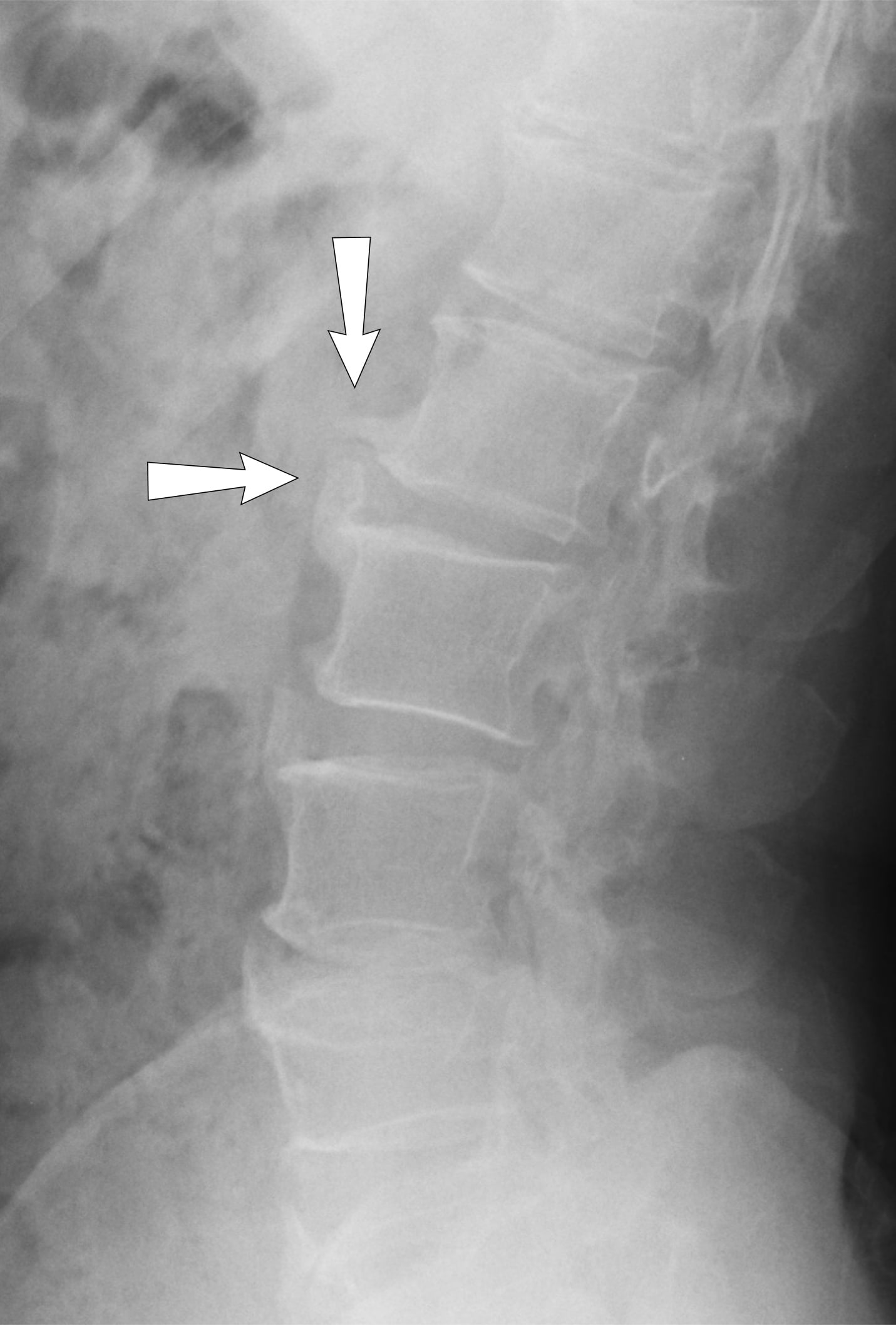
Radiographs. Standing AP and lateral films assess disc height loss, alignment and instability, with flexion-extension views if instability is suspected. Their sensitivity for early discogenic pain is limited.
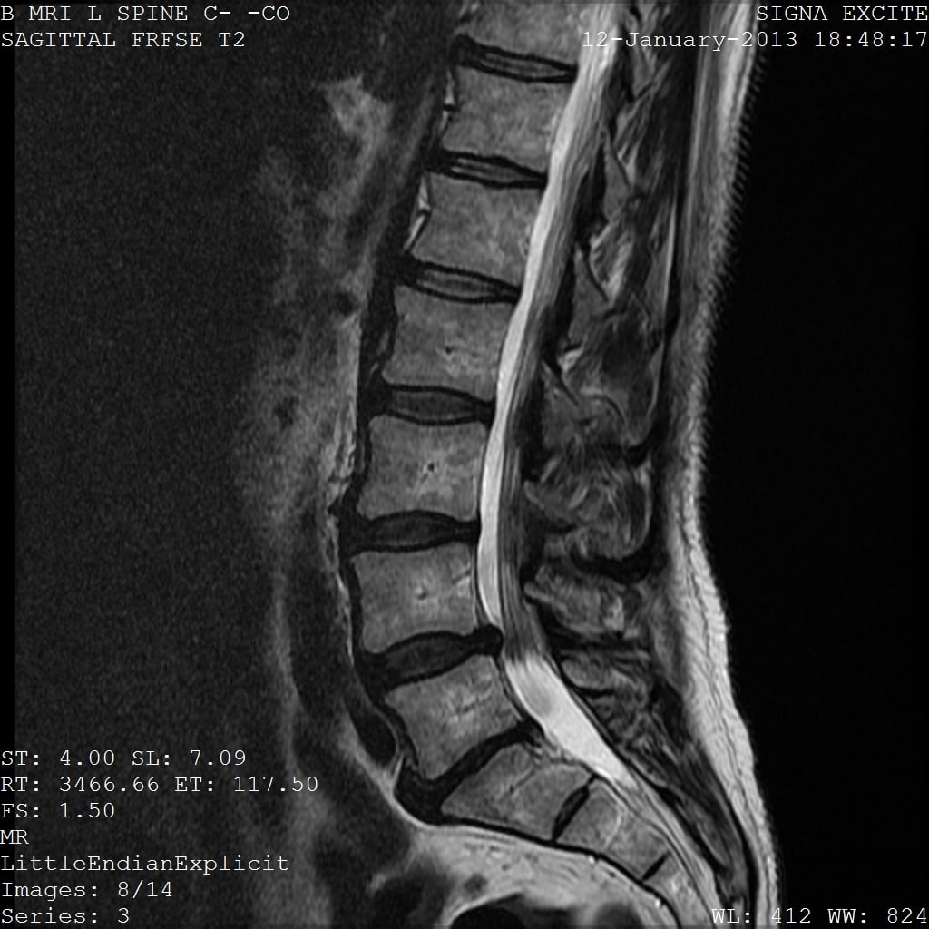
MRI is the gold standard. T1 and T2 sequences, read for disc morphology (the Pfirrmann grade), the high-intensity zone and Modic endplate changes. Sensitivity is 80-90% when combined with clinical correlation.
- Description
- 5-grade system based on T2 signal and disc height
- Clinical Correlation
- Grade 4-5 = advanced degeneration, Grade 3 = intermediate
- Sensitivity
- High sensitivity (90%) but low specificity (many asymptomatic have degeneration)
- Description
- Hyperintense focal area in posterior annulus on T2
- Clinical Correlation
- Annular tear - 80% correlation with discogenic pain
- Sensitivity
- 80% sensitivity, 80% specificity for concordant discography
- Description
- Hypointense T1, hyperintense T2 in vertebral endplate
- Clinical Correlation
- Marrow oedema, inflammation - correlates with pain
- Sensitivity
- 70% sensitivity for active discogenic pain
- Description
- Reduced disc space height compared to adjacent levels
- Clinical Correlation
- Advanced degeneration, may indicate instability
- Sensitivity
- Moderate correlation with pain (biomechanical loading)
Diagnostic injections. Facet blocks or sacroiliac joint injections exclude the alternative pain sources. Over 50% pain relief from either suggests a non-discogenic source.
Discography - Controversial Gold Standard
The test. Intradiscal contrast injection under fluoroscopy with pressure monitoring: opening pressure and pain response are recorded at each level. A concordant result reproduces the patient's exact clinical pain at low pressure (under 15 psi above opening) at the affected level, with the control levels negative. It is called the gold standard, and the label is contested because of the potential for disc injury.
Indications are highly selective:
- Failed 6+ months of conservative care
- Single-level MRI abnormality with clinical correlation
- Fusion or disc replacement under consideration
- Multi-level degeneration in which the pain generator must be identified
The false-positive rate is not a fixed property of the test; it is a property of the patient. This is the single most important thing to know about discography, and it comes from Carragee's work. Injecting 26 people who had no back pain at all and following them for a year: not one subject with normal psychometric testing developed persistent pain (0 of 11), whereas 6 of 15 with abnormal psychometric testing (40%) reported significant new low back pain. Broken down by group: 0% of the pain-free group, 20% of those with chronic neck pain, and 66% of those with somatisation disorder.
The consequence for practice. A positive discogram in a psychologically well patient means something; a positive discogram in a distressed, disabled or somatising patient may mean nothing at all, and quoting a single "20-40% false-positive rate" hides exactly the variable that decides it.
It is not a harmless test. Beyond the immediate risks of discitis and bleeding, the same body of work links discography to accelerated degeneration of the injected disc, and in the study above the injection itself produced new, persistent back pain lasting at least a year in susceptible people. Inter-rater reliability is poor, and the pressure and volume thresholds that define a "positive" result are themselves contested.
The practical position. Many surgeons no longer perform it, and it should never be a screening test. If used at all, it is to answer a specific question in a psychologically screened patient in whom a decision hinges on the answer, and a positive result is a permissive finding, not an indication to operate.
Management Algorithm
Conservative Management - First Line
Conservative care is the mainstay, because of the natural history set out above. It is a structured, multimodal programme run for a minimum of 6 months before any intervention is considered.
Conservative Treatment Pathway
The goals are pain control, activity modification and education. NSAIDs with paracetamol for analgesia; relative rest, avoiding bed rest and maintaining activity as tolerated; education on the natural history and prognosis; and postural advice to avoid prolonged sitting.
The drug question is examined more often than the surgical one, because the wrong answer here harms more patients.
- NSAIDs are the first-line analgesic and the only class with consistent (if modest) evidence in chronic low back pain. Use the lowest effective dose for the shortest period, with gastric and renal caution
- Paracetamol alone performs poorly in low back pain and should not be relied on as monotherapy
- Do NOT offer opioids for chronic low back pain. This is an explicit guideline position, not a preference. The evidence of benefit is short-term and small, it does not persist, and the harms - tolerance, dependence, opioid-induced hyperalgesia, and reduced likelihood of returning to work - accrue with duration. A patient on escalating opioids is a patient getting worse, and the escalation is frequently mistaken for evidence of a structural lesion needing surgery
- Gabapentinoids and antidepressants are for neuropathic pain, and discogenic axial pain is nociceptive. Do not offer gabapentin or pregabalin for non-radicular low back pain; duloxetine has some evidence in chronic musculoskeletal pain and is a reasonable option where mood and pain are entangled
- Do not offer benzodiazepines beyond the first few days for muscle spasm, and do not offer them at all for chronic pain
- The prescribing test to apply: if a drug has not produced a functional gain - not a pain-score gain, a functional one - within a defined trial period, stop it. Chronic back pain accumulates medications that were never formally discontinued
A structured physical therapy programme: core strengthening and spinal stabilisation exercises, a flexion-based (Williams) or extension-based (McKenzie) programme chosen for the individual, aerobic conditioning and functional restoration.
If the response is inadequate: epidural steroid injections (a limited role, with no direct effect on the disc), cognitive behavioural therapy for pain coping, weight loss if obese to reduce the biomechanical load, and smoking cessation, since smoking impairs disc nutrition.
Assess the response. Persistent severe disability despite compliance with the 6-month structured programme prompts surgical evaluation against the selection criteria below. Reassess the diagnosis at the same time: the source may not be discogenic.
A modern, minimally invasive option that specifically targets the endplate-mediated (vertebrogenic) component that the Modic changes above represent is basivertebral nerve (BVN) ablation (e.g. the Intracept procedure). The basivertebral nerve runs within the vertebral body and carries nociceptive fibres from the degenerate, inflamed endplates; a transpedicular radiofrequency probe ablates this nerve, denervating the painful endplates without any fusion or implant. The defined indication is chronic (over 6 months) axial low back pain with Modic Type 1 or Type 2 changes at L3-S1 that has failed conservative care — that is, it selects the same Modic-endplate subgroup identified earlier as the most painful. RCT and sham-controlled data (the SMART and INTRACEPT trials) show durable improvements in pain and disability, and it is motion-preserving (nothing is fused), making it a genuine alternative to fusion in the carefully-selected vertebrogenic patient. The exam point: when the pain generator is the Modic endplate rather than the disc itself, BVN ablation treats the nerve, not the joint.
A genuinely controversial but examinable idea is that some Modic Type 1 changes reflect a chronic low-grade disc infection by anaerobic, low-virulence skin commensals — principally Cutibacterium (formerly Propionibacterium) acnes — that seed the disc (for example after an annular tear or prior surgery) and drive the endplate inflammation. On this basis, prolonged oral antibiotics (long-course amoxicillin-clavulanate, around 3 months) — "Modic Antibiotic Spine Therapy (MAST)" — were proposed, and the original Albert RCT (2013) reported benefit in chronic low back pain with Modic Type 1 changes following a previous disc herniation. The status to convey in a viva is deliberately cautious: the hypothesis is biologically plausible and supported by some culture data, but it is confounded by skin-commensal contamination, has not been consistently reproduced, and prolonged antibiotics carry real harms and resistance concerns, so MAST is not standard care and remains investigational. The exam point: know that the C. acnes / Modic-1 antibiotic hypothesis exists and why it is unproven, rather than recommending antibiotics for back pain.
A succession of treatments has been offered directly into the disc. Almost none have survived contact with sham-controlled evidence, and being able to say so - with the reason - is worth more than being able to list them.
- IDET (intradiscal electrothermal therapy): a heating catheter is threaded around the inner annulus, the stated aims being to coagulate nociceptive nerve endings and to shrink annular collagen. It was widely adopted in the early 2000s on uncontrolled data. Sham-controlled trials then showed little or no benefit over placebo, and it has largely been abandoned. Radiofrequency biacuplasty is the same idea by a different heating method, with similarly unconvincing results.
- Intradiscal steroid: no durable benefit in most studies; a possible short-lived effect specifically in patients with Modic Type 1 change, which is consistent with that being the inflammatory phase, but not enough to recommend routinely.
- Intradiscal methylene blue: a single striking positive trial that has not been reproduced. Treat an unreplicated single-centre result as a hypothesis, not a treatment.
- Biologics - PRP, mesenchymal stem cells, growth factors: mechanistically attractive because the disc is avascular and cell-poor, and the target (restoring matrix) is the right one. The evidence is early-phase and largely uncontrolled. These belong in trials, and should be described that way rather than offered.
- Chemonucleolysis (chymopapain): historical, for herniation rather than axial pain, abandoned after anaphylaxis and neurological complications.
The pattern to notice and to state: intradiscal treatments repeat a cycle of a plausible mechanism, enthusiastic uncontrolled series, adoption, then a sham-controlled trial that finds nothing. Discogenic back pain has an unusually large placebo response and a strongly fluctuating natural history, which is exactly the combination that makes uncontrolled series look positive. Insist on sham control before believing an intradiscal result - and note that the one intervention currently surviving that standard, basivertebral nerve ablation, targets the endplate rather than the disc.
Interventional and Surgical Management
Who is offered surgery. A highly selected patient with failed conservative care and a confirmed discogenic source. Because the outcomes are less predictable than those of decompression (see Outcomes), patient selection is critical.
- MRI Findings
- Pfirrmann 3-4, HIZ+, Modic 1, preserved height
- Treatment
- Intensive PT × 6 months, then consider disc replacement
- Key Pearl
- Disc replacement preserves motion but needs intact facets
- MRI Findings
- Pfirrmann 4-5, Modic 1-2, moderate collapse
- Treatment
- PT × 6 months, then consider ALIF/TLIF fusion
- Key Pearl
- Fusion is the traditional gold standard - more predictable than disc replacement
- MRI Findings
- Pfirrmann 4-5, facet arthropathy, instability
- Treatment
- PT, epidurals, avoid surgery unless neurological deficit
- Key Pearl
- Multi-level fusion high morbidity - manage expectations
Surgical Patient Selection
The ideal candidate.
- Single-level disc degeneration (L4-5 or L5-S1)
- Failed 6+ months of structured conservative care, with documented compliance
- Severe disabling pain (VAS over 7/10) with significant functional impairment (unable to work, activities of daily living compromised)
- Concordant MRI findings (HIZ, Modic 1, Pfirrmann 4-5) matching the clinical picture
- Positive discography (if performed) - concordant pain
- Normal neurological examination - no radiculopathy
- Age under 60 where disc replacement is being considered
- Psychologically appropriate - realistic expectations, no catastrophising
- No litigation or workers' compensation claim
The contraindications are also the predictors of a poor result:
- Multi-level disease
- Facet arthropathy or spondylolisthesis (needs fusion)
- Psychosocial factors (depression, catastrophising)
- Smoking - doubles the pseudarthrosis risk
- Obesity
- Active litigation or compensation
- Inadequate conservative trial (under 6 months)
- Unclear pain generator (multi-source pain)
Complications
- Incidence
- 5-15% (higher in smokers)
- Risk Factors
- Smoking, obesity, multi-level, diabetes, NSAID use
- Management
- Revision fusion with bone grafting, biologics (BMP), address risk factors
- Incidence
- 15-30% at 10 years
- Risk Factors
- Pre-existing degeneration (MRI changes at index surgery), biomechanical stress, age
- Management
- Conservative care first, revision fusion if symptomatic with failed conservative care
- Incidence
- 1-5%
- Risk Factors
- Anterior approach, vessel mobilization
- Management
- Immediate vascular repair, access surgeon on standby, haemodynamic resuscitation
- Incidence
- 1-5% males
- Risk Factors
- Superior hypogastric plexus injury during L5-S1 approach
- Management
- Irreversible - counsel preoperatively, consider sperm banking
- Incidence
- 1-3%
- Risk Factors
- Neural retraction, instrumentation malposition
- Management
- Decompress if haematoma, remove hardware if malpositioned, observation if neuropraxia
- Incidence
- 3-10%
- Risk Factors
- Posterior approaches, revision surgery
- Management
- Primary repair, fibrin glue, bed rest, consider lumbar drain if persistent CSF leak
- Incidence
- 5-10%
- Risk Factors
- Osteoporosis, oversizing, endplate injury
- Management
- Observation if asymptomatic, revision to fusion if symptomatic with height loss
- Incidence
- 1-3% (higher with fusion)
- Risk Factors
- Obesity, diabetes, prolonged surgery, revision
- Management
- Antibiotics, washout and debridement if deep, hardware retention if stable fusion
Adjacent segment disease. The pre-existing degeneration that predicts it means MRI changes present at the index surgery. Many cases are asymptomatic radiographic degeneration; only 5-10% of patients need revision surgery. Disc replacement theoretically reduces it by preserving motion, but the long-term clinical benefit is unclear.
Outcomes and Prognosis
Fusion. 60-70% good to excellent outcomes at 2-5 years in well-selected patients. The results are less predictable than those of decompression for stenosis or herniation, and the predictors of a poor result are the contraindications listed under patient selection.
Disc replacement. 70-80% good to excellent outcomes at 5 years, similar to fusion. Adjacent segment disease is lower radiographically (15% vs 30%), but whether that is clinically significant is unclear. The failures are facet arthropathy, which develops in 10-15% and is itself a contraindication to the implant, and implant loosening or wear in the long term (over 10 years), which remains unknown.
Guidelines, Registries & Global Practice
Global Epidemiology
Low back pain is the single leading cause of years lived with disability worldwide; global disability from low back pain rose by 54% between 1990 and 2015, driven mainly by population growth and ageing, with the steepest rise in low- and middle-income countries (Hartvigsen, Lancet 2018). For the large majority of patients no specific nociceptive cause can be identified, and only a small minority have a clearly defined pathology such as fracture, malignancy or infection. Discogenic (internal disc disruption) pain is therefore a sub-diagnosis applied to a fraction of chronic axial low back pain, and degenerative MRI findings - including disc desiccation, anular tears, high-intensity zones and Modic changes - are common in entirely asymptomatic adults, rising in prevalence with age (Kjaer, Spine 2005). Physically demanding work, smoking, obesity, psychological distress and low socioeconomic status are the most consistent risk factors for reporting disabling low back pain.
Major Guidelines Side by Side
- Core recommendation
- Exercise programmes, manual therapy within a package, psychological (CBT) therapy; offer combined physical and psychological programme for persistent disabling pain
- Stance on fusion for discogenic / non-specific pain
- Do NOT offer spinal fusion for low back pain except as part of a randomised controlled trial; imaging only if it would change management
- Evidence strength
- Strong - based on RCTs including the MRC trial
- Core recommendation
- First-line non-pharmacological care (exercise, CBT, multidisciplinary rehabilitation); NSAIDs as first-line drug therapy
- Stance on fusion for discogenic / non-specific pain
- Fusion an option only in carefully selected patients after exhaustive non-operative care; outcomes less predictable than for radiculopathy or instability
- Evidence strength
- Moderate - conflicting RCTs (Fritzell positive, Brox and MRC neutral)
- Core recommendation
- Active rehabilitation and self-management; reserve surgery for a minority with concordant single-level disease
- Stance on fusion for discogenic / non-specific pain
- Arthroplasty acceptable in strictly selected single-level patients with intact facets and preserved height; otherwise non-operative or fusion
- Evidence strength
- Moderate - registry and IDE-trial informed
- Core recommendation
- Education, staying active and simple analgesia emphasised; advanced imaging and surgery scarce
- Stance on fusion for discogenic / non-specific pain
- Fusion and arthroplasty rarely available; conservative care is the default pathway
- Evidence strength
- Pragmatic - resource-driven
The world standard of care for chronic non-specific and discogenic low back pain is active rehabilitation, exercise and psychologically informed management, not surgery. NICE explicitly advises against fusion for low back pain outside a trial. Fusion may help a narrowly selected subgroup (single concordant level, exhausted conservative care), but RCTs that used genuine active rehabilitation (Brox, MRC) showed no clear surgical advantage, whereas the older Swedish trial (Fritzell) - which compared fusion with unstructured physiotherapy - favoured surgery. Quoting these three trials and their differing comparators is high-yield.
Registry and Device-Trial Evidence
- Arthroplasty IDE trials: The CHARITE and ProDisc-L FDA investigational device exemption RCTs underpin regulatory approval of lumbar total disc replacement, showing non-inferiority to fusion at 2 and 5 years with preserved segmental motion in selected single-level patients (Guyer, Spine J 2009).
- Spine registries (for example Sweden's Swespine, the British Spine Registry and several national arthroplasty/spine registries) collect patient-reported outcomes and revision data; they consistently show that outcomes of fusion for axial low back pain are less predictable than for decompression of radiculopathy or stenosis, and that careful patient selection drives results.
Practice Variation
Rates of lumbar fusion vary several-fold between and within countries for similar presentations, reflecting differences in reimbursement, surgeon preference, access to multidisciplinary rehabilitation and medicolegal climate rather than differences in disease. High-resource systems show rising arthroplasty and fusion volumes; lower-resource systems rely almost entirely on conservative care. This unexplained variation is itself an argument for adhering to guideline thresholds and shared decision-making.
Consent and Documentation (Globally Applicable)
- Approach-specific (ALIF): vascular injury (1-5%), retrograde ejaculation in males (1-5%), sympathetic and visceral injury
- General fusion / arthroplasty: pseudarthrosis (5-15%, doubled by smoking), adjacent segment disease (15-30% radiographic at 10 years), nerve injury (1-3%), infection (1-3%)
- Realistic outcome: roughly 60-70% good result, less predictable than decompression; persistent pain in a substantial minority
- Record the duration and modalities of failed conservative care (active rehabilitation, psychological therapy, analgesia)
- Confirm a concordant single pain generator and exclusion of facet, sacroiliac and psychosocial contributors
- Document shared decision-making and that guideline thresholds were met before offering surgery
- Identify poor prognostic factors (psychological distress, ongoing compensation/litigation) and address before surgery
Discogenic back pain surgery has high medicolegal risk due to less predictable outcomes compared to decompression surgery. Critical documentation includes:
- Informed consent: realistic expectations, 60-70% success rate, risk of persistent pain
- Conservative trial: minimum 6 months documented structured PT, medications, injections
- Patient selection: psychological screening, exclude catastrophizing, litigation, workers comp
- Surgical planning: concordant MRI findings, consider discography if multi-level degeneration
- Approach-specific risks: ALIF retrograde ejaculation (1-5% males), vascular injury (1-5%)
MCQ Practice Points
Q: Which Modic change type on MRI correlates BEST with active discogenic pain? A: Modic Type 1 (hypointense T1, hyperintense T2) represents marrow edema and inflammation, correlating most strongly with active discogenic pain. Type 2 (fatty replacement) is chronic and less painful. Type 3 (sclerosis) is end-stage.
Q: What is the High-Intensity Zone (HIZ) on MRI and what is its clinical significance? A: HIZ is a hyperintense focal area in the posterior annulus on T2-weighted MRI, representing an annular tear. It has 80% sensitivity and 80% specificity for concordant pain on discography. Correlates with full-thickness radial annular tears reaching pain-sensitive outer annulus.
Q: What percentage of patients with discogenic back pain improve with conservative care, and what is the minimum trial period before considering surgery? A: 80% of patients improve with structured conservative care over 6-12 months. Minimum 6 months of structured PT, activity modification, and multimodal analgesia should be completed before surgical consideration. Surgery is reserved for failed conservative care with concordant MRI findings.
Q: What are the ideal patient criteria for lumbar disc replacement vs fusion? A: Disc replacement ideal criteria: age under 60, single-level L4-5 or L5-S1, preserved disc height (over 5mm), intact facet joints (no arthropathy), normal alignment (scoliosis under 11 degrees), no spondylolisthesis, normal bone quality. Fusion preferred if any contraindication present (facet disease, multi-level, instability, older age).
Q: What do the randomised trials show regarding fusion versus conservative care for chronic discogenic low back pain? A: The evidence hinges on the comparator. The Swedish Lumbar Spine Study (Fritzell 2001) found fusion better than unstructured physiotherapy (back pain reduced 33% vs 7%). However, when surgery was compared with genuine active rehabilitation, the Brox RCT (2003) and the MRC Spine Stabilisation Trial (Fairbank 2005) found no clear advantage of fusion - the MRC ODI difference of -4.1 only just reached the minimal clinical difference. This underpins NICE NG59, which advises against fusion for low back pain outside a trial.
Q: What is the incidence of adjacent segment disease after lumbar fusion and what are the risk factors? A: Adjacent segment disease (ASD) develops in 15-30% of patients at 10 years post-fusion (radiographic). Only 5-10% require revision surgery. Risk factors include pre-existing degeneration on MRI at index surgery, biomechanical stress transfer from fused segment, and older age. Disc replacement theoretically reduces ASD by preserving motion (16% vs 28% in studies), but clinical significance unclear.
Exam Viva Scenarios
Practise clinical reasoning and management decisions out loud
“A 35-year-old office worker presents with 18 months of mechanical low back pain. Pain is worse with sitting and bending, better with standing. No leg pain. Failed physiotherapy and NSAIDs. MRI shows Pfirrmann Grade 4 degeneration at L5-S1 with High-Intensity Zone (HIZ) and Modic Type 1 changes. What is your diagnosis and management?”
“The patient from Scenario 1 has now completed 8 months of structured conservative care including PT, weight loss, and medications, but continues to have severe disabling pain (VAS 8/10). Discography performed shows concordant pain at L5-S1 at low pressure, negative at L4-5. Facet joints appear intact on MRI and CT. The patient is 35 years old, non-smoker, psychologically appropriate. Discuss your surgical options and decision-making.”
“You performed an ALIF fusion at L5-S1 for discogenic pain in a 45-year-old male. At 12 months post-op, the patient has persistent pain similar to preoperative levels. CT scan shows lucency around the interbody cage with no bridging bone. What is your assessment and management?”
Key Pathophysiology
- Internal disc disruption (IDD) = nucleus dehydration + annular tears WITHOUT frank herniation
- Inflammatory cytokines (IL-6, TNF-alpha) in annular tears sensitize nociceptors
- Nerve ingrowth into normally aneural inner annulus (NGF-mediated)
- Modic Type 1 (marrow edema) = most painful, Type 2 (fatty replacement) = chronic, Type 3 (sclerosis) = end-stage
MRI Classification
- Pfirrmann Grade 1-2 = normal to early degeneration (bright T2 signal)
- Pfirrmann Grade 3 = intermediate degeneration (grey T2 signal)
- Pfirrmann Grade 4-5 = advanced degeneration (dark T2 signal, collapsed)
- HIZ (High-Intensity Zone) = annular tear, 80% sensitivity/specificity for discogenic pain
- Modic Type 1 = hypointense T1, hyperintense T2 = inflammation = most painful
Diagnosis
- Diagnosis of EXCLUSION - rule out facet, SI joint, myofascial sources
- Mechanical axial pain worse with sitting/flexion, better with standing/walking
- Normal neurological exam - NO radiculopathy (distinguishes from herniation)
- Provocative discography = concordant pain reproduction. Its false-positive rate depends on the PATIENT, not the test: 0 of 11 psychologically normal subjects developed pain after injection, against 40% with abnormal psychometrics and 66% with somatisation (Carragee)
Treatment Algorithm
- Conservative FIRST - minimum 6 months structured PT, core strengthening, multimodal analgesia
- 80% improve with conservative care - surgery is last resort
- Surgery if: failed 6+ months conservative, concordant MRI findings, single-level, no psychosocial red flags
- Fusion vs disc replacement: ADR if age under 60, preserved height, intact facets, single-level
Surgical Pearls
- ALIF preferred for L5-S1 (large footprint, lordosis restoration, direct access)
- TLIF for L4-5 or if need decompression (unilateral approach, single position)
- Disc replacement: requires REPLACE criteria (Range, Early, Posterior facets, Level, Age, Coronal, Endplates)
- Supplement with posterior pedicle screws for stability (fusion rate over 95% vs 70-80% stand-alone)
Complications
- Pseudarthrosis 5-15% (doubles in smokers) - revise with posterior fusion + bone graft + BMP
- Adjacent segment disease 15-30% radiographic at 10 years (5-10% need revision surgery)
- ALIF specific: vascular injury 1-5%, retrograde ejaculation 1-5% (counsel males, sperm banking)
- Outcomes: 60-70% success with fusion, less predictable than decompression surgery
Evidence Base and Key Trials
Pfirrmann MRI Grading of Disc Degeneration - Original Classification
- Reliability study deriving a 5-grade T2 MRI classification of lumbar disc degeneration from 300 discs in 60 patients
- Grades defined by nucleus signal and structure, nucleus-annulus distinction and disc height
- Intra-observer agreement kappa 0.84-0.90 and inter-observer kappa 0.69-0.81 (substantial to excellent)
- Complete agreement in 83.8% of discs, a one-grade difference in 15.9%
- Now the most widely used grading system for disc degeneration in research and practice
Modic Vertebral Endplate Changes - Original Description
- 474 consecutive lumbar MRI studies. Type 1 change (hypointense T1, hyperintense T2) in 20 patients (4%); Type 2 (hyperintense T1, iso- or slightly hyperintense T2) in 77 patients (16%). Every case had degenerative disc disease at the same level
- The histology is what makes this a classification rather than a signal description: Type 1 showed DISRUPTION AND FISSURING OF THE ENDPLATES WITH VASCULARISED FIBROUS TISSUE, and Type 2 showed yellow marrow replacement
- Natural history, from 16 patients followed longitudinally: Type 1 converted to Type 2 in 5 of 6 patients over 14 months to 3 years, whereas Type 2 remained stable over 2 to 3 years
- NOTE ON TYPE 3: this paper described only Types 1 and 2. The sclerotic Type 3 (hypointense on both T1 and T2) is a later addition to the literature and should not be attributed to the 1988 paper
- The changes were interpreted as a spectrum of marrow response to degenerative disc disease rather than as separate diseases