Bicolumnar Concept | Anatomic Reduction Essential | ORIF vs TEA
- Bicolumnar anatomy - triangular architecture must be reconstructed
- Anatomic articular reduction is critical for good outcomes
- Dual plate fixation (90-90 or parallel) is standard for Type C fractures
- Olecranon osteotomy provides best articular visualization
- Total elbow arthroplasty is an option in elderly, low-demand patients with comminution
- “Columns form an inverted triangle - both must be stabilized
- “Articular step more than 2mm increases arthritis risk significantly
- “Identify and protect the ulnar nerve for medial plating; whether to transpose it is discretionary - an RCT found no difference
- “TEA contraindicated in young active patients - reserved for elderly
Overview and Epidemiology
Distal humerus fractures make up 2-6% of all fractures and a significant proportion of elbow fractures, and about 30% of them are intra-articular. The Edinburgh series (Robinson) puts the adult incidence at 5.7 per 100,000 per year, with a bimodal age distribution and a near-equal male-to-female ratio overall. They are difficult injuries: the anatomy is complex, it has to be reconstructed, and the result depends on meticulous surgical technique.
Who. The two peaks are different patients. Young males aged 20-40 break the distal humerus in high-energy trauma (road traffic accidents, falls from height, sport). Elderly females aged 65 and over break it in a low-energy fall onto osteoporotic bone.
Mechanism. A fall directly onto the elbow is the most common. The others:
- Fall on the outstretched hand with the elbow flexed
- High-energy direct trauma
- Sports injuries (football, rugby)
The osteoporotic elbow. Achieving stable fixation in osteoporotic bone is challenging. The old "bag of bones" approach of leaving these fractures alone has largely been replaced by ORIF or primary total elbow arthroplasty, which give better functional outcomes; the place it still has is set out under Management.
Anatomy and Pathophysiology
The two columns. The distal humerus is an inverted triangle. The medial column supports the medial trochlear ridge, the lateral column supports the capitellum and the lateral trochlea, the apex is where the columns meet at the metaphysis, and the base is the articular surface of trochlea and capitellum. The columns form a triangular arch that carries the joint, and surgical reconstruction has to restore that arch: both columns must be independently fixed to the shaft for the elbow to be stable.
The joint. The trochlea is spool-shaped and articulates with the ulna; the capitellum is hemispherical and articulates with the radial head. In front, the coronoid fossa accepts the coronoid in flexion; behind, the olecranon fossa accepts the olecranon in extension. The bone between the two fossae is very thin.
Normal angles.
- Carrying angle: 10-15 degrees of valgus with the arm extended and the palm forward
- Baumann angle: mean about 71 degrees, but the normal range in healthy children is wide (54-90 degrees, PMID 42024825); it is a comparison against the uninjured elbow, not a threshold
- Anterior humeral line: passes through the middle third of the capitellum
The nerves and the artery. The radial nerve travels in the spiral groove and passes anterior at the level of the lateral column. The brachial artery lies anterior to the joint and is at risk in anterior approaches, and the median nerve travels with it.
The ulnar nerve runs in the cubital tunnel behind the medial epicondyle and must be identified, mobilised and protected before any medial plate goes on - that part is not negotiable. What you do with it at the end of the case is discretionary. A multicentre RCT (Dehghan / COTS, 58 patients, 8 centres) randomised in-situ replacement against anterior subcutaneous transposition after bicolumnar plating and found no difference at any time point in the Gabel & Amadio neuropathy score, Mayo score, DASH or 2-point discrimination. Its conclusion is explicit: either strategy is acceptable, at the surgeon's discretion.
Two things follow. First, do not describe transposition as the protection against ulnar neuropathy - it has been tested and it does not reduce it. Second, warn the patient properly: ulnar function was poor in both arms early on, and recovery is slow but real, improving significantly out to 12 months. Early dysfunction is not evidence that you chose wrong.
Classification Systems
The AO/OTA system is the working language, and the letter tells you what the operation has to achieve. A is extra-articular and is plated without an osteotomy; B is partial articular, one column with its share of the joint; C is complete articular, the articular block separated from both columns, so the joint has to be rebuilt and then fixed stably to the shaft. Type C is the most common pattern requiring surgical reconstruction, and C3, comminuted at both the joint and the metaphysis, is the most complex.
- Description
- Extra-articular
- Pattern
- Supracondylar
- Description
- Apophyseal avulsion
- Pattern
- Epicondyle fracture
- Description
- Simple metaphyseal
- Pattern
- Transverse or oblique
- Description
- Multifragmentary metaphyseal
- Pattern
- Comminuted supracondylar
- Description
- Partial articular
- Pattern
- Unicondylar
- Description
- Lateral sagittal
- Pattern
- Lateral condyle
- Description
- Medial sagittal
- Pattern
- Medial condyle
- Description
- Coronal (capitellar/trochlear)
- Pattern
- Shear fractures
- Description
- Complete articular
- Pattern
- Bicolumnar
- Description
- Simple articular, simple metaphyseal
- Pattern
- T or Y pattern
- Description
- Simple articular, comminuted metaphyseal
- Pattern
- Articular simple, column comminution
- Description
- Comminuted articular and metaphyseal
- Pattern
- Most complex
13-ABCAO Distal Humerus Classification
Hook:13-ABC: A is Away, B is a Bit, C is Complete.
Clinical Presentation and Assessment
History. Ask about:
- Mechanism (fall, direct trauma, high or low energy)
- Age and activity level
- Hand dominance
- Pre-injury function and comorbidities
- Occupation, which bears directly on whether a TEA could be considered
Examination. Gross swelling and deformity confirm the fracture; the rest of the examination is a search for the findings that change the plan.
- Significance
- Fracture confirmed
- Action
- Splint, ice, elevate
- Significance
- Impending open fracture
- Action
- Urgent reduction, consider early surgery
- Significance
- Open fracture
- Action
- Antibiotics, debridement, staged treatment
- Significance
- Nerve injury
- Action
- Document, monitor, consider early exploration
- Significance
- Vascular injury
- Action
- Urgent reduction, angiography
- Significance
- Impending compartment syndrome
- Action
- Urgent fasciotomies
The ulnar nerve before surgery. Pre-operative ulnar nerve injury occurs in 15-20% of distal humerus fractures. Document it carefully before surgery. Most are neurapraxias and recover; persistent or worsening symptoms may require exploration.
Neurovascular examination. Test and record each of the three nerves and the circulation:
- Ulnar nerve: sensation over the little finger, first dorsal interosseous strength, Froment's sign
- Radial nerve: wrist and finger extension, sensation over the dorsal first web
- Median nerve: sensation over the thumb and index, thenar strength
- Brachial artery pulse and capillary refill
Associated injuries.
- Ipsilateral forearm fractures (floating elbow)
- Olecranon fractures
- Proximal ulna fractures
Differential diagnosis. The painful, swollen elbow after trauma has a short list, and the radiograph separates most of it; the coronal shear fracture is often missed on plain films.
- Distinguishing features
- Supracondylar tenderness, bicolumnar deformity, articular crepitus
- Key investigation
- AP/lateral plus traction views; CT for articular pattern
- Distinguishing features
- Anterior elbow pain, block to flexion, double-arc sign on lateral
- Key investigation
- CT essential - often missed on plain films
- Distinguishing features
- Lateral tenderness, painful rotation, positive fat-pad sign
- Key investigation
- AP/lateral plus radiocapitellar view
- Distinguishing features
- Posterior tenderness, loss of active extension, palpable gap
- Key investigation
- Lateral radiograph
- Distinguishing features
- Gross instability, dislocation on imaging
- Key investigation
- CT plus stress views
- Distinguishing features
- Obvious deformity, olecranon prominent posteriorly
- Key investigation
- AP/lateral pre- and post-reduction
- Distinguishing features
- Child, extension-type deformity, anterior interosseous nerve at risk
- Key investigation
- AP/lateral - assess anterior humeral line, Baumann angle
- Distinguishing features
- Effusion with normal alignment, raised fat pad
- Key investigation
- Repeat film at 10-14 days or CT/MRI if suspicion persists
Investigations
Radiographs. Two views, and each answers a different question:
- AP elbow: column involvement and the carrying angle
- Lateral elbow: anterior or posterior displacement and articular involvement
- Oblique views: may help delineate the fracture pattern
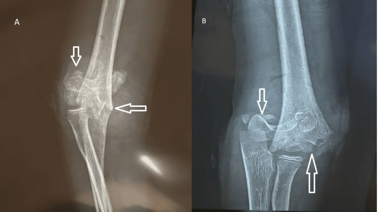
Traction views. AP and lateral films taken with longitudinal traction reduce the overlap of the fragments and define the pattern better, which makes them useful for surgical planning.
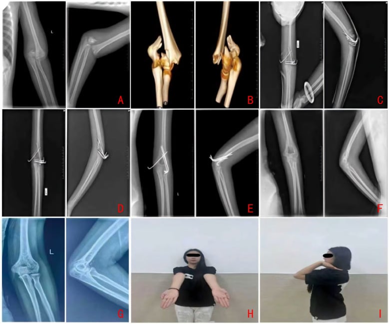
CT with 3D reconstruction is essential for surgical planning in all Type C fractures. It defines the articular involvement, finds the small fragments and lets you plan the fixation. Do not operate without adequate imaging. Request it for:
- All intra-articular fractures (Type B and C)
- Surgical planning for ORIF
- Coronal shear fractures (capitellar/trochlear)
- Comminuted patterns
MRI is rarely indicated acutely. It may be useful for soft-tissue assessment in delayed presentations.
Management Algorithm
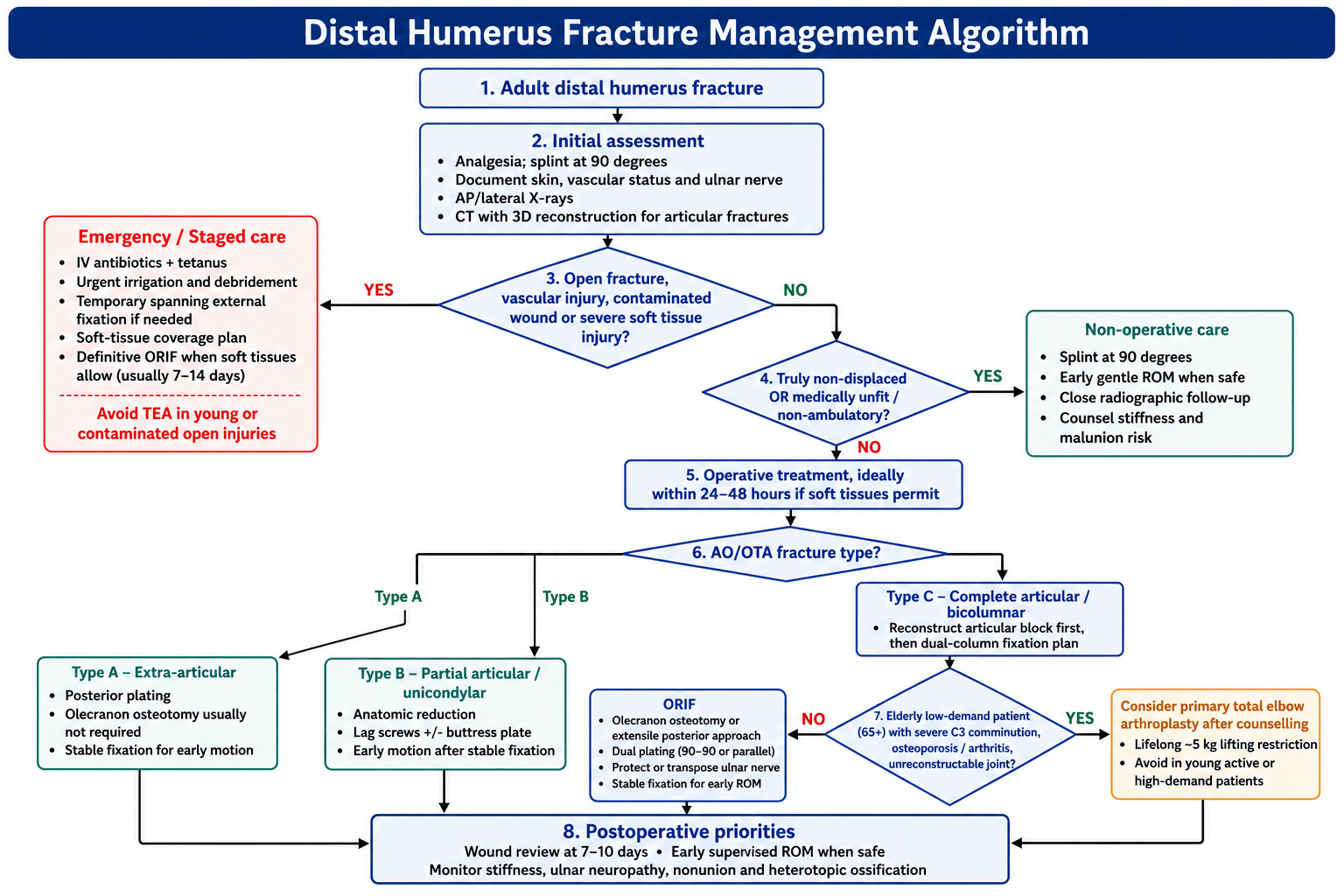
The decision. Virtually all displaced distal humerus fractures are treated operatively, to restore the anatomy and allow early mobilisation. The fracture type sets the operation; the patient sets whether that operation is fixation or replacement.
- Patient Factors
- Any age
- Treatment
- Posterior plating, no osteotomy needed
- Patient Factors
- Any age
- Treatment
- Lag screws plus or minus buttress plate
- Patient Factors
- Young, active, good bone
- Treatment
- Dual plate ORIF via olecranon osteotomy
- Patient Factors
- Young patient
- Treatment
- Dual plate ORIF - accept some complexity
- Patient Factors
- Elderly (age 65 plus), low demand
- Treatment
- Consider primary TEA
- Patient Factors
- Any patient
- Treatment
- Staged: debridement, spanning ex-fix, then definitive
Operative indications.
- Virtually all displaced distal humerus fractures
- Any articular involvement (Type B, C)
- Open fractures
- Associated vascular injury
- Polytrauma with planned early mobilisation
Non-operative treatment. Conservative management has a very limited role in displaced fractures: the truly undisplaced fracture, the non-ambulatory patient, and the patient whose comorbidities preclude surgery. If it is chosen, splint at 90 degrees, start gentle motion early, and accept deformity and functional limitation, because it often results in stiffness and malunion.
"Bag of bones". This is deliberate non-operative management of a comminuted fracture: a brief period of immobilisation followed by early active motion, accepting that the elbow may heal as a malunion or as a painless, mobile fibrous pseudarthrosis, the "functional flail" elbow, rather than as an anatomically reduced joint. It is for the frail, very elderly, non-ambulatory or severely demented patient, or one medically unfit for the prolonged anaesthesia of a complex ORIF or TEA, in whom the morbidity of surgery outweighs the functional gain, especially if the arm is mainly a helper or is used with a frame or wheelchair.
The case for it is that it avoids the substantial wound, infection and hardware complications of operating on osteoporotic comminution in a fragile patient, and that surprisingly acceptable function and good pain relief are reported when early motion is allowed, because even a lax elbow can serve low demands. The price is reduced strength, some instability and deformity. It is a demand-matched compromise rather than a reconstruction, so reserve it for the genuinely high-risk patient and counsel accordingly; for the fit elderly patient with an unreconstructable joint, primary TEA remains the better functional option.
Timing. Operate within 24-48 hours if the soft tissues permit. The elbow tolerates delay poorly: swelling, stiffness and surgical difficulty all increase with time, and delay beyond 2-3 weeks increases the difficulty significantly.
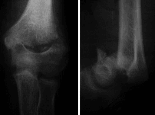
Staged surgery. Where the soft tissues will not take a definitive operation, a temporary external fixator holds the fracture while they recover, and reconstruction follows; it is appropriate for open or severely comminuted fractures. Stage for:
- Open fractures (Gustilo II, III)
- Severe soft-tissue injury
- Contaminated wounds
- Vascular injury requiring repair
- Polytrauma requiring damage control
Surgical Technique
Choosing the approach. The choice depends on the fracture pattern and the soft tissues.
Olecranon osteotomy is the gold standard exposure for Type C fractures because it gives the best view of the articular surface. The steps:
- Posterior midline incision
- Identify and protect the ulnar nerve
- Chevron (apex distal) or transverse osteotomy 2cm from the tip
- Predrill for the later fixation; pre-drilling before the cut ensures an accurate reduction
- Reflect the olecranon proximally with the triceps attached
Fixing the osteotomy. The osteotomy has traditionally been fixed with a tension band wire or a plate, and plate fixation may have lower hardware-removal rates. The comparative evidence on this page (Dumartinet-Gibaud) goes further: tension-band wiring carries a high hardware-removal and nonunion burden, and rigid fixation with screws or a plate reduces reoperation. So fix it with screws or a plate rather than a tension band.
The alternatives.
- Triceps-splitting: a direct posterior split; adequate for Type A, with a limited articular view
- Bryan-Morrey (paratricipital): the triceps is elevated off the columns and its continuity maintained; limited articular view
- TRAP: the triceps is reflected with the anconeus as a continuous pedicle flap, which maintains its blood supply and keeps the extensor mechanism in continuity; the anconeus maintains soft-tissue coverage, the posterior capsule and stability are preserved, and there is no osteotomy
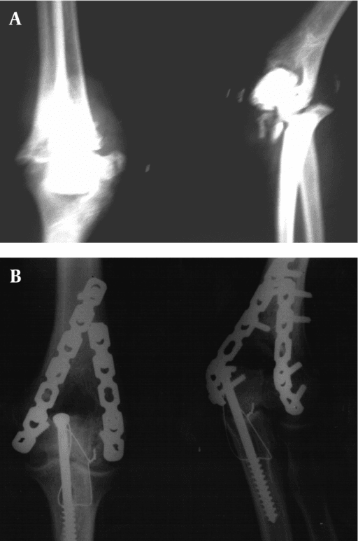
Even a perfectly reduced, solidly fixed distal humerus can be mechanically blocked if hardware intrudes into the olecranon, coronoid or radial fossae, the thin bare area between the columns where the olecranon must seat in extension and the coronoid and radial head in flexion. A screw tip or a plate contoured too far distally that crosses the olecranon fossa blocks terminal extension, because the olecranon hits the hardware, and an intra-articular screw into the trochlear notch causes pain and arthrosis. Hardware or a too-long screw filling the coronoid or radial fossa blocks flexion, because the coronoid or radial head cannot seat.
Avoiding it is a matter of technique. Contour the plates to the supracondylar ridges and stop short of the fossae; choose distal screw lengths and trajectories that pass between or around the fossae, so the interdigitating articular screws run within the bone of the columns and trochlea rather than across the empty fossae; and check a full flexion-extension arc and orthogonal fluoroscopy before closing. If there is a mechanical block, reposition the offending screw.
This is a mechanical cause of stiffness, distinct from capsular or heterotopic-ossification stiffness, fully preventable, and a common reason an early arc is lost despite "good" fixation.
Complications
- Incidence
- 15-50%
- Prevention/Management
- Most common complication; see below
- Incidence
- 10-15%
- Prevention/Management
- Gentle handling and protection during exposure
- Incidence
- 5-15%
- Prevention/Management
- Prophylaxis and early motion; see below
- Incidence
- 2-10%
- Prevention/Management
- Stable fixation, bone graft if needed
- Incidence
- 3-5%
- Prevention/Management
- Adequate fixation, protected loading
- Incidence
- 1-3%
- Prevention/Management
- Prophylactic antibiotics, good soft tissue handling
- Incidence
- 5-10%
- Prevention/Management
- Adequate fixation, consider plate over TBW
- Incidence
- 10-20%
- Prevention/Management
- Anatomic reduction, minimise step-off
Stiffness is the most common complication, at 15-50%. The functional arc for most activities of daily living is 30-130 degrees of flexion and 50 degrees of pronation-supination; that is the minimum acceptable outcome, and patients should be counselled that some stiffness is expected. Prevention is stable fixation that allows early motion, and continuous passive motion may help. Established stiffness is treated with physiotherapy, dynamic splinting, and arthroscopic or open release.
Ulnar neuropathy after surgery runs at 10-15%, and new symptoms may develop post-operatively. Most neurapraxias recover over 6-12 months. Persistent symptoms may require revision transposition.
Heterotopic ossification is more common with delayed surgery, head injury and severe trauma. Prophylaxis is indomethacin 75mg/day for 2 weeks or single-dose radiotherapy. Ossification that limits motion may need excision, after waiting 12 months or more.
Arthritis. An articular step of more than 2mm increases the risk of post-traumatic arthritis significantly, which is the reason for the emphasis on anatomic reduction.
Postoperative Care and Rehabilitation
The principle. The goal is early motion to prevent stiffness, and stable fixation that allows early movement produces better outcomes than rigid immobilisation. If the fixation is not stable enough for motion, it is not adequate. The programme balances motion against healing, and often needs a long course of therapy.
- Posterior splint at 90 degrees
- Elevation, ice
- Wound check at 48 hours
- Gentle finger motion
- Remove splint for supervised motion
- Begin active assisted ROM
- Focus on flexion-extension first
- Continue finger and wrist motion
- Progressive active ROM
- Night splinting if developing flexion contracture
- Dynamic splinting if significant stiffness
- No resistive exercises
- Begin gentle strengthening
- Progressive loading as tolerated
- Continue ROM exercises
- Full strengthening
- Return to most activities
- Heavy lifting/sport when healed
Outcomes and Prognosis
Union. In the Edinburgh series 90.6% of fractures had united by 12 weeks, and about half of the remainder healed by 24 weeks without further surgery. The patterns that risk union problems are the "low" transcondylar fractures (A2.3, A3) and the simple intercondylar fracture (C1.3); the AO grade alone does not predict it.
What to expect by treatment.
- Good/Excellent Outcomes
- 90-95%
- Key Points
- Anatomic reduction, early motion
- Good/Excellent Outcomes
- 75-85%
- Key Points
- More stiffness, higher complication rate
- Good/Excellent Outcomes
- 85-90%
- Key Points
- Reliable pain relief, restrictions required
- Good/Excellent Outcomes
- 50-60%
- Key Points
- Stiffness, malunion, poor function
What predicts it.
- Patient age and bone quality
- Fracture complexity (C3 worse than C1)
- Quality of reduction (articular step-off)
- Adequacy of fixation
- Associated injuries
- Compliance with rehabilitation
Say which endpoint you mean - McKee's two disagree. At 2 years the Mayo score favoured TEA and stayed there (86 vs 73, p=0.015), while DASH had equalised (34 vs 38, p=0.6) after favouring TEA early. Reoperation was 12% TEA vs 27% ORIF but did not reach significance (p=0.2) in a trial of 40 patients - a real difference of that size would need several hundred. So the defensible claim is better and more predictable Mayo function with TEA, not fewer reoperations, and not equivalent outcomes.
The finding that decides cases is the one people forget: 5 of 21 patients randomised to ORIF (24%) reached theatre, were opened, and could not be fixed - they were converted to TEA on the table. In this fracture, in this age group, the operation you planned is not reliably the operation you will perform. Consent for both, and have the implant available.
Guidelines, Registries & Global Practice
Global epidemiology (PubMed-backed):
- Figure
- 5.7 per 100,000 per year
- Source
- Robinson, Edinburgh (PMID 12499966)
- Figure
- Bimodal; near-equal male:female overall
- Source
- Robinson (PMID 12499966)
- Figure
- Simple fall
- Source
- Robinson (PMID 12499966)
- Figure
- 90.6%
- Source
- Robinson (PMID 12499966)
The elderly, low-energy ("fragility") fracture is the fastest-growing subgroup worldwide as populations age, and these patients drive the ORIF-versus-TEA debate.
Guideline and consensus positions (side-by-side):
- Position on distal humeral fractures
- Anatomic articular reduction plus dual-column plate fixation (parallel or perpendicular) for displaced bicolumnar fractures; early motion
- Evidence level
- Expert consensus / technique
- Position on distal humeral fractures
- Open fractures: early IV antibiotics, combined ortho-plastics debridement, staged definitive fixation with soft-tissue cover
- Evidence level
- Consensus standard
- Position on distal humeral fractures
- Operative fixation for displaced/articular fractures; primary TEA reserved for selected elderly with unreconstructable comminution
- Evidence level
- Limited / consensus
- Position on distal humeral fractures
- Supports shared decision-making between ORIF and TEA in the elderly; distal humeral hemiarthroplasty considered investigational
- Evidence level
- Consensus
No single high-level society guideline mandates ORIF versus TEA; the McKee RCT (PMID 18823799) remains the anchor evidence and all guidance is consistent with selective primary TEA.
Registry evidence:
- Finding
- TEA survival 92% / 81% / 71% / 61% at 5/10/15/20 years
- Source
- Krukhaug (PMID 29332662)
- Finding
- Post-traumatic sequelae and uncemented ulnar fixation are the main revision risks
- Source
- Krukhaug (PMID 29332662)
National joint registries (Norwegian Arthroplasty Register, AOANJRR, UK NJR) consistently show elbow arthroplasty durability below that of hip and knee replacement, which underpins the lifelong activity restriction and the avoidance of TEA in younger, higher-demand patients.
Practice variation:
- High-volume elbow/upper-limb units perform more primary TEA and more complex C3 reconstructions; smaller units more often transfer or default to ORIF.
- Parallel-plate use is more common in North American practice, perpendicular (90-90) in parts of Europe and Asia; functional outcomes are equivalent (TRON study, PMID 36527504), with the main difference being symptomatic hardware removal.
- In ageing health systems worldwide, the rising fragility-fracture burden is increasing demand for both ORIF and selective primary TEA; national registries including the AOANJRR track elbow arthroplasty outcomes.
Be ready to argue ORIF versus TEA with their indications, contraindications and the supporting evidence (McKee RCT, Norwegian Register). Show you can apply the same principles across health systems rather than quoting any single national tariff.
MCQ Practice Points
Q: What is the bicolumnar concept in distal humerus fractures? A: The distal humerus forms an inverted triangular architecture with medial and lateral columns that support the articular surface (trochlea and capitellum). Both columns must be reconstructed and fixed to the shaft for stable elbow function.
Q: Which surgical approach provides the best visualization of the distal humerus articular surface? A: Chevron olecranon osteotomy. This elevates the olecranon with the triceps attached, providing direct visualization of the entire articular surface. Alternative approaches (triceps-splitting, paratricipital) have limited articular visualization.
Q: In 90-90 plating of the distal humerus, where are the plates positioned? A: Medial plate on the medial column and posterolateral plate on the lateral column, positioned approximately 90 degrees apart. Alternative is parallel plating, one plate directly medial and one directly lateral, in the same plane.
Q: What are the indications for primary TEA in distal humerus fractures? A: Age 65 plus years, low-demand patient, severe articular comminution (C3 pattern) that cannot be reconstructed, and pre-existing arthritis (especially rheumatoid). Contraindicated in young, active patients.
Q: What is the most common complication following distal humerus fracture fixation? A: Elbow stiffness (15-50%). The goal is to achieve a functional arc of 30-130 degrees. Prevention includes stable fixation allowing early motion and appropriate physiotherapy.
Viva Scenarios
Practise clinical reasoning and management decisions out loud
“A 45-year-old right-hand dominant carpenter falls from a ladder, landing on his elbow. X-rays show a Type C2 distal humerus fracture (simple articular, comminuted metaphysis). The ulnar nerve is intact. Describe your management.”
“A 78-year-old woman with osteoporosis and rheumatoid arthritis falls at home. CT shows a Type C3 distal humerus fracture with severe articular comminution. She lives alone but is independent. What are your treatment options?”
“A 35-year-old male motorcyclist has a Type C3 distal humerus fracture with severe soft tissue injury and skin loss posteriorly. The wound is contaminated. The hand is well perfused but he has ulnar nerve palsy. How do you manage this?”
BICOLUMNAR CONCEPT
- Inverted triangular architecture
- Medial column plus lateral column support articular surface
- Both columns must be fixed to shaft for stability
- Interdigitating screws in articular block
CLASSIFICATION
- AO Type A: extra-articular (supracondylar)
- AO Type B: partial articular (unicondylar)
- AO Type C: complete articular (bicolumnar)
- C3 equals comminuted articular and metaphyseal equals most challenging
SURGICAL APPROACHES
- Olecranon osteotomy: best articular visualization (gold standard)
- Triceps-splitting: simple patterns, limited view
- Paratricipital: TEA, some ORIF, no osteotomy
- TRAP: TEA, preserves triceps blood supply
PLATE CONFIGURATION
- 90-90: medial plus posterolateral plates (90 degrees apart)
- Parallel: directly medial plus directly lateral plates (same plane)
- Both configurations acceptable
- Minimum 2 screws per column into shaft
TEA INDICATIONS
- Age 65 plus (ideally age 75 plus)
- Low-demand patient
- Severe articular comminution (C3)
- Pre-existing arthritis (especially RA)
- Contraindicated in young, active patients
TRAPS AND PEARLS
- Always get CT for surgical planning
- Ulnar nerve: identify and protect for medial plating; transposition is discretionary (RCT: no difference)
- Early motion is essential to prevent stiffness
- Functional arc: 30-130 degrees flexion
- TEA requires lifetime 5kg lifting restriction
Evidence Base
McKee et al. - ORIF vs TEA in the Elderly (landmark RCT)
- Multicentre RCT (42 patients randomised) comparing ORIF with primary semiconstrained TEA in patients older than 65 with displaced OTA 13C fractures
- TEA had significantly better Mayo Elbow Performance Score at 2 years (86 vs 73, p=0.015) and faster early DASH recovery
- 5 of 21 patients randomised to ORIF (24%) were converted to TEA intra-operatively because fixation could not be achieved
- Reoperation rates (TEA 12% vs ORIF 27%) were not statistically different (p=0.2)
O'Driscoll - Principle-Based Parallel-Plate Fixation
- Describes the parallel-plate (both columns plated medially and laterally) principle-based approach to distal humeral fixation
- Distal screws should be as long as possible, pass through plates, engage as many articular fragments as possible, and interdigitate to lock the columns together
- Each screw should pass through a plate and engage a fragment on the opposite side that is also fixed to a plate
- Stability is sufficient to permit immediate intensive rehabilitation, with bone graft rarely required
Dumartinet-Gibaud et al. - Olecranon Osteotomy Fixation
- Retrospective comparison of 39 transolecranon exposures fixed with double screws versus tension-band wiring (TBW) for complex distal humeral fractures
- Significantly fewer all-cause revisions with double screws than with TBW (3/14 versus 14/25, p=0.049)
- Symptomatic implant removal was the commonest reason for revision and was more frequent after TBW
- Olecranon nonunion occurred only in the TBW group (4/25 versus 0/14)
Yokoyama et al. (TRON) - Parallel vs Perpendicular Plating
- Multicentre study of 383 distal humeral fractures, propensity-matched to 50 perpendicular versus 50 parallel double-plate cases
- No significant difference in Mayo Elbow Performance Score at any time point between the two plate configurations
- Implant removal was significantly more frequent after parallel plating (50% versus 26.5%, p=0.023)
- Medial-lateral parallel plates lie under thin subcutaneous tissue, increasing symptomatic prominence
Ring, Jupiter & Gulotta - Articular (Coronal Shear) Fractures
- Defined that apparent isolated capitellar fractures are often complex multi-fragment articular injuries with up to five components (capitellum-lateral trochlea, lateral epicondyle, posterior lateral column, posterior trochlea, medial epicondyle)
- All 21 fractures healed after open reduction and fixation with implants buried beneath the articular surface
- Mayo Elbow Performance Index excellent or good in 16 of 21; mean ulnohumeral arc 96 degrees
- Ten patients required a second operation, most commonly for contracture release
Robinson, Court-Brown et al. - Epidemiology & Outcomes (Edinburgh)
- Consecutive series of 320 adult distal humeral fractures from a defined catchment population; incidence 5.7 per 100,000 per year
- Bimodal age distribution with an almost equal male-to-female ratio; simple falls were the commonest mechanism overall
- Most fractures were extra-articular (OTA type A) or complete articular (OTA type C); 90.6% united within 12 weeks
- Union complications were higher after high-energy injury, open fractures and nonoperative treatment
Krukhaug et al. - Norwegian Arthroplasty Register (TEA survivorship)
- 838 primary total elbow arthroplasties reported to the Norwegian Arthroplasty Register 1994-2016
- Implant survival 92% at 5 years, 81% at 10 years, 71% at 15 years and 61% at 20 years
- A diagnosis of post-traumatic sequelae and uncemented ulnar fixation were the main risk factors for revision
- Aseptic loosening was the commonest revision cause; survivorship was poorer than for hip and knee arthroplasty
Dehghan / Canadian Orthopaedic Trauma Society - Ulnar Nerve In Situ vs Anterior Transposition (RCT)
- Multicentre RCT across 8 Canadian centres: 58 patients having bicolumnar plate fixation, randomised at the end of the case to in-situ replacement in the cubital tunnel (31) or anterior subcutaneous transposition (27)
- No difference at any time point in the Gabel and Amadio ulnar neuropathy score, Mayo Elbow Performance Score, DASH or 2-point discrimination
- Ulnar nerve function was poor in BOTH groups early, with significant recovery by 12 months (6.0 to 7.8, p<0.001)
- Authors' conclusion: either strategy is acceptable and may be used at the surgeon's discretion