Radial Head Fracture + IOM Disruption + DRUJ Instability | Longitudinal Forearm Instability | Do NOT Excise Radial Head
- Never excise radial head without replacement in Essex-Lopresti injury
- DRUJ must be assessed in ALL comminuted radial head fractures
- IOM central band is primary longitudinal stabilizer of forearm
- Proximal migration of radius indicates longitudinal instability
- Chronic cases extremely difficult to salvage - early diagnosis critical
- “Mechanism: Axial load through extended wrist (FOOSH)
- “Missed diagnosis leads to irreversible proximal migration
- “Radial head replacement is essential - not optional
- “IOM reconstruction techniques are salvage procedures
Overview
The Essex-Lopresti injury is longitudinal instability of the whole forearm: a radial head fracture, disruption of the interosseous membrane (IOM) and instability of the distal radioulnar joint (DRUJ). Peter Essex-Lopresti described it in 1951, and the pattern represents complete disruption of the forearm's longitudinal stabilising structures.
How rare, and how often missed. It accounts for less than 1% of all radial head fractures, and the true incidence is likely underestimated because the injury is frequently missed. Most patients are working-age adults of 20-50 years, with a male predominance that reflects the high-energy mechanism: high-energy trauma, a fall from height, a motor vehicle accident or an industrial injury.
Mechanism. A fall onto the outstretched hand loads the extended wrist axially, and the force passes from the carpus through the radius to the radial head, which fractures under compression. The force then continues proximally, the central band of the IOM ruptures, and the DRUJ is disrupted as the final stabiliser fails.
Associated injuries. Carpal fractures, capitellar injury and elbow dislocation may accompany the triad.
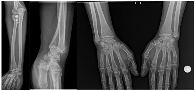
Anatomy and Pathophysiology
Forearm Longitudinal Stability
Longitudinal stability of the forearm rests on the interosseous membrane and the radial head, with the DRUJ the third link in the chain. The injury takes out all three, and the treatment is organised around which of them can be restored.
The membrane. The IOM is a fibrous sheet connecting the radius and ulna, its fibres running obliquely from distal-ulnar to proximal-radial, and it contains several distinct bands. It transmits force from the radius to the ulna and holds the radioulnar relationship while still allowing pronation and supination.
The central band. The thickest and strongest part of the membrane, at the junction of the middle and proximal thirds, providing approximately 60% of longitudinal stability. It is the structure that ruptures in an Essex-Lopresti injury. The other components - the proximal or accessory band, the distal band and the distal oblique bundle - lie around it.
Why the figure is quoted two ways. The 71% often heard is Hotchkiss's cadaver measurement of longitudinal stiffness, made after the radial head had been excised and cited in the evidence section below. The two figures are the same dominance from different measurements, and either answers the question of which structure matters most.
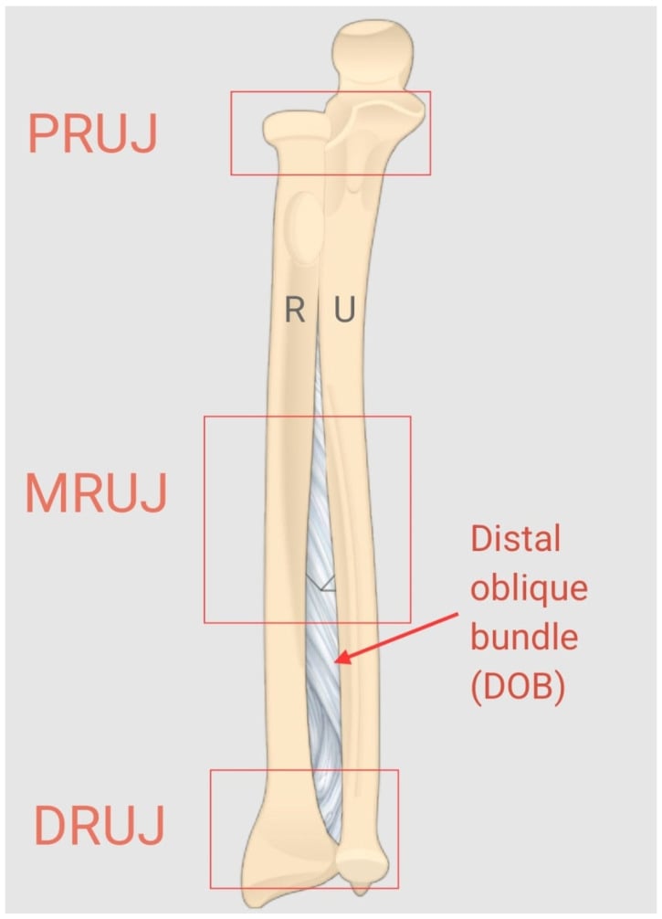
The radial head. It articulates with the capitellum and the proximal ulna, is covered circumferentially by articular cartilage and is stabilised by the annular ligament. It transfers axial load to the humerus and blocks proximal migration of the radius, the secondary longitudinal restraint at approximately 30% of longitudinal stability.
Why excision is safe in one forearm and ruinous in another. With the membrane intact, load still reaches the ulna and the radial head can be excised safely. With the central band torn, the radial head becomes the primary stabiliser and the only restraint left, so excising it allows catastrophic proximal migration of the radius. A head lost when the membrane is incompetent must be replaced.
The DRUJ. The ulnar head articulates with the sigmoid notch of the radius and the joint allows forearm rotation. Its primary stabiliser is the TFCC, supported by the dorsal and palmar radioulnar ligaments and the ulnocarpal ligaments, with the interosseous membrane a secondary stabiliser. DRUJ disruption is the third component of the injury and is usually dorsal instability; it may reduce once the radial head has been replaced, or it may need direct stabilisation, which is why stability is assessed intraoperatively after replacement.
At the wrist, the radius carries about 80% of the axial load and the ulna only about 20% (through the TFCC). That load is transferred proximally from the radius to the ulna across the interosseous membrane, mainly the central band, so that at the elbow the radial head (radiocapitellar joint) carries roughly 60% and the ulnohumeral joint about 40%.
The distal oblique bundle - a distal IOM thickening present in around 40% of forearms, running from the distal radius to the ulna - is an additional distal radioulnar joint stabiliser worth noting when planning reconstruction.
Classification
Classification
The triad, component by component. All three must be present for the diagnosis, and each is established differently:
- Radial head fracture - usually Mason type III (comminuted), sometimes type IV (with dislocation), occasionally a type II with significant comminution
- IOM disruption - central band rupture, which may extend to the proximal band, diagnosed clinically and by longitudinal instability
- DRUJ disruption - a TFCC tear with dorsal and/or palmar radioulnar ligament rupture, producing DRUJ instability
RIDEssex-Lopresti Triad
Hook:RID yourself of missing this diagnosis - always check all three components!
Time since injury is the other axis of classification, and early diagnosis and treatment significantly improves outcomes.
- Acute, within 2 weeks - best prognosis with appropriate treatment; radial head replacement essential
- Subacute, 2-6 weeks - some proximal migration may occur, with still reasonable outcomes after replacement
- Chronic, beyond 6 weeks - established proximal radial migration, IOM reconstruction may be required, and generally poor outcomes
Migration, measured as ulnar variance. The degree of proximal migration correlates with the difficulty of treatment and with the outcome, so it is graded against the other wrist:
- Minimal, under 2mm positive ulnar variance - may respond to radial head replacement alone, better prognosis
- Moderate, 2-5mm - replacement plus consideration of IOM reconstruction, intermediate prognosis
- Severe, over 5mm - complex reconstruction, poor prognosis, and may need salvage procedures
- Fracture Pattern
- Marginal, non-displaced
- Essex-Lopresti Risk
- Low
- Management
- Non-operative, check DRUJ
- Fracture Pattern
- Marginal, displaced
- Essex-Lopresti Risk
- Moderate
- Management
- Fix or replace, check DRUJ
- Fracture Pattern
- Comminuted, unreconstructable
- Essex-Lopresti Risk
- HIGH
- Management
- Replace, high index of suspicion
- Fracture Pattern
- With elbow dislocation
- Essex-Lopresti Risk
- HIGH
- Management
- Replace, very high suspicion
Differential Diagnosis
The key discriminator is whether the forearm is longitudinally unstable (Essex-Lopresti) or stable. Isolated injuries at one end of the forearm and the other classic fracture-dislocations must be excluded.
- Distinguishing feature
- Radial head fracture + IOM (central band) tear + DRUJ disruption with longitudinal instability
- Key test / finding
- Positive ulnar variance vs other side; positive radial pull test
- Distinguishing feature
- Elbow injury only; IOM and DRUJ intact, forearm longitudinally stable
- Key test / finding
- Neutral ulnar variance; negative radial pull test; no wrist pain
- Distinguishing feature
- Wrist instability without radial head fracture or proximal migration
- Key test / finding
- DRUJ ballottement positive; normal elbow and ulnar variance
- Distinguishing feature
- Distal-third radial shaft fracture with DRUJ disruption
- Key test / finding
- Radial shaft fracture on radiograph; radial head intact
- Distinguishing feature
- Proximal ulnar fracture with radial head dislocation
- Key test / finding
- Ulnar shaft fracture; radiocapitellar line disrupted
- Distinguishing feature
- Combined radial and ulnar shaft fracture with longitudinal instability (Artiaco group)
- Key test / finding
- Both shafts fractured; dynamic instability on stressing
History
Symptoms. Elbow pain from the radial head fracture is what the patient complains of; forearm pain is often overlooked, and wrist pain is the clue to the DRUJ. Weakness of grip and pain with forearm rotation complete the picture.
Red flags. Pain at both the elbow and the wrist after a fall onto the outstretched hand should raise suspicion.
- High-energy mechanism
- Pain at both elbow and wrist
- Significant swelling along the entire forearm
- Inability to supinate or pronate
Elbow Examination
Inspection and palpation. Swelling over the lateral elbow and ecchymosis are the usual findings, and a deformity may indicate a dislocation. The radial head is tender, crepitus is felt with rotation, and the associated injuries are looked for at the same time.
Movement. Flexion and extension are limited by pain and pronation and supination are painful. A mechanical block suggests loose bodies.
Stability. Valgus stress testing and the posterolateral rotatory drawer test check for associated ligament injury.
DRUJ Examination
Inspection and palpation. Compare with the other side, looking for prominence of the ulnar head and swelling at the wrist. Palpate for tenderness over the DRUJ, the TFCC and the ulnar styloid.
Stability tests. Each is compared with the contralateral side.
- Piano key sign - dorsal prominence of the ulnar head with pressure
- Ballottement test - anteroposterior translation in neutral rotation
- Radial pull test - longitudinal instability
- Forearm squeeze test - pain at the DRUJ on squeezing the mid-forearm, which indicates IOM disruption
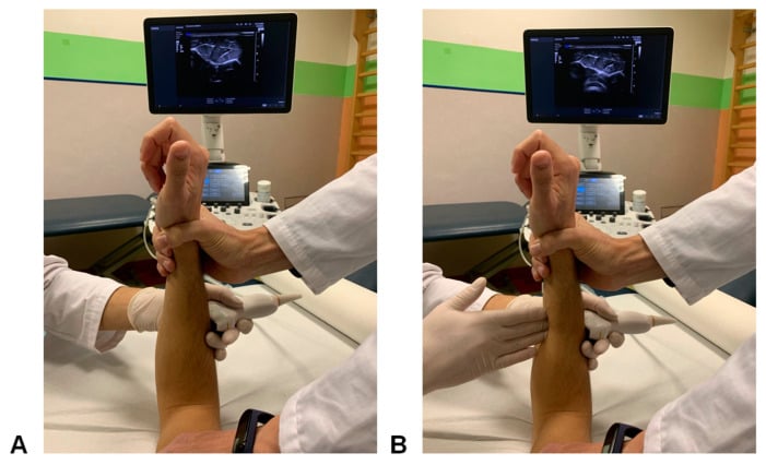
The hernia sign. With the elbow flexed 90 degrees and the forearm neutral, an ultrasound probe is placed dorsally over the middle third of the forearm while the examiner loads the membrane. Dynamic bulging through an IOM defect supports chronic membrane injury, and it supplements rather than replaces whole-forearm radiographs and DRUJ assessment.
The radius (radial) pull test is the key intraoperative test for an IOM/longitudinal injury and is increasingly examined. After the radial head fragments are removed (or before replacement), longitudinal traction of about 9 kg (20 lb) is applied to the proximal radius while the relative proximal migration of the radius is measured under fluoroscopy:
- Migration of under 3 mm = intact IOM (the longitudinal restraint is competent).
- 3 to 6 mm = partial IOM injury.
- Over 6 mm = complete IOM disruption (a true Essex-Lopresti pattern) - the radial head must be replaced and longitudinal stability restored, never left excised.
Preoperatively, comparative wrist radiographs (ulnar variance versus the other side) plus the forearm squeeze and DRUJ ballottement tests raise suspicion, but the intraoperative radius pull test gives a direct, quantified answer about whether the forearm is longitudinally stable.
ALWAYS examine the DRUJ in patients with comminuted radial head fractures. The Essex-Lopresti injury is frequently missed because the wrist is not examined. Pain at both elbow AND wrist should raise immediate suspicion.
Investigations
Imaging Studies
Radiographs of the forearm, not of the elbow. Dedicated elbow AP and lateral views with a 45° oblique radial head view show the fracture and its comminution. Full-length forearm AP and lateral films that include both the wrist and the elbow are what demonstrate radioulnar dissociation, and PA and lateral wrist views allow ulnar variance to be measured against the contralateral side.
What the films show. Four findings matter, and the last of them appears only once the injury is old.
- Comminuted radial head fracture
- Positive ulnar variance compared with the other wrist, which suggests proximal migration of the radius
- DRUJ widening or subluxation
- Ulnar impaction changes if the injury is chronic
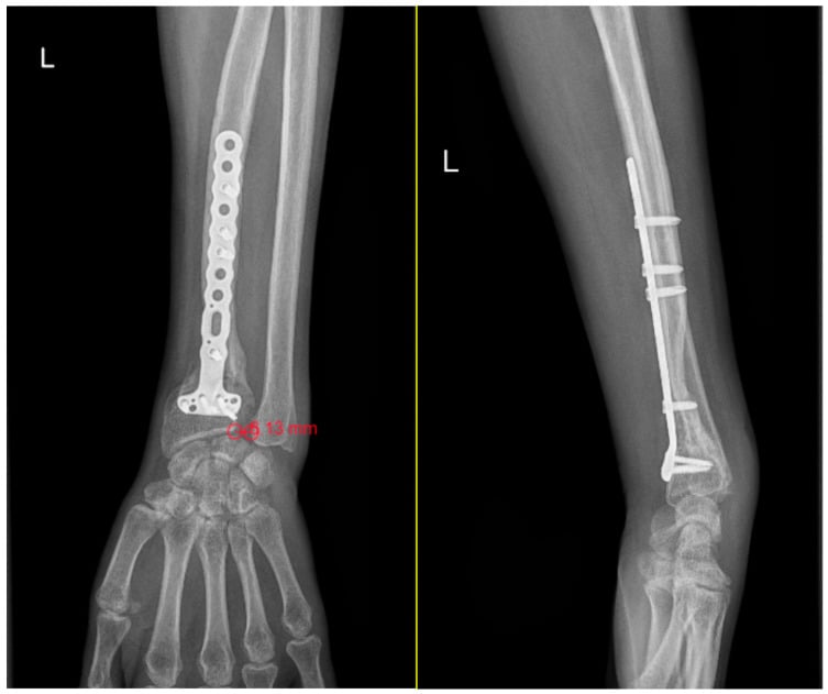
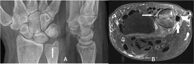
CT. It is the planning study: whether to fix or replace the radial head, and what the chronic bone changes amount to.
- Elbow: fragment number and size, articular surface involvement, associated capitellum injury
- Wrist: DRUJ congruency, ulnar impaction changes, sigmoid notch morphology
MRI. A central band tear appears as high signal through the membrane, and the study also shows TFCC tears, oedema along the forearm and associated ligament injuries. Its place is the chronic case being planned for IOM reconstruction. It is not always needed acutely, clinical examination is often sufficient, and it may delay treatment.
Management Algorithm
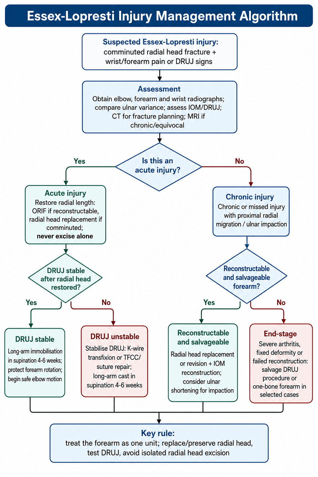
Treatment Decision Making
The decision. Surgery is the standard of care for the acute injury, and the order is fixed: the radial head first, then the DRUJ tested on the table, then a decision about whether the DRUJ needs holding.
- IOM Status
- Intact
- DRUJ
- Stable
- Treatment
- Standard radial head management
- IOM Status
- Disrupted
- DRUJ
- Unstable
- Treatment
- Radial head replacement + DRUJ stabilization
- IOM Status
- Disrupted
- DRUJ
- Unstable
- Treatment
- RH replacement + possible IOM reconstruction
- IOM Status
- Disrupted
- DRUJ
- Unstable
- Treatment
- Salvage: IOM reconstruction, ulnar shortening
- IOM Status
- Disrupted
- DRUJ
- Destroyed
- Treatment
- Consider one-bone forearm or arthroplasty
Step one, the radial head. Internal fixation if the head is reconstructable, which is rare in this injury, and replacement with a metal prosthesis when it is comminuted. It is never excised without replacement.
Step two, the DRUJ. With the head dealt with, test DRUJ stability with the ballottement test in neutral rotation and compare with the contralateral side.
Step three, holding the DRUJ. A stable joint is immobilised in supination for 4-6 weeks. An unstable one is transfixed with K-wires or repaired with sutures and then held in a long-arm cast in supination. Elbow motion can begin earlier than forearm rotation, which stays protected, and early motion once the elbow is stable is what prevents stiffness.
Timing and counselling. The goals are to replace the radial head, restore length, stabilise the DRUJ and protect the repair while it heals. Surgery within 2 weeks is ideal, earlier is better, and delayed surgery is associated with worse outcomes. Counsel the patient that outcomes may be limited.
Chronic injury. Once migration is established the options run from reconstruction to salvage:
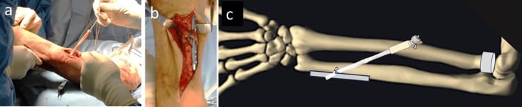
- IOM reconstruction - various techniques described, with bone-tendon-bone grafts (Achilles, patellar tendon) or synthetic options, and technically demanding
- Ulnar shortening - addresses the ulnar impaction but does not restore longitudinal stability, so it is often combined with another procedure
- Radial lengthening - rarely performed, technically difficult, and risks nonunion
- Salvage - Sauve-Kapandji procedure, Darrach resection (avoid if possible), one-bone forearm as extreme salvage
The errors that do the damage. Excising the radial head without replacement, missing the DRUJ injury, immobilising inadequately and failing to assess longitudinal stability all end in the same place: irreversible proximal migration, ulnar impaction syndrome, and chronic pain and disability.
Surgical Technique
Operative Procedures
Setting up. Supine with the arm on a hand table, a tourniquet on the upper arm and fluoroscopy available.
The approach. A Kocher or lateral approach, between anconeus and extensor carpi ulnaris. Protect the lateral ulnar collateral ligament, and identify and preserve the annular ligament.
The implant. A metallic prosthesis, titanium or cobalt-chrome, modular or monoblock, cemented or press-fit. Match the diameter of the native head and restore its length: overlengthening wears the capitellum, underlengthening fails to stop the migration.
The steps. Work through them in order, with fluoroscopy for the part that matters most, which is length.
- Excise the radial head fragments, preserving annular ligament remnants
- Ream the medullary canal and trial the implant, confirming length under fluoroscopy
- Seat the final implant and close the annular ligament if possible
- Restore radiolunate alignment and the lateral ulnohumeral joint space
- Check a full range of movement without capitellar impingement, and stable tracking
- Test DRUJ stability
When sizing the radial head prosthesis, the lateral ulnohumeral joint should be restored. Overlengthening causes capitellar wear and pain; underlengthening fails to prevent proximal migration.
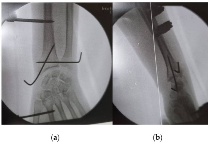
DRUJ stabilisation. A DRUJ that is stable after replacement needs a long-arm cast in supination for 4-6 weeks, with protected movement after 6 weeks. An unstable one is transfixed with two parallel K-wires, or repaired directly at the TFCC.
- Position the forearm in supination
- Pass two 1.6mm K-wires from the ulna to the radius, above the level of the DRUJ
- Cut the wires below the skin
- Remove them at 6 weeks
IOM reconstruction. Reserved for chronic injury with established migration, failed acute treatment or persistent instability. The graft may be an Achilles tendon allograft, patellar tendon autograft or allograft, hamstring tendons, or a synthetic, the last of which remains controversial. The principles are to recreate the orientation of the central band through bone tunnels in the radius and ulna, tension the graft appropriately, and protect it with immobilisation.
What it achieves. Results are variable in the literature. Reconstruction may reduce migration without eliminating it, it is often combined with other procedures, and it is generally considered a salvage.
Complications
Potential Complications
Missed diagnosis is the most common and the most devastating complication. The DRUJ is not examined, an isolated radial head excision is performed, and the radius migrates proximally, bringing ulnar impaction syndrome, wrist pain, grip weakness and chronic forearm instability.
Problems with the prosthesis. Two of the four are sizing errors, which is why length is confirmed on the table.
- Overlengthening - capitellar wear and erosion, elbow pain, limited movement, and may require revision
- Underlengthening - continued proximal migration, ulnar impaction and treatment failure
- Loosening - progressive stem loosening with pain, and may require revision
- Instability - subluxation or dislocation of the prosthesis, usually indicating ligament injury, and may need revision or stabilisation
Persistent longitudinal instability. Migration may progress despite radial head replacement when the membrane fails to heal, and the response is IOM reconstruction, ulnar shortening or a combination of procedures. At end stage, with DRUJ arthritis and ulnocarpal impaction, salvage is what remains, including a one-bone forearm in severe cases.
Radial head excision without replacement in Essex-Lopresti injury leads to inevitable proximal radial migration and chronic disability. This error is essentially irreversible - prevention through proper diagnosis is key.
Postoperative Care
Rehabilitation Protocol
Phase 1, protection (0-6 weeks). The point is to protect DRUJ stability, let the soft tissues heal and prevent proximal migration. A long-arm cast or splint holds the forearm in supination with the elbow at 90 degrees.
- Elbow flexion and extension out of the cast, if stable
- No forearm rotation
- Shoulder and hand exercises
- If the DRUJ was pinned, the K-wires come out at 6 weeks under local anaesthesia
Phase 2, motion (6-12 weeks). Forearm rotation is restored and elbow movement progressed while longitudinal stability is still protected.
- Weeks 6-8: gentle supination and pronation, active-assisted motion, continuing elbow exercises
- Weeks 8-12: progress rotation, light functional activities, avoiding heavy gripping or loading
- Throughout: no axial loading, no heavy lifting, no forceful gripping
Phase 3, strengthening (12 weeks onwards). Strength and function, with migration still being watched for.
- Weeks 12-16: progressive strengthening including grip, and functional activities
- Week 16 onwards: return to work on light duties first, sport-specific training, full activities as tolerated
- Monitoring: serial radiographs for migration, grip strength, and comparison with the contralateral side
Outcomes
Expected Results
Acute treatment. Early diagnosis and treatment gives fair to good outcomes in 60-80%, and restoration of stability is achievable. Movement usually reaches 80-90% of the contralateral side and grip strength 70-80%, with return to work variable and often with restrictions.
- Migration despite treatment: 10-30%
- Revision surgery: 15-25%
- Chronic pain: 20-40%
Chronic treatment. Outcomes are generally poor once migration is established, with a high revision rate and chronic symptoms common, and significantly worse than after acute treatment. IOM reconstruction gives variable results in the literature and may slow but not stop migration. Salvage procedures give limited functional outcomes with variable pain relief and may require multiple procedures.
End-stage. A one-bone forearm eliminates rotation and fusion procedures leave limited function; both are accepted for pain relief.
What predicts the result. Time to diagnosis is the most important prognostic factor.
- Favourable: diagnosis within 2 weeks, appropriate acute treatment, a successful radial head replacement, a stable DRUJ afterwards
- Unfavourable: delayed diagnosis, a radial head excision, established proximal migration, multiple failed procedures
- Long term: prosthesis longevity is uncertain, revision surgery may be needed, and chronic disability is common
Guidelines, Registries & Global Practice
Global Practice, Guidelines and Evidence
Essex-Lopresti injury (also termed longitudinal radioulnar dissociation) is a rare, globally relevant pattern. There is no internationally agreed definition and no high-level (RCT) guideline, so practice rests on cadaver biomechanics, case series and expert consensus reviews. The world standard of care is consistent: in a comminuted radial head fracture, actively exclude longitudinal instability, restore radial length, stabilise the DRUJ, and avoid radial head excision.
Epidemiology (global)
- Accounts for less than 1% of radial head fractures and is widely under-recognised; mechanism is axial load through an extended wrist (fall onto the outstretched hand, fall from height, motor-vehicle and industrial trauma).
- Typically affects working-age adults with a male predominance, reflecting the high-energy mechanism.
- A PRISMA systematic review found only 266 reported patients across 47 studies, and most studies did not document the interosseous-membrane component - the literature base is small and heterogeneous (Kooistra et al., EFORT Open Rev 2022).
Guidance and consensus, side by side
- Position
- Reconstructable radial head: fix; comminuted with longitudinal instability: replace, never excise in isolation; assess and stabilise the DRUJ
- Evidence level
- Expert consensus
- Position
- Use an intraoperative radial pull test to detect IOM injury; treat the forearm as one functional unit; classification-guided algorithm
- Evidence level
- Level V review
- Position
- Restore radial length plus temporary DRUJ stabilisation acutely; approach acute radial head excision with caution; acute IOM repair indications unclear
- Evidence level
- Level V review
- Position
- No accepted definition; radial head fracture with wrist/forearm pain should trigger comparative imaging and a dynamic stability test
- Evidence level
- Level III systematic review
- Position
- No condition-specific guideline exists - this injury is too rare for a formal guideline
- Evidence level
- Not addressed
Registry evidence
Joint and trauma registries (NJR, AJRR, AOANJRR, SHAR, Norwegian and NZJR) do not separately capture Essex-Lopresti injury or radial head arthroplasty for it, so there is no registry-level implant-survival or revision data to quote. Outcome evidence is therefore limited to single-centre case series and biomechanical studies, which is itself an examinable point about the quality of the evidence base.
Practice variation
- High-resource settings: ready access to modular radial head prostheses, intraoperative fluoroscopy and CT, and to bone-ligament-bone central band reconstruction for chronic cases.
- Limited-resource settings: where modern prostheses are unavailable, surgeons may have to choose between accepting the consequences of radial head loss and improvised stabilisation; this drives the global teaching point to preserve or replace, never simply excise, the radial head.
- Worldwide, the dominant cause of poor outcome is the same - a missed acute diagnosis leading to fixed proximal migration - so the universal priority is recognition, not a particular implant or technique.
Viva Scenarios
Viva Scenarios
Clinical Decision Scenarios
Practise clinical reasoning and management decisions out loud
“A 35-year-old man falls from a ladder onto his outstretched hand. He has a comminuted radial head fracture. How would you assess for an Essex-Lopresti injury?”
“You have confirmed an Essex-Lopresti injury in the patient from the previous scenario. What is your surgical plan?”
“A patient presents 6 months after a radial head excision for what was thought to be an isolated comminuted radial head fracture. He now has wrist pain and grip weakness. Radiographs show 8mm positive ulnar variance. What has happened and how would you manage this?”
“Describe the anatomy of the interosseous membrane and its role in forearm stability.”
MCQ Practice
High-Yield Exam Points
Q: What is the most important examination to perform in a patient with a comminuted radial head fracture?
A: DRUJ stability assessment is CRITICAL in all comminuted radial head fractures to rule out Essex-Lopresti injury. Use ballottement test, piano key sign, and always compare to the contralateral side. Missing this examination leads to missed diagnosis with catastrophic consequences.
Q: In an acute Essex-Lopresti injury, what is the most important surgical intervention and why?
A: Radial head replacement is essential. The radial head is the only remaining longitudinal stabilizer after IOM rupture. Excision without replacement leads to inevitable proximal radial migration, ulnar impaction, and chronic disability. This is a NEVER excise scenario.
Q: What percentage of forearm longitudinal stability is provided by the IOM central band versus the radial head?
A: The IOM central band provides approximately 60-70% of forearm longitudinal stability, while the radial head provides approximately 30%. When both are disrupted, catastrophic instability results with proximal migration of the radius.
Q: What is the most common reason Essex-Lopresti injuries are missed, and how can this be prevented?
A: Failure to examine the DRUJ is the most common reason these injuries are missed. The wrist is simply not examined in patients presenting with elbow injuries. Prevention: ALWAYS perform DRUJ examination in ALL comminuted radial head fractures and obtain full-length forearm radiographs.
Q: A patient had radial head excision 6 months ago. Now has wrist pain and 7mm positive ulnar variance. What happened and what are the management options?
A: This is a missed Essex-Lopresti injury with established proximal migration. Management options include: 1) Radial head replacement + IOM reconstruction (best salvage option), 2) Ulnar shortening osteotomy + DRUJ procedure, 3) One-bone forearm (extreme salvage). All have poor outcomes compared to acute treatment - this emphasizes the critical importance of early diagnosis.
Q: How do you assess DRUJ stability intraoperatively after radial head replacement?
A: Test DRUJ stability in neutral rotation using ballottement test. Compare to contralateral side. If stable, immobilize in supination for 6 weeks. If unstable, perform K-wire transfixion with 2 parallel wires from radius to ulna, forearm in supination, or consider direct TFCC repair.
Key facts
- Triad: Radial head fracture + IOM disruption + DRUJ instability
- NEVER excise radial head without replacement
- Examine DRUJ in ALL comminuted radial head fractures
- IOM central band provides 60% of longitudinal stability
- Radial head provides 30% of longitudinal stability
- Positive ulnar variance indicates proximal migration
- Acute treatment within 2 weeks gives best outcomes
- Chronic cases have poor outcomes regardless of treatment
Surgical steps
- Kocher approach to radial head
- Excise unreconstructable fragments
- Size and insert radial head prosthesis
- Check length restoration with fluoroscopy
- Test DRUJ stability after RH replacement
- If unstable: K-wire transfixion in supination
- Long-arm cast 6 weeks protecting DRUJ
- Remove K-wires at 6 weeks
Common pitfalls
- Missing diagnosis by not examining DRUJ
- Excising radial head without replacement
- Inadequate prosthesis sizing (over/underlengthening)
- Not assessing DRUJ intraoperatively
- Inadequate immobilization post-operatively
- Delayed diagnosis leading to chronic migration
- Treating as isolated radial head fracture
Examiner favourites
- What examination is mandatory in comminuted RH fractures?
- Why can't you excise the radial head in Essex-Lopresti?
- Describe the anatomy and function of the IOM
- What is the treatment for acute Essex-Lopresti?
- How do you assess DRUJ stability intraoperatively?
- What are the consequences of missed diagnosis?
- How would you manage chronic Essex-Lopresti with migration?
Evidence Base
Key Studies
Essex-Lopresti - Original Description
- First description of the injury pattern (two cases)
- Comminuted radial head fracture with radial head excision followed by proximal radial migration
- Recognised the link between radial head loss, forearm length and distal radioulnar joint disruption
- Emphasised the need to recognise the whole forearm injury, not just the elbow
Hotchkiss et al. - IOM Biomechanics (landmark)
- Anatomic and mechanical cadaver study (12 specimens) of the interosseous membrane
- Central band identified in all specimens, approximately twice the thickness of adjacent membrane
- Central band provided 71% of forearm longitudinal stiffness after radial head excision; the TFCC contributed 8%
- Silicone radial head implants were far less stiff than the intact membrane - central band injury is key to proximal radial migration
Skahen et al. - Central Band Reconstruction (cadaver)
- Cadaver study (12 forearms) of central band strain and reconstruction
- Central band strain greatest in pronation and increased significantly after radial head excision
- Both central band and TFCC are important to axial forearm stability
- Flexor-carpi-radialis-based central band reconstruction prevented complete radial migration but did not fully restore longitudinal stability
Marcotte and Osterman - LRUD Treatment Algorithm
- Concepts in anatomy, biomechanics, diagnosis and treatment of longitudinal radioulnar dissociation
- Reports encouraging results for chronic injury treated by bone-ligament-bone (patellar tendon) central band reconstruction
- Provides a treatment algorithm for acute and chronic injury
- Frames the forearm as a single functional unit requiring length and stability restoration
Sheth et al. - Narrative Review
- Contemporary narrative review of Essex-Lopresti and longitudinal radioulnar instability
- Acute management centres on restoring radial length and temporary DRUJ stabilisation
- Radial head excision in the acute setting should be approached with caution - migration may develop over time
- Indications for acute IOM repair/reconstruction remain unclear; central band reconstruction can restore load transfer in chronic cases
Kooistra et al. - Definition Heterogeneity (systematic review)
- PRISMA systematic review: 47 clinical studies, 266 patients
- 39 of 47 studies did not even mention an IOM lesion as part of the injury
- Threshold for significant positive ulnar variance ranged from more than 1 mm to more than 12 mm across studies
- Only 9 studies used any dynamic intraoperative test of longitudinal stability
Meglic et al. - IOM Role and Radial Pull Test
- ESSKA instructional review of the interosseous membrane in post-traumatic forearm instability
- Describes the intraoperative radial pull test to improve detection of IOM injury
- Reframes the forearm as a functional unit and proposes the Artiaco classification of forearm fracture-dislocations
- Provides a management flowchart for acute and chronic forearm instability