Systemic inflammatory cascade following long bone trauma
Critical Must-Knows
- Classic Triad: Hypoxia, Confusion, Petechiae
- Petechial rash is pathognomonic but late/transient
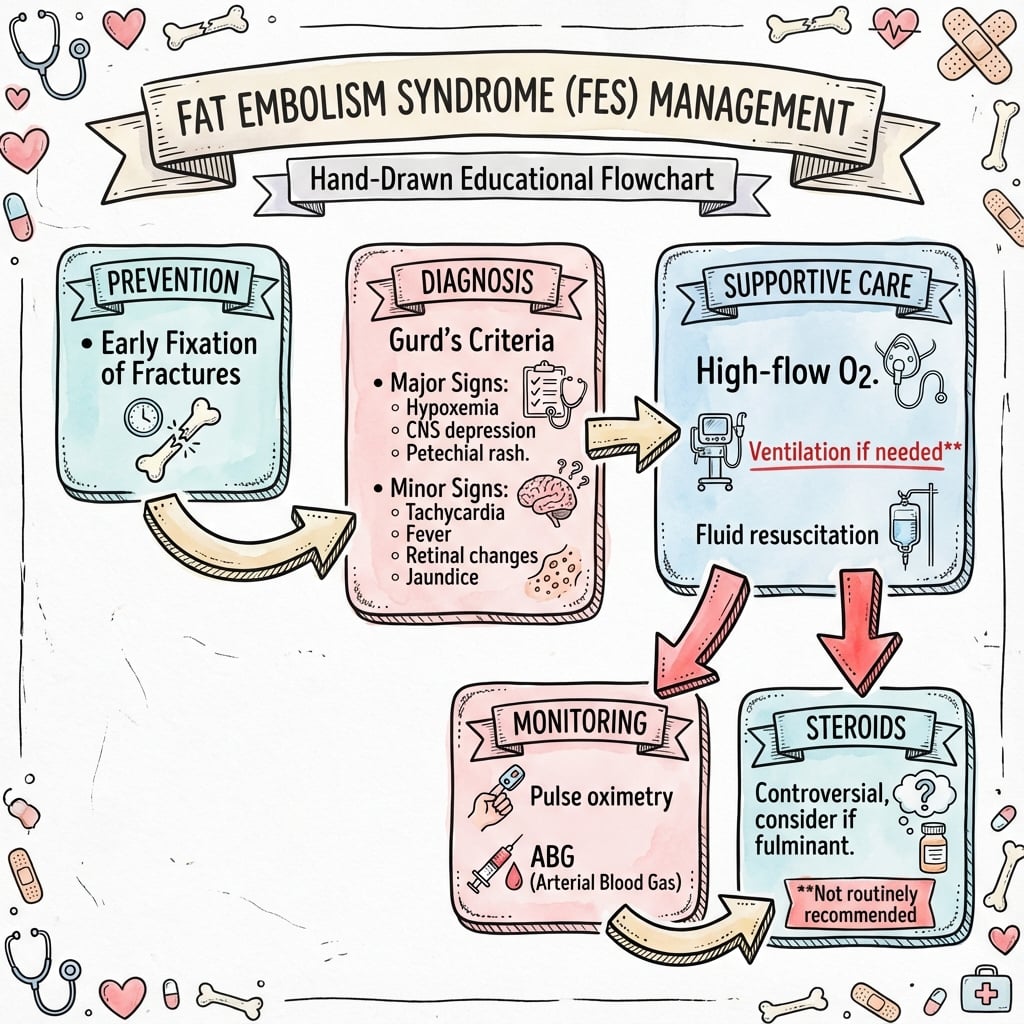
- Prevention: Early fixation within 24h
Clinical Pearls
- "Diagnosis of exclusion - Rule out PE
- "Steroids are controversial (reduce incidence, not mortality)
Exam Red Flags
Diagnostic Trap
Do NOT wait for the rash. Petechiae are specific but late (24-48h) or absent in 50%.
FES vs PE
Distinction. FES: 24-72h, "Snowstorm" CXR, Starfield MRI. PE: Vascular occlusion, lobar defects.
At a Glance
Key Facts
R-C-PGurd's Major Criteria
| R | Respiratory insufficiency Hypoxia (PaO2 less than 60mmHg) |
| C | Cerebral involvement Confusion, agitation, coma |
| P | Petechial rash Pathognomonic rash (axilla/chest) |
| R | Respiratory insufficiency Hypoxia (PaO2 less than 60mmHg) |
| C | Cerebral involvement Confusion, agitation, coma |
| P | Petechial rash Pathognomonic rash (axilla/chest) |
Hook:Royal College of Physicians (RCP)
LONGRisk Factors for FES
| L | Long bone fractures Femur and Tibia (especially reamed) |
| O | Open vs Closed Closed fractures generate higher intramedullary pressure |
| N | Non-operative Delayed fixation increases risk |
| G | Gradient of force High-energy trauma in young males |
| L | Long bone fractures Femur and Tibia (especially reamed) | N | Non-operative Delayed fixation increases risk |
| O | Open vs Closed Closed fractures generate higher intramedullary pressure | G | Gradient of force High-energy trauma in young males |
Hook:Long bones break
SPOTFES Management
| S | Supportive care Oxygen, fluids, ventilator support (Mainstay) |
| P | Prevention Early fixation within 24 hours |
| O | Oxygenation Maintain SaO2 over 90% |
| T | Theatre Damage control (Ex-Fix) if unstable |
| S | Supportive care Oxygen, fluids, ventilator support (Mainstay) | O | Oxygenation Maintain SaO2 over 90% |
| P | Prevention Early fixation within 24 hours | T | Theatre Damage control (Ex-Fix) if unstable |
Hook:Spot the rash
Overview
Fat Embolism Syndrome (FES) is a serious, life-threatening clinical manifestation of fat emboli entering the systemic circulation. It is characterized by a "multisystem dysfunction" primarily involving the lungs, brain, and skin. While the presence of fat in the circulation (fat embolism) is a common occurrence following long bone fractures (detected in over 90% of cases using transesophageal echocardiography), the development of the systemic inflammatory syndrome (FES) is much rarer, affecting only 1-5% of patients. [1,2]
Epidemiology
The incidence varies depending on the injury profile and management:
- Incidence:
- Isolated long bone fractures: 0.5% to 2%. [3]
- Polytrauma patients: 5% to 10%. [3]
- Bilateral femoral fractures: Up to 33% in historical series without early fixation. [4]
- Demographics: Most common in young men (10-40 years) due to higher exposure to high-energy trauma (MVA, falls). It is less common in the elderly (despite hip fractures) and rare in children (due to different marrow composition - more hematopoietic, less fatty).
Risk Factors
- Injury Pattern: Multiple long bone fractures (femur, tibia, pelvis).
- Fixation Timing: Delayed stabilization (over 24 hours) is the single biggest modifiable risk factor.
- Technique: Intramedullary reaming has been shown to causing transient showers of emboli, though its clinical significance in stable patients is debated.
Pathophysiology
The exact mechanism is debated, but the "Two-Hit Theory" is widely accepted, combining mechanical obstruction with a delayed biochemical inflammatory storm.
1. Mechanical Theory (The First Hit)
Trauma disrupts venules in the marrow sinusoids. Elevated intramedullary pressure (often exceeding venous pressure) forces liquid fat, bone marrow debris, and inflammatory factors from the marrow into the venous circulation. These globules travel to the right heart and lodge in the pulmonary capillaries.
- Effect: Ventilation-perfusion (V/Q) mismatch and transient hypoxemia. This explains the early symptoms but not the delayed systemic syndrome (e.g., rash, confusion). Large emboli can cause acute cor pulmonale, but FES is usually micro-embolic. [5]
2. Biochemical Theory (The Second Hit)
This explains the latency (24-72 hours). Lodged fat globules are hydrolyzed by pneumocytes (producing lipase) into Free Fatty Acids (FFAs).
- Local Toxicity: FFAs are directly toxic to the pneumocytes and capillary endothelium, causing acute lung injury (ALI), vasogenic edema, hemorrhage, and inactivation of surfactant. This pathological picture closely resembles ARDS (Acute Respiratory Distress Syndrome).
- Systemic Inflammation: The release of inflammatory mediators (CRP, IL-6, TNF-alpha) causes a systemic inflammatory response syndrome (SIRS). This biochemical storm affects other organs, leading to the delayed systemic features (petechiae from capillary permiability, confusion from cerebral inflammation, and coagulopathy). [5,6]
Classification
Diagnosis is primarily clinical. There is no single "gold standard" test. Several criterion-based systems exist to increase diagnostic consistency.
Gurd and Wilson's Criteria (1974)
This is the most widely cited system. Diagnosis requires 1 Major + 4 Minor signs. [9]
Classification
| Grade/Type | Description | Management |
|---|---|---|
| MAJOR SIGNS | Respiratory Insufficiency (PaO2 less than 60 mmHg) | ~96% |
| Cerebral Involvement (Unexplained agitation, confusion) | ~59% | |
| Petechial Rash (Vest, axilla) | ~33% | |
| MINOR SIGNS | Tachycardia (over 110 bpm) | Variable |
| Pyrexia (over 38.5°C) | Variable | |
| Retinal changes (Fat on fundoscopy) | Rare | |
| Jaundice | Rare | |
| Renal signs (Anuria/Oliguria) | Rare | |
| Thrombocytopenia (Platelets under 150k) | Variable | |
| Anemia (Sudden drop in Hb) | Variable | |
| High ESR (over 71 mm/hr) | Variable |
Clinical Assessment
The clinical presentation is dynamic. The hallmark of FES is the Classical Triad, presenting typically 24 to 72 hours after injury (a "lucid interval"). Note that fulminant FES can present acutely on the table or in recovery.
History
- Mechanism: Recent long bone fracture (especially femur) or major polytrauma.
- Timing: Symptom onset usually 24-72 hours post-injury. Immediate collapse suggests massive Macro-embolism (mechanical block) rather than FES syndrome.
- Symptoms: Deteriorating respiratory status (shortness of breath) despite initial stability. New confusion, restlessness, or agitation (often misdiagnosed as alcohol withdrawal or head injury).
Physical Examination
- Respiratory Insufficiency (96%):
- Usually the earliest and most consistent sign.
- Spectrum: Mild hypoxemia and tachypnea → Severe dyspnea → Frank respiratory failure (ARDS).
- Exam: Tachypnea, dyspnea, cyanosis, accessory muscle use. Auscultation reveals crackles (pulmonary edema).
- Cerebral Involvement (59%):
- Ranges from mild disorientation, acute confusion, and drowsiness to seizures, rigid hemiplegia, and coma.
- Why? Micro-emboli to the brain or toxic FFA effect crossing the blood-brain barrier.
- Key Clue: Neurological deficit purely from FES is often reversible.
- Petechial Rash (20-50%):
- Pathognomonic feature. Usually appears on day 2 or 3 and resolves by day 5-7.
- Location: Non-dependent areas → Anterior axillary folds, chest wall, lateral neck, and subconjunctiva (Head & Neck distribution).
- Mechanism: Occlusion of dermal capillaries by fat emboli leading to extravasation of erythrocytes, compounded by thrombocytopenia. [8]
Investigations
Workup aims to support the diagnosis (as no specific test exists) and rule out common differentials like Pulmonary Embolism (PE), Pneumothorax, and Pneumonia.
Laboratory
- ABG (Arterial Blood Gas): Hypoxia (PaO2 less than 60mmHg) with an increased A-a gradient. Respiratory alkalosis initially (due to tachypnea).
- FBC: Unexplained anemia (drop in Hb) and thrombocytopenia (platelets under 150k) occur in 37-67% of cases due to consumption coagulopathy and alveolar hemorrhage.
- Coagulation: may show disseminated intravascular coagulation (DIC) in severe cases.
- Biochemistry: Lipase may be elevated (non-specific, also rises in trauma).
- Urine: Fat globules may be seen (Lipuria) using Sudan staining. Historically emphasized but low sensitivity and specificity.
- Bronchoalveolar Lavage (BAL): If performed, lipid-laden macrophages (over 30%) are suggestive of FES.
Imaging
-
Chest X-ray (CXR):
- Often normal initially (lag period).
- Progression to "Snowstorm appearance" (diffuse bilateral fluffy alveolar infiltrates) usually after 24-48 hours. Distinct from the wedge-shaped infarct of PE. [11]
-
CT Chest:
- More sensitive than CXR. Shows ground-glass opacities, intralobular septal thickening ("crazy paving"), and patchy consolidation.
- Main role is to rule out large central Pulmonary Embolism (PE). Note that FES itself produces micro-emboli not visible as filling defects on CTPA.
-
MRI Brain:
- Highly sensitive for cerebral FES (better than CT Brain, which is often normal).
- Starfield pattern: Multiple punctate hyperintensities on T2-weighted and Diffusion-Weighted Imaging (DWI) sequences representing micro-infarcts in white matter (water-shed areas). [12]
Differential Diagnosis
The post-injury hypoxic, confused patient has a wide differential. FES is a diagnosis of exclusion.
| Diagnosis | Typical timing | Discriminating features |
|---|---|---|
| Fat embolism syndrome | 24-72 h (lucid interval) | Triad of hypoxia, confusion, petechiae; thrombocytopenia; snowstorm CXR; starfield MRI; CTPA without large filling defect |
| Pulmonary embolism (thrombotic) | Usually after day 3-5; can be earlier | Pleuritic pain, unilateral leg signs, large filling defect on CTPA, right-heart strain; no petechiae |
| Pneumonia / aspiration | Days, often later | Fever, productive cough, focal consolidation and raised inflammatory markers; positive cultures |
| Pulmonary contusion | Immediate (at injury) | History of chest trauma; opacities match impact site on initial CT; no lucid interval |
| Hypovolaemic / haemorrhagic shock | Immediate / early | Tachycardia and hypotension responsive to blood; falling Hb with a bleeding source, not petechiae |
| Head injury / raised ICP | Variable | Focal neurology, pupillary signs, abnormal CT brain; FES brain CT is usually normal |
| Drug / alcohol withdrawal | 2-4 days | Autonomic features, tremor, history of use; normal oxygenation and CXR |
Management
Supportive Care (Mainstay)
Because there is no specific antidote to dissolve fat emboli, treatment is purely supportive while the body clears the lipid load.
- Oxygenation: Goal is prevent hypoxemia. Maintain PaO2 over 60 mmHg or SaO2 over 90%. Start with High-flow nasal cannula or CPAP.
- Ventilation: Early intubation and Mechanical ventilation (low tidal volume, high PEEP) is required if ARDS develops or GCS drops.
- Hemodynamics: Aggressive fluid resuscitation is crucial to prevent shock, but careful balance is needed to avoid worsening pulmonary edema ("wet lungs"). Albumin has been proposed to bind serum FFAs but evidence is weak. [14]
- Blood Products: Correct anemia and thrombocytopenia if present.
Surgical Technique
Intraoperative Techniques to Reduce Embolic Load
The act of reaming a femoral canal acts like a piston, generating high intramedullary pressures (often exceeding 800 mmHg) which forces marrow contents into the venous system.
- Venting: Drilling a vent hole in the proximal or distal femur before reaming acts as a relief valve. It reduces intramedullary pressure spikes, allowing fat/blood to exit the cortex rather than entering the venous circulation.
- Reamer-Irrigator-Aspirator (RIA): This advanced reaming system provides continuous irrigation and suction of marrow contents during reaming. It significantly reduces embolic load to the lungs compared to standard reaming. It is strongly indicated for femoral nailing in patients with concurrent chest injuries (AIS Thorax greater than 3).
- Unreamed Nails: Smaller diameter nails inserted without reaming. While they generate less pressure than reamed nails, they have lower union rates. Current preference is Reamed nails with RIA or Venting in high-risk patients.
Complications
- ARDS (Acute Respiratory Distress Syndrome): The major cause of morbidity and mortality. Requires prolonged ventilation and ICU stay.
- Cerebral deficits: Usually reversible as the edema and micro-infarcts resolve. Permanent neurological sequelae are rare but possible in severe cases.
- Right Heart Failure: Acute cor pulmonale can occur from massive mechanical blockage, leading to obstructive shock (rare, distinct from classic FES syndrome).
- Death: Usually from fulminant respiratory failure or refractory shock.
Postoperative Care
- Monitoring: Continuous pulse oximetry for at least 24-48 hours in all high-risk fracture patients.
- Hydration: Aggressive but balanced resuscitation to maintain preload and cardiac output.
- Vigilance: Frequent GCS checks to detect early cerebral involvement (e.g., confusion, restlessness).
- Thromboprophylaxis: Mechanical and chemical prophylaxis (once bleeding risk acceptable) to prevent superimposed VTE, which is hard to distinguish from FES.
Prognosis
- Mortality: Historically 10-20%, now estimated at 5-10% with modern intensive care support. [2]
- Resolution: Most cases are self-limiting. Clinical signs usually resolve spontaneously within 3-7 days with appropriate supportive care.
- Long-term: Respiratory function usually recovers fully. Rare minor cognitive deficits have been reported in severe cerebral FES.
Evidence
Early vs Delayed Fixation
- Prospective RCT of 178 adults with acute femoral fractures
- In multiply-injured patients, delayed stabilisation raised pulmonary complications (ARDS, fat embolism, pneumonia)
- Delayed fixation also lengthened hospital and ICU stay
Corticosteroids in FES
- Meta-analysis of 7 RCTs (389 patients) of prophylactic corticosteroids in long-bone fractures
- Reduced risk of FES by 78% (NNT 8) and reduced hypoxia
- No difference in mortality and no increase in infection rate
Damage Control Orthopaedics
- Synthesis of database, animal and prospective data on fixation strategy in polytrauma
- Early prolonged surgery and reamed femoral nailing raise ARDS risk when cofactors (e.g. chest injury) are present
- A validated grading system identifies 'borderline' patients in whom staged temporary external fixation reduces systemic complications
Reamed vs Non-reamed Nailing (SPRINT)
- Multicentre RCT (29 sites, 1200 patients) of reamed vs non-reamed tibial shaft nailing
- Primary outcome was reoperation to promote healing, treat infection or preserve the limb
- Largest trial of the reaming question; supports reaming in closed tibial fractures with no excess of clinically significant embolic harm
Pathophysiology & Management Review
- Authoritative review of incidence, aetiology, pathophysiology, diagnosis and treatment of FES
- Clinical syndrome is rare (under 1% in retrospective series) whereas marrow fat embolisation is near-universal after long-bone fracture
- Multiple mechanical and biochemical factors interact; current treatment is supportive with generally good outcome
Clinical Decision Scenarios
Use these scenarios to practise clinical reasoning and management decisions
"What is your differential diagnosis and immediate management?"
"Summarize the evidence for corticosteroids in FES."
"How do you explain the risk and your mitigation strategy?"
Key Points
Diagnosis is Clinical
Do not rely on lipiduria or fat in sputum (low sensitivity/specificity). Use Gurd's criteria or Schonfeld Index.
Petechiae are Specific
The rash is pathognomonic but transient. Look in the axilla and subconjunctiva.
Prevention is Key
Once established, treatment is supportive. Early fixation (or DCO) is the only proven preventive measure.
Steroids
Prophylaxis only. No role in treatment.
Controversies & Areas of Uncertainty
- No diagnostic gold standard: Gurd and Schonfeld criteria predate modern imaging, were derived from small cohorts, and have never been prospectively validated against a reference test. Sensitivity and specificity are uncertain, and clinical FES is frequently under- or over-diagnosed.
- Corticosteroid prophylaxis: Meta-analysis suggests prophylactic steroids reduce FES incidence without affecting mortality, but the constituent trials are small and old. There is no consensus on dose, timing or which (if any) high-risk subgroup should receive them, so routine use remains contested.
- Reaming and embolic load: Reaming demonstrably raises intramedullary pressure and embolic showers, yet large trials (e.g. SPRINT) show no clinically meaningful excess of pulmonary harm in stable patients. Whether RIA or venting changes patient-centred outcomes (vs surrogate embolic markers) is unproven.
- Timing in the borderline patient: The ETC vs DCO decision in physiologically borderline polytrauma is individualised; the optimal thresholds and the role of serial biomarkers (lactate, IL-6) remain debated.
- Cerebral FES: MRI "starfield" changes are sensitive but non-specific, and the prognosis of isolated cerebral fat embolism is variable and poorly quantified.
Guidelines, Registries & Global Practice
Global epidemiology
- Clinical FES complicates under 1% of long-bone fractures in retrospective series, rising to 5-10% in polytrauma; subclinical marrow fat embolisation is near-universal (detectable in over 90% by transoesophageal echo during nailing).
- Predominantly affects young men after high-energy trauma; rare in young children (haematopoietic, less fatty marrow).
Side-by-side guidance
| Body | Position on FES-relevant practice |
|---|---|
| AO Foundation / OTA | Early appropriate care; reamed IM nailing standard for stable femoral shaft fractures; staged DCO (external fixation) for unstable/borderline polytrauma. |
| BOA / BOAST (UK) | Damage-control principles and physiology-led fixation timing in major trauma; early stabilisation of long bones in the multiply injured. |
| AAOS / ACS-TQIP (US) | Trauma quality programmes emphasise early fracture stabilisation and lung-protective ventilation if ARDS develops; no specific FES drug therapy endorsed. |
| Consensus (global) | No society recommends routine corticosteroid prophylaxis; treatment is supportive. Diagnosis remains clinical (Gurd / Schonfeld). |
Registry & practice variation
- Trauma registries (e.g. TARN in the UK, the German TraumaRegister DGU) track timing of fixation and pulmonary complications, reinforcing early stabilisation in polytrauma.
- High-resource settings: ready access to CTPA, MRI ("starfield" pattern), ICU/ARDS support, and Reamer-Irrigator-Aspirator (RIA) or canal venting for femoral nailing in patients with significant chest trauma to limit embolic load.
- Limited-resource settings: diagnosis leans on clinical criteria, pulse oximetry and chest radiography; external fixation is a pragmatic, low-cost damage-control option, and earlier definitive fixation may be constrained by theatre and implant availability.
Clinical summary
Key Stats
- •90% have emboli, only 1-5% have syndrome
- •Mortality 5-10%
- •Onset 24-72 hrs
Triad
- •Hypoxia
- •Confusion
- •Petechiae (Axilla)
Treatment
- •O2 Support
- •Early Fixation
- •No Heparin
References
- Sevitt S, Gallagher N. Fat embolism and its clinical significance. Br J Surg. 1961;48:475-81.
- Mellor A, Soni N. Fat embolism. Anaesthesia. 2001;56(2):145-54. doi:10.1046/j.1365-2044.2001.01724.x. PMID: 11167474.
- Stein PD et al. Incidence and clinical characteristics of fat embolism syndrome. Am J Med Sci. 2008;336(6):472-7.
- Talucci RC et al. Early intramedullary nailing of femoral shaft fractures: a cause of fat embolism syndrome. Am J Surg. 1983;146(1):107-11.
- Eriksson EA et al. The pathophysiology of fat embolism syndrome. J Trauma Acute Care Surg. 2016.
- Pape HC et al. The timing of fracture treatment in polytrauma patients: relevance of damage control orthopedics. Am J Surg. 2002.
- Gupta A et al. Fat embolism syndrome: a review. Int J Crit Illn Inj Sci. 2011;1(2):148-51.
- Adams CB. The retinal manifestations of fat embolism. Injury. 1971;2:221.
- Gurd AR, Wilson RI. The fat embolism syndrome. J Bone Joint Surg Br. 1974;56B(3):408-16.
- Schonfeld SA et al. Fat embolism prophylaxis with corticosteroids. Ann Intern Med. 1983;99(4):438-43.
- Feldman F et al. The fat embolism syndrome. Radiology. 1975;114:635.
- Butterworth JF et al. Morgan & Mikhail's Clinical Anesthesiology. 5th Ed. McGraw-Hill; 2013.
- Bone LB, Johnson KD, Weigelt J, Scheinberg R. Early versus delayed stabilization of femoral fractures. A prospective randomized study. J Bone Joint Surg Am. 1989;71(3):336-40. PMID: 2925704.
- Robinson CM. Current concepts of respiratory insufficiency following femoral shaft fractures. J Bone Joint Surg Br. 2001;83(6):785-91.
- Bederman SS, Bhandari M, McKee MD, Schemitsch EH. Do corticosteroids reduce the risk of fat embolism syndrome in patients with long-bone fractures? A meta-analysis. Can J Surg. 2009;52(5):386-93. PMID: 19865573.