Post-Traumatic Embolic Phenomenon | Gurd Criteria | Preventable with Early Fixation
- Fat emboli from marrow enter venous circulation after long-bone or pelvic fractures
- Classic triad: hypoxia, petechiae, altered mental status appearing 24-72 hours post-injury
- Gurd criteria require one major + four minor features for clinical diagnosis
- Early definitive fixation of fractures within 24 hours reduces incidence
- Management is entirely supportive; no specific pharmacological therapy alters course
- “Petechial rash in axillae or conjunctivae is virtually diagnostic when present
- “Differential includes pulmonary embolism, pneumonia, fat vs thrombus embolus
- “Prophylactic steroids or heparin have no proven benefit in prevention
- “Monitor for ARDS progression and right heart strain in severe cases
Overview and Epidemiology
Fat embolism syndrome is a leading cause of preventable morbidity and mortality in young trauma patients with long-bone fractures. It is rare in absolute terms, complicating about 1-5% of long-bone fractures, and almost entirely preventable by timely fracture stabilisation. The danger lies in the interval: the syndrome declares itself 24-72 hours after injury. The earliest signs may appear at 12 hours, but the classic presentation is on day 2 to 3, which is when the patient has to be re-examined.
Risk factors. The injuries and interventions that matter are those that expose a large marrow volume or disturb the fracture repeatedly:
- Long-bone fractures, the femur, tibia and humerus most commonly; the femur is the highest-risk single long-bone injury
- Pelvic and acetabular fractures, which expose a large marrow volume and release large volumes of fat
- Multiple fractures, the risk additive with each further bone
- Closed fractures, which hold a higher intramedullary pressure than open injuries
- Young adults, whose marrow has a high fat content
- Intramedullary nailing, where reaming and instrumentation force fat into the veins
- Repeated manipulation, since each reduction attempt embolises more fat
- Delayed fixation, beyond 24 hours
What it costs. Respiratory failure progresses to ARDS in severe cases, and the neurological injury ranges from cognitive impairment through focal deficits to coma. Recognition within the 24-72 hour window lets supportive measures start before fulminant respiratory failure or irreversible neurological injury.
Pathophysiology
From marrow to lung. Fat globules from the bone marrow enter the venous circulation through torn medullary veins at the fracture site, under raised intramedullary pressure, and travel to the pulmonary capillary bed. There they injure the lung in two ways. Mechanical obstruction of the capillaries raises pulmonary vascular resistance; lipase then releases free fatty acids from the trapped fat, and these damage the endothelium directly and inactivate surfactant, a chemical pneumonitis. The inflammatory cascade that follows produces capillary leak and an ARDS picture, ventilation-perfusion mismatch produces refractory hypoxia, and a massive embolus load strains the right heart.
Why the delay. The 24-72 hour latency reflects the time required for fat mobilisation, embolisation and the secondary inflammatory lung injury. The cascade runs in three phases:
- Embolisation, immediate to hours: fat globules enter the venous system, often subclinically
- Pulmonary phase, 24-48 hours: mechanical block plus chemical injury, giving hypoxia, tachypnoea and infiltrates
- Systemic phase, 48-72 hours: paradoxical emboli and inflammation, giving petechiae and confusion
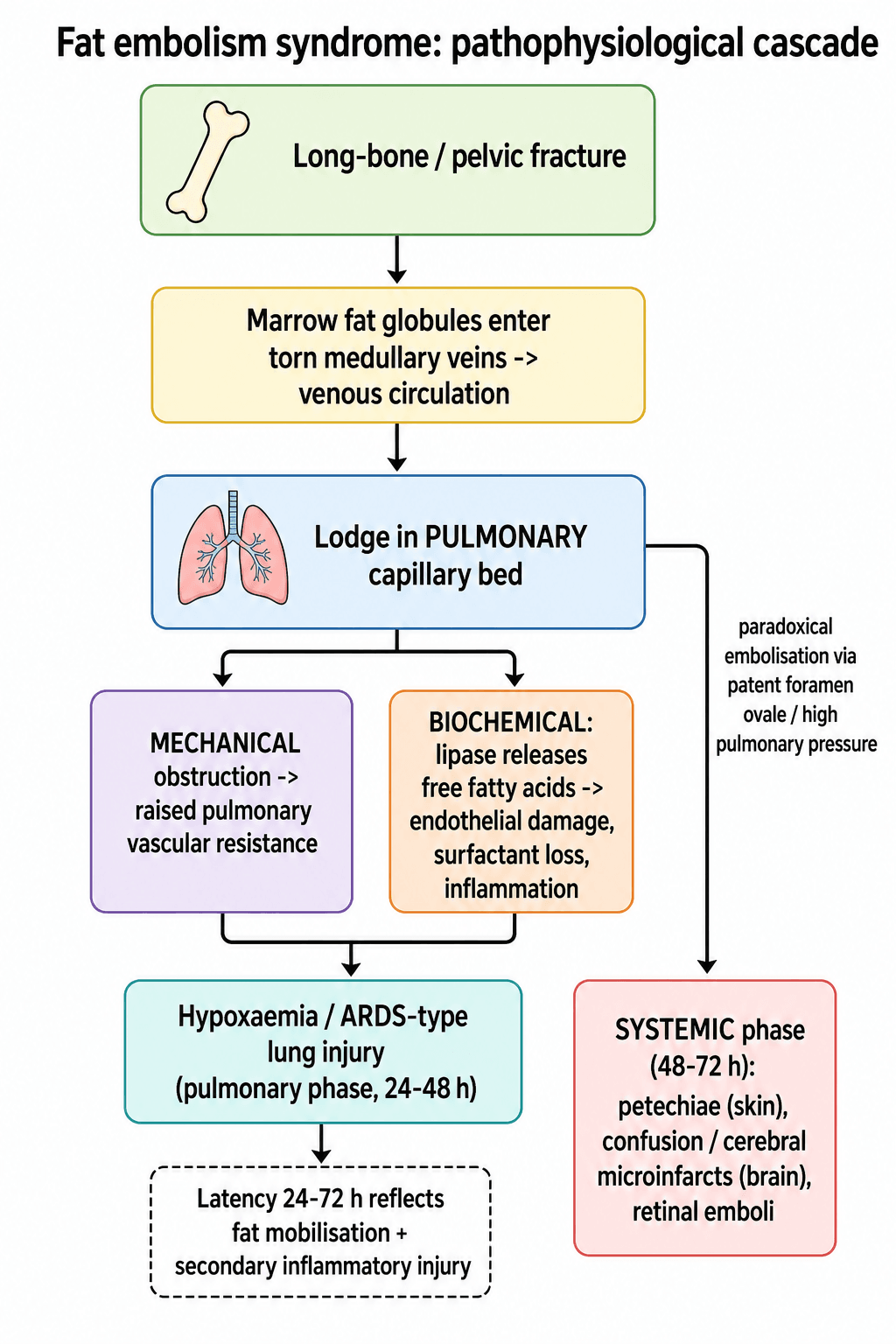
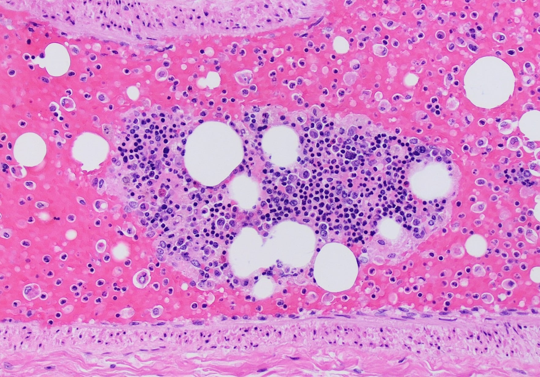
Venous source, arterial disease. Marrow fat lodges in the pulmonary capillaries, yet the cerebral, cutaneous and retinal features are in systemic, arterial territories. For venous fat to reach them it must cross to the arterial side, and how it does so is a stock examiner question. There are two routes:
- Transpulmonary passage, the dominant route. Fat globules are small and deformable, so they pass through the pulmonary capillary bed, or through precapillary arteriovenous channels that open as pulmonary pressure rises, into the left heart and out to the systemic arteries. No intracardiac defect is required, which is why most patients with cerebral or cutaneous features have a structurally normal heart.
- Paradoxical embolism through a patent foramen ovale. A PFO is present in roughly a quarter of adults. The pulmonary fat load raises right-heart and pulmonary-artery pressure, which can open a right-to-left shunt and let venous fat bypass the lungs entirely. This is invoked particularly for florid or early cerebral fat embolism, and for cerebral involvement out of proportion to the lung injury.
The second hit at each end organ. Once globules reach a capillary bed, the free-fatty-acid injury amplifies the local damage. In the brain that means cerebral oedema and microinfarcts; in the skin, dermal capillary occlusion and leak, together with the thrombocytopenia, produce the petechial rash; in the retina, emboli may be seen on fundoscopy as Purtscher retinopathy.
Bone Cement Implantation Syndrome: The Other Peri-operative Fat Embolism
Reaming pistons marrow fat into the venous system at high intramedullary pressure, and the same mechanism operates, often more dramatically, during cemented arthroplasty.
Bone cement implantation syndrome (BCIS). Pressurising PMMA cement into a prepared canal and impacting the prosthesis generates high intramedullary pressure that embolises marrow fat, cement monomer and air to the lungs. Within minutes of cementation or stem insertion this produces hypoxia, hypotension, pulmonary hypertension with right-heart strain, arrhythmia and, in severe cases, cardiac arrest.
The classic and most lethal setting is a cemented hemiarthroplasty for a hip fracture in a frail elderly patient, the opposite demographic to fracture-related FES, which is the young, high-energy male. The timing is intra-operative, on cementing or stem insertion, not the 24-to-72-hour lucid interval of classic FES. Getting the demographic and the timing the right way round is the mark.
Risk reduction. Warn the anaesthetist before cementing, and ensure euvolaemia and optimised haemodynamics. Lower the intramedullary pressure at the source:
- Lavage and dry the canal
- Use a cement restrictor
- Use a suction or venting catheter
- Insert the cement retrograde with a cement gun
Consider uncemented fixation in the very highest-risk patients, balanced against the higher periprosthetic-fracture rate of uncemented stems in osteoporotic bone.
Classification and Types
Gurd criteria. The diagnosis is clinical: one major plus four minor criteria. Of the major criteria, petechiae are the most specific finding.
- Feature
- Respiratory distress
- Clinical Note
- Hypoxia, PaO2 less than 60 mmHg on room air
- Feature
- Petechial rash
- Clinical Note
- Axilla, chest, conjunctiva classic
- Feature
- Neurological change
- Clinical Note
- Confusion most common, focal signs possible
- Feature
- Tachycardia greater than 110
- Clinical Note
- Nonspecific but supportive
- Feature
- Pyrexia greater than 38.5 C
- Clinical Note
- Common in trauma, low specificity
- Feature
- Thrombocytopenia
- Clinical Note
- Platelets less than 150 times 10 to the 9
- Feature
- Fat macroglobulinaemia
- Clinical Note
- Fat globules in urine or sputum
- Feature
- Renal involvement
- Clinical Note
- Oliguria, fat in urine
- Feature
- Jaundice
- Clinical Note
- Hepatic involvement
- Feature
- Retinal emboli
- Clinical Note
- Purtscher retinopathy on fundoscopy
Severity. The picture runs from subclinical to fulminant, and severity correlates with the embolus load and the host inflammatory response. Early fixation dramatically reduces progression to fulminant disease.
- Features
- Mild hypoxia, no rash, minimal confusion
- Management Setting
- Ward with close monitoring
- Mortality Risk
- Very low
- Features
- Significant hypoxia, rash present, confusion
- Management Setting
- High-dependency or ICU
- Mortality Risk
- Low to moderate
- Features
- Refractory ARDS, coma, cardiovascular collapse
- Management Setting
- ICU with advanced support
- Mortality Risk
- High (10-20 percent)
Other criteria. The Gurd (and Gurd and Wilson) criteria are the most widely used, but they are not the only system, and examiners often ask what else exists. All are clinical, all overlap, and none has been validated as superior; they are best understood as different ways of weighting the same features. If asked for one alternative, the Lindeque criteria are the most useful answer: they make the point that a sustained, unexplained respiratory deterioration after a long-bone fracture can itself be sufficient to diagnose fat embolism syndrome, even before petechiae or confusion appear.
- Basis
- Major (respiratory distress, petechiae, cerebral signs) plus minor (tachycardia, pyrexia, thrombocytopenia, fat macroglobulinaemia, retinal/renal/jaundice changes)
- Threshold / key point
- One major plus four minor features; the most cited system
- Basis
- Respiratory parameters alone in a long-bone-fracture patient
- Threshold / key point
- Sustained PaO2 under 60 mmHg, PaCO2 over 55 mmHg or pH under 7.3, respiratory rate over 35 despite sedation, and increased work of breathing - argues FES can be diagnosed on respiratory deterioration alone
- Basis
- Weighted point score of clinical features
- Threshold / key point
- Petechiae 5, diffuse infiltrates 4, hypoxaemia 3, then confusion, fever, tachycardia and tachypnoea 1 each; a cumulative score of 5 or more suggests FES
Clinical Assessment
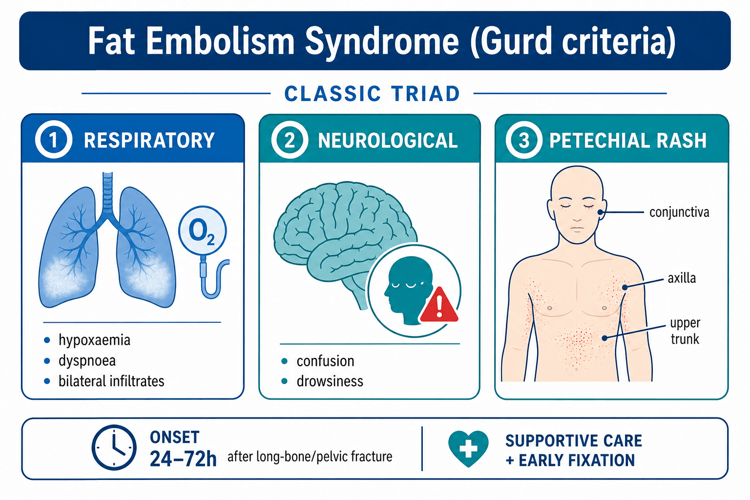
History. Establish the injury pattern (long-bone, pelvic or multiple fractures), the exact time of injury and of any surgery, and the precise onset of hypoxia, confusion or rash. Then the risk modifiers: age, closed versus open fracture, and whether the canal was reamed.
Examination. Reassess every multiply-injured patient on day 2 to 3 for new hypoxia or confusion, and look at four things:
- Respiratory: tachypnoea, hypoxia, bilateral crackles
- Skin: a diligent search for axillary or conjunctival petechiae
- Neurological: GCS, level of confusion, focal deficits
- Fracture sites: stability, swelling, any ongoing manipulation
The rash. Examine the axillae, upper chest and conjunctivae under good light. The petechiae are fine, 1-2 mm in diameter, often in crops, and may be transient, which is why they are missed unless deliberately sought; they typically resolve within 5-7 days. With hypoxia and neurological change after a long-bone fracture the rash makes the diagnosis virtually certain, but its absence does not exclude it. In any long-bone-fracture patient who becomes hypoxic or confused on day 2 or 3, examine the skin and eyes before attributing the change to atelectasis or head injury.
Differential diagnosis. Post-traumatic hypoxia and confusion have several causes, and the latency and the rash are what separate them.
- Distinguishing Features
- 24-72 h latency, petechiae, long-bone fracture
- Key Discriminator
- Axillary petechiae + Gurd criteria
- Investigation
- Clinical diagnosis, CT chest, echo
- Distinguishing Features
- Earlier onset possible, risk factors for VTE
- Key Discriminator
- No petechiae, D-dimer, CT pulmonary angiogram
- Investigation
- CTPA
- Distinguishing Features
- Focal signs, fever pattern, aspiration history
- Key Discriminator
- Unilateral findings, sputum culture
- Investigation
- Chest X-ray, sputum
- Distinguishing Features
- Immediate onset, focal neurology, external signs
- Key Discriminator
- No latency, CT head positive
- Investigation
- CT brain
- Distinguishing Features
- Within 6 hours of transfusion
- Key Discriminator
- Transfusion history, no fracture relation
- Investigation
- Clinical timing
Investigations
What investigations are for. The diagnosis is clinical, on the Gurd criteria. Investigations quantify the hypoxia, support the minor criteria and exclude the treatable differentials; imaging supports the diagnosis but is never pathognomonic. Do not delay supportive care while awaiting advanced imaging, and base fixation decisions on fracture pattern and physiological stability, not on imaging confirmation of emboli.
Arterial blood gas, immediately, on any suspicion of hypoxia or tachypnoea. It measures the hypoxia against the Gurd threshold, and shows a widened A-a gradient and, initially, a respiratory alkalosis; it quantifies severity and guides oxygen therapy.
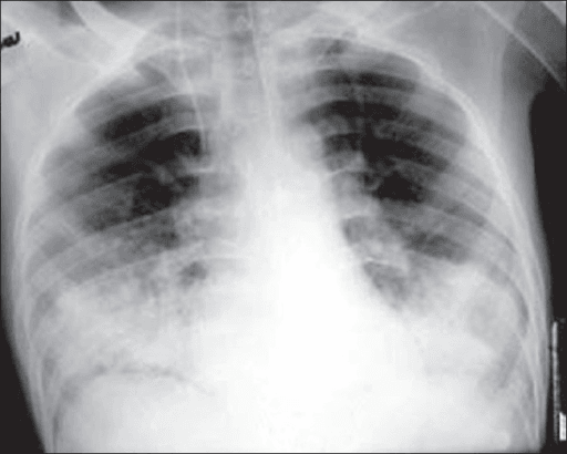
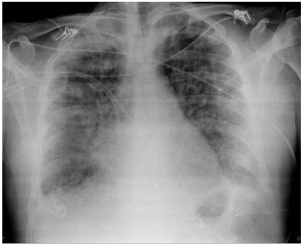
Chest radiograph, PA or AP erect. The classic finding is bilateral diffuse infiltrates resembling ARDS or pulmonary oedema; the "snowstorm" appearance is classic but uncommon. The film may be normal early, and its changes lag behind the clinical hypoxia.
Laboratory tests. Full blood count for the thrombocytopenia, coagulation, renal and liver function, and lipase if available. Fat globules in the urine or sputum have low sensitivity. These results support the minor Gurd criteria rather than make the diagnosis.
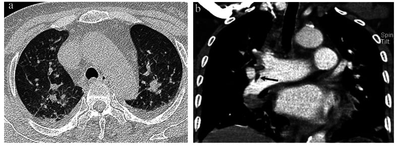
CT of the chest. High-resolution CT may demonstrate ground-glass opacities and centrilobular nodules. CT pulmonary angiography is ordered in the severe case to exclude thromboembolic pulmonary embolism, not to find fat: fat emboli are not visible on CTPA, although a filling defect of negative attenuation, as in the figure below, favours macroscopic fat over thrombus. Both rule out treatable differentials and guide ICU escalation.
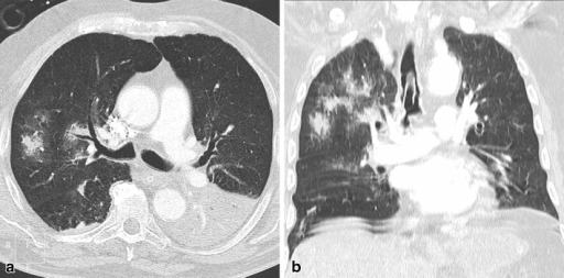
Echocardiography, in the severe case, looks for right ventricular strain, right ventricular dilatation suggesting a massive embolus load, or a patent foramen ovale facilitating paradoxical emboli.
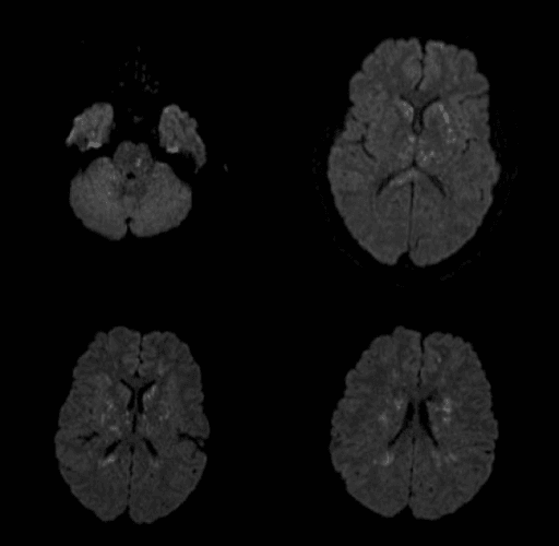
Brain imaging. CT or MRI is indicated for focal signs or persistent coma once head injury has been excluded. MRI can reveal diffuse white-matter hyperintensities on T2-weighted sequences consistent with embolic microinfarcts, which supports the diagnosis and helps prognostication; the diffusion-weighted pattern is described in the next section.
Cerebral Fat Embolism
Presentation. A subset of patients present with neurological features predominating, sometimes with little or no respiratory compromise. Consciousness fluctuates, with confusion, drowsiness, seizures or focal deficits and occasionally coma, and the neurological phase may precede or occur without the pulmonary phase. It is under-recognised because the chest can look well, and it is easily mistaken for traumatic brain injury, sedation, hyponatraemia or sepsis, so it must be actively considered in a long-bone-fracture patient who becomes confused or drowsy on day 2 to 3.
MRI is the key test. Diffusion-weighted imaging shows numerous small foci of restricted diffusion scattered through the white matter, the starfield pattern, reflecting microembolic infarcts, with T2 and FLAIR white-matter hyperintensities. The diffusion pattern is more specific than the T2 changes alone and helps separate cerebral fat embolism from diffuse axonal injury.
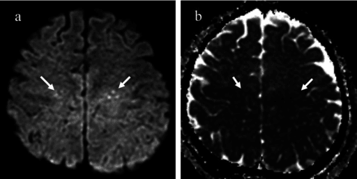
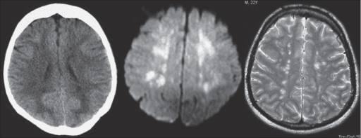
Prognosis. Despite a dramatic presentation, neurological recovery with supportive care is often good, which is the argument for aggressive supportive management rather than nihilism.
Management Algorithm
Prevention through early fixation. The goal is to stabilise the fracture before ongoing embolisation from a mobile fracture site adds to the load. Multiple studies demonstrate that definitive fixation within 24 hours of injury significantly reduces the incidence of the syndrome compared with delayed fixation, by over 50%. Timing of surgery is the modifiable risk factor. The protocol runs in three steps:
- Pre-hospital and emergency department, 0-6 hours. Splint fractures to reduce motion and further embolisation; avoid excessive manipulation, log-roll carefully and limit reduction attempts; book theatre within 24 hours if the patient is physiologically stable
- Damage control, first 24 hours. External fixation or traction for the unstable patient; correct acidosis, hypothermia and coagulopathy; serial observations for early signs of the syndrome
- Definitive fixation, within 24 hours. Early total care, converting to an intramedullary nail or plate when safe; unreamed nails or gentle reaming in high-risk patients; continued vigilance for 72 hours after surgery
In the polytrauma patient too unstable for early total care, damage-control external fixation followed by conversion within 5-7 days still lowers the risk compared with prolonged traction.
Reaming. Reaming raises intramedullary pressure and produces a transient shower of emboli. Whether that becomes clinically important fat embolism syndrome is another matter: the Controversies section below records that large trials show no significant difference between reamed and unreamed nails when fixation occurs early, and that the choice of nail is dictated by fracture pattern and surgeon preference. The advice here is therefore graded: ream gently or use an unreamed nail in the high-risk patient, and avoid reaming where possible once the syndrome is established.
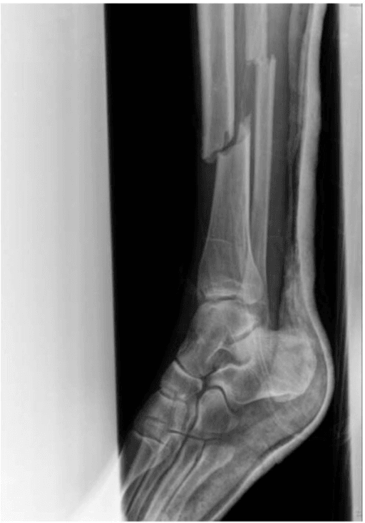
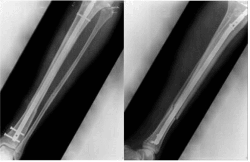
Supportive care in established FES. No pharmacological agent, whether steroids, heparin or statins, has proven benefit in altering the course of established fat embolism syndrome, so treatment is supportive: maintain oxygenation, prevent secondary organ injury, and support the patient through the inflammatory phase. Deterioration to ARDS can be rapid, so observe closely with serial blood gases and clinical examination, rule out pulmonary embolism, pneumonia and cardiac events, and run the care jointly between orthopaedics, intensive care and anaesthetics.
Airway and breathing. Target an SpO2 over 92%, with high-flow nasal oxygen or non-invasive ventilation initially. Intubate for refractory hypoxia, fatigue, a GCS under 8 or impending respiratory arrest. Ventilate with low tidal volumes (6 mL/kg) and titrated PEEP, avoiding over-distension.
Circulation. Balanced resuscitation, avoiding the overload that worsens pulmonary oedema, and avoiding hypotension and hypoxia, which cause secondary injury. Vasopressors are for a septic-like inflammatory response or right heart failure. Monitor lactate, urine output and central venous pressure.
Neurological. Check the GCS frequently; deterioration may indicate worsening embolisation or cerebral oedema. Seizure prophylaxis is not routine unless seizures occur. Maintain normocapnia and normoglycaemia to avoid secondary brain injury.
Fracture care. Complete stabilisation once the patient is physiologically optimised, using unreamed implants where possible, with daily liaison between the orthopaedic and intensive care teams.
Complications
Mortality rises sharply once the syndrome progresses to fulminant ARDS or coma, and once it is established management is supportive only. The single most effective intervention against the complications below is early definitive fixation, whenever the patient's physiological state permits.
- Incidence
- Up to 30 percent of moderate-severe cases
- Risk Factors
- Delayed fixation, large marrow volume, young age
- Management
- Mechanical ventilation, prone positioning if refractory
- Incidence
- 10-20 percent of those with CNS involvement
- Risk Factors
- Severe or prolonged hypoxaemia, large embolus load
- Management
- Neurorehabilitation, supportive care
- Incidence
- Rare but life-threatening
- Risk Factors
- Massive pulmonary embolus load, pre-existing pulmonary hypertension
- Management
- Inotropes, pulmonary vasodilators, ECMO in extreme cases
- Incidence
- Common minor criterion
- Risk Factors
- Consumptive process from emboli
- Management
- Supportive, avoid unnecessary transfusions
- Incidence
- 5-20 percent depending on severity
- Risk Factors
- Fulminant presentation, delayed recognition, comorbidities
- Management
- Prevention is the only effective strategy
Outcomes and Prognosis
What to expect. With fixation within 24 hours most patients have minimal long-term sequelae. Moderate disease treated with oxygen, monitoring and fracture fixation recovers fully in 70-80%, with possible mild cognitive or respiratory limitation. Fulminant disease needing ventilation and advanced therapies carries the mortality given in the severity table, and its survivors may be left with deficits and often need neurorehabilitation.
Prognostic factors. The timing of fracture fixation is the only evidence-based intervention that meaningfully alters incidence, and therefore outcome. Beyond that:
- Best prognosis: early recognition, prompt supportive care, fracture stabilisation achieved, a young patient without comorbidities
- Poor prognosis: diagnosis delayed beyond 72 hours, progression to refractory ARDS, pre-existing lung disease, a coexisting massive head injury
Guidelines, Registries & Global Practice
- Long-bone fractures account for the majority of FES cases worldwide, with femur fractures carrying the highest risk
- Incidence ranges from 1-3 percent after isolated femur fracture to 5-10 percent in polytrauma with multiple long bones
- Young adults (15-40 years) predominate because of higher marrow fat content and greater exposure to high-energy trauma
- Resource-limited settings see higher rates when early fixation is delayed by operating theatre access or transfer logistics
- High-resource centres: 24-hour trauma theatres, damage-control protocols, immediate ICU availability, routine early total care
- Limited-resource environments: Prolonged traction or external fixation common, delayed definitive nailing, higher FES incidence
- Universal principle: The earlier fracture stability is achieved, the lower the embolus load, regardless of implant type
- Registry data: No dedicated FES registry exists; incidence inferred from trauma databases and orthopaedic registries
- Diagnosis emphasis
- Clinical Gurd criteria; imaging supportive only
- Prevention priority
- Early definitive fixation within 24 hours when safe
- Supportive care
- Oxygenation, ventilation, fluid balance; no specific drugs
- Diagnosis emphasis
- High index of suspicion in long-bone fractures on day 2-3
- Prevention priority
- Damage-control orthopaedics followed by early conversion
- Supportive care
- ICU-led supportive care; avoid over-resuscitation
- Diagnosis emphasis
- Gurd criteria remain diagnostic standard
- Prevention priority
- Early stabilisation reduces pulmonary complications
- Supportive care
- Mechanical ventilation per ARDSNet protocol if required
- Diagnosis emphasis
- Clinical diagnosis; petechiae highly specific
- Prevention priority
- Timing of surgery is the key modifiable risk factor
- Supportive care
- Multidisciplinary trauma-ICU-ortho collaboration essential
No international registry specifically tracks fat embolism syndrome. Incidence and outcomes are derived from large trauma databases (e.g., NTDB, TARN) and orthopaedic registries (NJR, AJRR) that capture fracture fixation timing and pulmonary complications. Evidence consistently shows that early fracture stabilisation is the only intervention with a measurable preventive effect.
Record in every long-bone fracture patient:
- Time of injury and time of definitive fixation
- Daily respiratory observations and oxygen requirements on days 1-3
- Presence or absence of axillary or conjunctival petechiae
- Neurological status on day 2-3
- Gurd criteria checklist if any suspicion arises
Missed or delayed diagnosis of fat embolism syndrome in a young trauma patient is a recurring source of morbidity and occasional mortality worldwide. Early fixation and vigilant monitoring on days 2-3 are the global standard of care.
Controversies & Areas of Uncertainty
Reaming increases intramedullary pressure and produces a transient shower of emboli. However, large trials show no significant difference in clinically important fat embolism syndrome rates between reamed and unreamed nails when fixation occurs early. Choice is dictated by fracture pattern and surgeon preference rather than FES prevention.
Older trials suggested high-dose methylprednisolone might reduce inflammatory response, but modern systematic reviews find no improvement in clinical outcomes or mortality. Current guidelines do not recommend routine prophylactic steroids for FES prevention.
The precise threshold at which a borderline polytrauma patient should undergo conversion from damage-control external fixation to definitive nailing remains debated. Physiological parameters (lactate clearance, base deficit, temperature, coagulation) guide individualised decisions rather than a fixed time cut-off.
Routine preoperative echocardiography to detect patent foramen ovale is not justified. In established fulminant FES with right heart strain, echocardiography guides haemodynamic management but does not alter the supportive treatment paradigm.
MCQ Practice Points
Q: What is the primary route by which fat enters the circulation in long-bone fractures? A: Torn medullary veins at the fracture site allow marrow fat to enter the venous system under elevated intramedullary pressure. Reaming and instrumentation further increase this pressure and embolus load.
Q: What are the three major Gurd criteria required for clinical diagnosis of fat embolism syndrome? A: Respiratory distress (hypoxia, tachypnoea, bilateral infiltrates), petechial rash (typically axillary or conjunctival), and neurological abnormality (confusion, drowsiness, focal deficit). One major plus four minor criteria establish the diagnosis.
Q: What is the typical latency period between injury and onset of fat embolism syndrome? A: 24-72 hours after injury. Earliest manifestations may appear at 12 hours, but the classic presentation occurs on the second or third day. Onset after 96 hours is unusual unless fracture sites remain unstable.
Q: What is the single most effective intervention to reduce the incidence of fat embolism syndrome? A: Early definitive fracture fixation within 24 hours of injury whenever the patient's physiological condition permits. Damage-control external fixation followed by conversion within days is an acceptable alternative in unstable polytrauma patients.
Q: What is the mainstay of treatment once fat embolism syndrome is diagnosed? A: Entirely supportive care: oxygen or mechanical ventilation to maintain oxygenation, careful fluid management, fracture stabilisation, and prevention of secondary organ injury. No pharmacological agent has proven benefit.
Q: What is the approximate mortality rate in fulminant fat embolism syndrome progressing to ARDS? A: 10-20 percent. Survivors of severe cases may have residual cognitive or respiratory impairment requiring rehabilitation. Prevention through early fixation remains far more effective than treatment.
Exam Viva Scenarios
Practise clinical reasoning and management decisions out loud
“A 25-year-old motorcyclist sustains an isolated closed midshaft femur fracture. He undergoes intramedullary nailing 18 hours after injury. On the second postoperative day he develops tachypnoea, hypoxia requiring 6 L oxygen, and mild confusion. Axillary petechiae are noted on examination. What is your diagnosis and immediate management?”
“A 32-year-old with bilateral femur fractures and an unstable pelvic ring injury undergoes damage-control external fixation on day 1 because of hypotension and acidosis. On day 3 he is still in the ICU, now with refractory hypoxia on 100 percent oxygen, bilateral infiltrates, petechial rash, and GCS 10. The orthopaedic team asks whether to proceed with conversion to definitive fixation. What is your recommendation?”
Key Pathophysiology
- Fat from marrow enters venous circulation via torn medullary veins after long-bone or pelvic fracture
- Latency 24-72 hours reflects embolisation plus secondary chemical pneumonitis from free fatty acids
- Pulmonary capillary obstruction plus inflammation produces ARDS-like picture
- Paradoxical emboli through PFO or high pulmonary pressure cause systemic petechiae and cerebral effects
Gurd Diagnostic Criteria
- Major (one required): respiratory distress, petechial rash, neurological change
- Minor (four required): tachycardia, pyrexia, thrombocytopenia, fat globules, renal involvement, jaundice, retinal emboli
- Petechiae in axillae or conjunctivae are the most specific physical finding
- Diagnosis is clinical; imaging and labs support but do not confirm
Prevention Strategy
- Early definitive fixation within 24 hours reduces incidence by greater than 50 percent
- Damage-control external fixation in unstable patients followed by conversion within days
- Avoid excessive fracture manipulation and prolonged traction
- No pharmacological prophylaxis (steroids, heparin) has proven benefit
Supportive Management
- Oxygen or mechanical ventilation targeting SpO2 greater than 92 percent
- Low-tidal-volume ventilation (6 mL/kg) if ARDS develops
- Careful fluid balance to avoid worsening pulmonary oedema
- Fracture stabilisation once physiologically optimised
Prognosis and Key Thresholds
- Mortality 10-20 percent in fulminant cases progressing to ARDS or coma
- Most moderate cases recover fully with supportive care
- Early fixation is the only evidence-based intervention that alters incidence
- Residual cognitive or respiratory impairment possible in severe survivors
Evidence Base and Key Trials
Fat embolism: an aid to diagnosis
- Original description of clinical diagnostic criteria still used today
- Major criteria (respiratory, neurological, petechial) plus minor laboratory and clinical features
- Emphasised the 24-72 hour latency and association with long-bone fractures
- Highlighted that petechiae are the most specific physical sign
Early versus delayed stabilization of femoral fractures. A prospective randomized study.
- Prospective randomised study of 178 patients comparing early versus delayed femoral-fracture stabilisation
- In multiply-injured patients, DELAYED fixation gave a higher incidence of pulmonary complications (ARDS, fat embolism, pneumonia), longer hospital stay and more ICU days
- Hospital cost was significantly higher with delayed treatment for all patients
- Established the principle that early fracture stabilisation improves pulmonary outcomes in polytrauma
Fat embolism in patients with multiple injuries
- Early surgical stabilisation in polytrauma patients markedly reduced fat embolism incidence
- Benefit of prompt fixation demonstrated in patients with multiple long-bone injuries
Post-traumatic fat embolism syndrome
- Clinical presentation and management of post-traumatic FES in a modern case series
- Supports Gurd criteria and supportive care approach in resource-variable settings