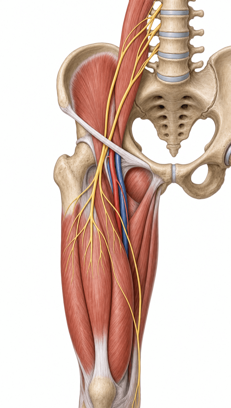
Largest Branch of the Lumbar Plexus
- The femoral nerve arises from the posterior divisions of the anterior rami of L2, L3 and L4 - the largest branch of the lumbar plexus.
- It descends in the groove between psoas major and iliacus, then passes UNDER the inguinal ligament LATERAL to the femoral artery (outside the femoral sheath).
- Lateral-to-medial at the groin: NAVEL - Nerve, Artery, Vein, Empty space (canal), Lymphatics. The NERVE is most lateral.
- Motor: iliacus, pectineus, sartorius and the quadriceps femoris (the knee extensor).
- Sensory: anterior thigh (cutaneous branches) and, via the saphenous nerve, the medial leg, ankle and foot.
- Femoral nerve palsy causes quadriceps weakness (knee buckling), a reduced/absent patellar reflex, and anteromedial sensory loss.
- “NAVEL from lateral to medial places the femoral nerve outside (lateral to) the femoral sheath - which contains only artery, vein and canal.
- “In direct anterior approach THA, the femoral nerve is closest to the acetabular rim at the 'three o'clock' (90°) position - avoid a retractor there.
- “An iliacus/retroperitoneal haematoma (anticoagulation, haemophilia) is a classic non-surgical cause of femoral nerve palsy.
The nerve is vulnerable in anterior-approach THA (retractor over the anterior acetabular rim), acetabular/pelvic fracture surgery, inguinal and pelvic procedures, and from excessive leg lengthening. Cadaveric work shows it lies closest to the anterior rim at the 90° position.
Iliacus or retroperitoneal haematoma (anticoagulation, haemophilia) compresses the nerve in the iliopsoas groove; lithotomy positioning and prolonged hip hyperflexion can also stretch it. These are classic, examinable non-operative causes.
Origin & Course
Origin
- The femoral nerve forms from the posterior divisions of the anterior rami of L2, L3 and L4.
- It is the largest branch of the lumbar plexus.
- It forms within the substance of psoas major and emerges from its lateral border.
Motor & Sensory Supply
The femoral nerve supplies the muscles that extend the knee and assist hip flexion: iliacus, pectineus, sartorius, and the quadriceps femoris (rectus femoris, vastus lateralis, medialis and intermedius).
- Hip flexion is assisted (iliacus, pectineus, sartorius, rectus femoris) - but psoas major also contributes via direct lumbar plexus branches, so hip flexion is only partly affected by a femoral lesion below the inguinal ligament.
- Knee extension depends on the quadriceps - the key deficit in femoral nerve palsy (knee gives way).
- Sensory: anterior thigh via the medial and intermediate cutaneous nerves of the thigh; and the saphenous nerve (the terminal sensory branch of the posterior division) supplies the medial leg, ankle and medial foot.
Clinical Correlations
Deficit
- Weak knee extension (quadriceps) - the patient describes the knee buckling/giving way, especially on stairs.
- Reduced or absent patellar (knee-jerk) reflex.
- Sensory loss over the anterior thigh and the medial leg (saphenous territory).
- Hip flexion is relatively preserved (psoas spared if the lesion is distal).
NAVELFemoral Nerve at the Groin
Hook:Lateral to medial: NAVEL - the femoral Nerve is most lateral.
Evidence Base
Femoral Nerve & the Anterior Acetabular Rim (Cadaveric)
- Cadaveric study of 84 hips mapping the femoral nerve's distance from the anterior acetabular rim
- Minimum distance ranged 16.6-33.2 mm; the nerve was closest to the rim at the 90 degree (anterior, 'three o'clock') position
- Iliopsoas thickness and femoral length correlated with the distance at 90 degrees
- Recommends avoiding retractor placement at 90 degrees to the anterior rim to reduce femoral nerve injury
Femoral Nerve Palsy after Direct Anterior THA
- Retrospective review of 273 primary direct-anterior-approach THAs; femoral nerve palsy incidence 1.1%
- Suspected causes: improper anterior acetabular retractor positioning and excessive leg lengthening
- All three palsies recovered completely within a year
- No significant relationship between palsy and the surgeon's direct-anterior experience
Viva Scenarios
Practise clinical reasoning and management decisions out loud
“Two days after a direct anterior approach THA, a patient's knee gives way and they cannot extend it; there is numbness over the anteromedial thigh and medial leg. What is the diagnosis and your management?”
Guidelines, Registries & Global Practice
Global Practice Picture
Femoral nerve anatomy underpins safe anterior-based hip exposure worldwide. With the international growth of the direct anterior approach, awareness of femoral nerve palsy (incidence around 1% in reported series) and the anatomical "danger zone" at the anterior acetabular rim has become standard teaching, alongside recognition of iliacus haematoma as a non-operative cause.
Side-by-Side Synthesis
- Detail
- L2-L4 posterior divisions (largest lumbar plexus branch)
- Detail
- Psoas-iliacus groove → under inguinal ligament, lateral to artery
- Detail
- NAVEL (nerve most lateral, outside sheath)
- Detail
- Iliacus, pectineus, sartorius, quadriceps (knee extension)
- Detail
- Anterior thigh + saphenous (medial leg/foot)
- Detail
- Weak knee extension, absent patellar reflex, anteromedial sensory loss
- Detail
- Anterior THA retractor / lengthening; iliacus haematoma; lithotomy
Anatomy
- L2-L4 posterior divisions
- Largest lumbar plexus branch
- Psoas-iliacus groove → under inguinal ligament
- Lateral to femoral artery (NAVEL), outside sheath
Supply
- Motor: iliacus, pectineus, sartorius, quadriceps
- Knee extension = key function
- Sensory: anterior thigh + saphenous (medial leg)
- Saphenous = terminal sensory branch
Clinical
- Palsy: weak knee extension, knee buckling
- Reduced/absent patellar reflex
- Risks: anterior THA retractor, leg lengthening
- Iliacus haematoma (anticoagulation)