CAM vs Pincer vs Mixed | Alpha Angle Measurement | FADIR Test Gold Standard | Hip Arthroscopy
- CAM impingement from aspherical femoral head with decreased head-neck offset
- Pincer impingement from acetabular overcoverage (focal or global)
- Alpha angle greater than 55 degrees diagnostic for CAM morphology on MRI
- FADIR test (flexion-adduction-internal rotation) has 94-99% sensitivity
- Mixed morphology present in 85% of symptomatic FAI cases
- Labral tears in 86% of FAI patients requiring surgery
- “CAM = femoral problem, Pincer = acetabular problem, Mixed = both
- “Alpha angle measured on lateral femoral head-neck junction
- “Crossover sign indicates anterior acetabular overcoverage (pincer)
- “Hip arthroscopy gold standard for treatment - open surgery declining
Overview and Epidemiology
Femoroacetabular impingement (FAI) is a mechanical hip disorder: abnormal contact between the femoral head-neck junction and the acetabular rim during hip motion. Repeated impingement damages the labrum and the articular cartilage, and may progress to early hip osteoarthritis.
Two morphologies, usually together. The abnormal contact comes from the femoral side, the acetabular side, or both:
- Cam - an aspherical femoral head with decreased head-neck offset
- Pincer - acetabular overcoverage, focal or global
- Mixed - both components present, and the pattern in 85% of symptomatic FAI
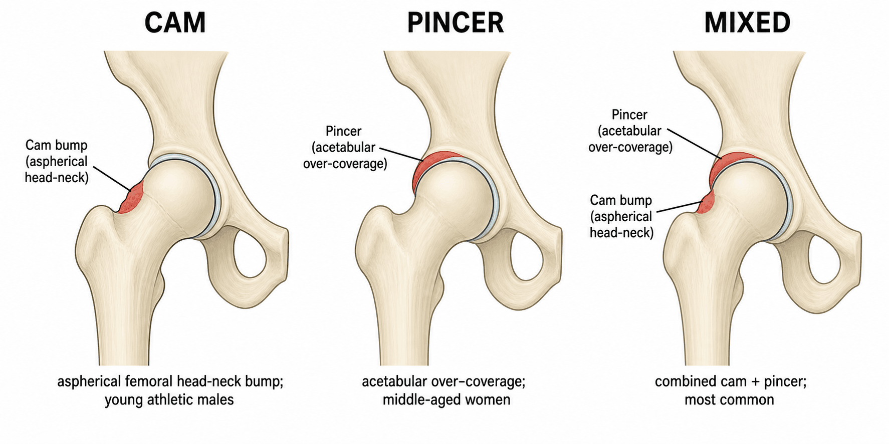
Who. Symptoms peak at 20-40 years. Cam predominates in males (3:1), pincer is more common in middle-aged females, and prevalence is higher in certain sports (soccer, hockey, dancing). The morphology itself is found in 10-15% of the general population and is often asymptomatic.
Why it matters. FAI is increasingly recognised as a major cause of early hip osteoarthritis in young active patients and may account for 10-15% of early hip OA. By the time of surgery, 86% have a labral tear and 70% have chondral lesions at arthroscopy. The activity limitation is significant in this group and can be career-ending for an athlete if untreated; early recognition and intervention may prevent or delay osteoarthritis, though the long-term data are still emerging.
Risk factors. Symptomatic FAI is more likely with:
- High-impact sport during skeletal development
- Repetitive hip flexion (dancers, martial artists)
- Underlying hip dysplasia or acetabular retroversion
- Previous Perthes disease or slipped capital femoral epiphysis (SCFE)
- Genetic predisposition (familial clustering is observed)
Pathophysiology and Mechanisms
The normal hip. A ball-and-socket joint with exceptional range of motion, built from four parts that matter here:
- Femoral head - spherical, covered by articular cartilage except at the fovea
- Femoral neck - offset from the head, creating a concave waist at the anterosuperior junction
- Acetabulum - a horseshoe-shaped socket covering about 40% of the head
- Labrum - a fibrocartilaginous rim that deepens the socket and seals the joint
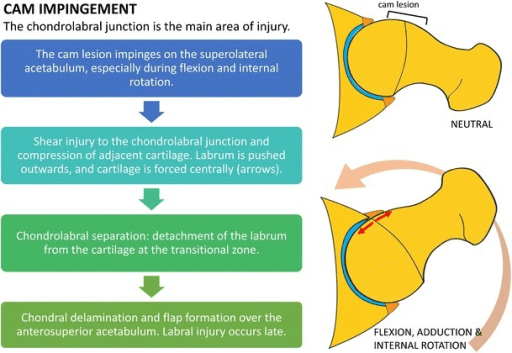
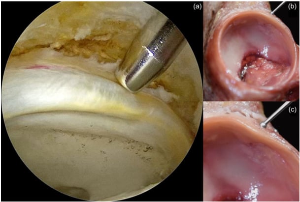
Why the offset matters. During flexion and internal rotation the anterosuperior head-neck offset is what lets the femoral neck clear the acetabular rim. Lose it and the neck meets the rim with every deep flexion, producing abnormal contact and shear on the anterosuperior labrum and cartilage. In the normal hip the cartilage sees compression only and the labrum works as a suction seal; in the impinging hip each flexion adds shear microtrauma, the cartilage delaminates progressively, and the end point is full-thickness cartilage loss.
Cam. The head is aspherical (the pistol-grip deformity) and the anterior offset is decreased or absent, so the alpha angle exceeds 55°. Contact lands on the anterosuperior acetabulum, and the damage runs outside-in: the aspherical head strikes the rim first, then shears into the labrum and cartilage like a snowplough, delaminating the cartilage as it goes.
Pincer. The acetabulum covers too much of the head, focally (retroversion) or globally (coxa profunda). Normal acetabular version is 15-20° of anteversion; a retroverted socket produces the crossover sign on the AP pelvis. Contact starts at the anterosuperior rim, but the damage runs inside-out: the rim strikes the femoral neck and levers the head, and the result is a posteroinferior contre-coup labral tear. Chronic rim impingement may also ossify into an os acetabuli.
Position. Cam impinges in flexion and internal rotation, the FADIR position. Pincer is worse in flexion, abduction and external rotation, the FABER position. Mixed hips hurt in more than one position.
Femoral Version: The Other Side of the Impingement Equation
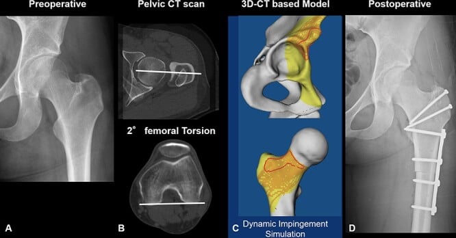
Clearance depends on the femur too. Reduced femoral anteversion or frank femoral retroversion brings the neck closer to the anterior acetabular rim in flexion and internal rotation, worsening (or even causing) anterior impingement when the alpha angle is only borderline, and it typically reduces internal rotation on examination. Excessive anteversion does the opposite: more anterior clearance, but a predisposition to posterior extra-articular impingement and anterior microinstability.
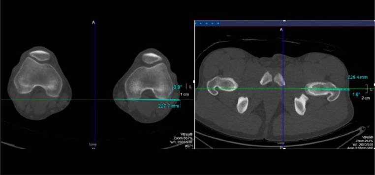
Read the two together. The McKibbin combined index is the sum of femoral version and acetabular version. A low combined value indicates global anterior under-clearance and a high value the reverse, which is why neither number is read in isolation: assess femoral version on a CT or MRI rotational profile alongside the acetabular crossover and centre-edge findings. Femoral retroversion is an often-missed driver of persistent anterior impingement and of failure after an isolated cam osteoplasty, and in marked cases a femoral derotation osteotomy, not arthroscopy, is the correct operation.
Post-SCFE and Post-Perthes Cam: When the Deformity Is Not Idiopathic
Most of what follows concerns the idiopathic cam, a subtle loss of head-neck offset laid down at the anterosuperior junction during adolescent physeal closure. A post-SCFE cam is a different animal, and treating the two as interchangeable is the commonest conceptual error here.
The mechanism is inverted. In an idiopathic cam the head is aspherical. After a slip the head is largely spherical but has moved: the epiphysis translates posteriorly and inferiorly, leaving the anterosuperior metaphysis projecting into the joint. The offending structure is therefore residual metaphyseal prominence, not head asphericity, and it strikes the acetabular rim directly. That is why alpha angles after a slip sit far above the idiopathic range and why internal rotation in flexion is lost so early. It also explains the physical sign, obligate external rotation as the hip is flexed (the Drehmann sign), which reflects the metaphysis having to swing out of the way.
- Idiopathic cam
- Aspherical anterosuperior head
- Post-SCFE cam
- Residual anterosuperior metaphyseal prominence after epiphyseal translation
- Idiopathic cam
- Modest elevation above the sixty-degree region
- Post-SCFE cam
- Markedly higher; correction of twenty to thirty-seven degrees is what surgery achieves
- Idiopathic cam
- Anterior impingement test positive, internal rotation reduced
- Post-SCFE cam
- Internal rotation lost early; obligate external rotation on flexion (Drehmann sign)
- Idiopathic cam
- Usually isolated
- Post-SCFE cam
- Femoral retroversion, coxa vara and shortening frequently coexist
- Idiopathic cam
- Arthroscopic osteochondroplasty in most
- Post-SCFE cam
- Osteochondroplasty for the prominence; proximal femoral osteotomy where the deformity is severe or multiplanar
Arthroscopic osteochondroplasty for FAI secondary to SCFE: systematic review
Correcting a healed slip is not the same problem as correcting an idiopathic cam, and the danger is vascular. Do not osteotomise through the old slip site: the retinacular supply to the epiphysis runs there and the avascular necrosis risk stays high even years after the physis has closed. Where the deformity is too severe for osteochondroplasty alone, the safer default is an intertrochanteric (Imhauser) flexion-rotation osteotomy, which corrects away from the physis. Anatomical realignment by surgical hip dislocation (modified Dunn) does address the deformity at its source, but it belongs in dedicated hip-preservation centres because it depends on protecting the medial femoral circumflex artery through a retinacular flap.
Post-Perthes hips fail differently. Here the problem is rarely a discrete bump. Coxa magna, coxa breva and relative trochanteric overgrowth produce extra-articular impingement between the greater trochanter and the pelvis alongside any intra-articular cam. An arthroscopic osteochondroplasty that ignores the trochanteric side of the equation will not restore clearance, which is why these hips more often need relative femoral neck lengthening and trochanteric transfer.
Classification Systems
The morphology names the side of the joint that is wrong and therefore the side that surgery corrects.
- Location
- Femoral head-neck
- Radiographic Signs
- Alpha angle greater than 55°
- Labral Pattern
- Anterosuperior tear
- Treatment
- Femoral osteoplasty
- Location
- Acetabular rim
- Radiographic Signs
- Crossover sign, coxa profunda
- Labral Pattern
- Posteroinferior tear
- Treatment
- Acetabular rim trimming
- Location
- Both femur and acetabulum
- Radiographic Signs
- Combined signs
- Labral Pattern
- Anterosuperior + posterior
- Treatment
- Combined osteoplasty
Look for both. The primary mechanism may be cam or pincer, but mixed morphology is the rule rather than the exception, and addressing only one component leaves persistent symptoms. A complete assessment evaluates both the femoral and the acetabular morphology.
Clinical Presentation and Assessment
History. Deep anterior groin pain in 85-90%, activity-related and insidious in onset. Ask what provokes it:
- Prolonged sitting (the theatre sign), getting out of a car, squatting, climbing stairs
- Cutting and pivoting sports, where performance falls off
- Clicking, catching or giving way, which point to a labral tear
Cam and pincer present differently.
- Cam
- Young males, athletes (soccer, hockey)
- Pincer
- Middle-aged females, endurance athletes
- Cam
- Deep groin pain with high-impact activity
- Pincer
- Anterior groin pain with prolonged activity; theatre sign after sitting with the hip flexed
- Cam
- Hip flexion with internal rotation
- Pincer
- Hip flexion, especially in adduction
- Cam
- Gradual worsening over months to years; reduced ability to change direction quickly
- Pincer
- Slower progression than cam
- Cam
- May have a labral tear causing clicking
- Pincer
- Posterior labral tears from levering
Examination. The impingement tests reproduce the patient's pain.
- Technique
- Flexion 90°, adduction, internal rotation
- Positive Finding
- Anterior groin pain
- Sensitivity
- 94-99%
- Technique
- Flexion, abduction, external rotation
- Positive Finding
- Groin or lateral pain
- Sensitivity
- 70-80%
- Technique
- Prone, hip extended, measure IR
- Positive Finding
- Less than 20° suggests FAI
- Sensitivity
- Variable
- Technique
- Resisted hip flexion at 30°
- Positive Finding
- Anterior groin pain
- Sensitivity
- 60-70%
- Technique
- Supine, passive hip rotation
- Positive Finding
- Groin pain or clicking
- Sensitivity
- Variable
FADIR. Flexion-adduction-internal rotation is the single most sensitive clinical test, at 94-99%. Supine, flex the hip to 90°, then adduct across the body and internally rotate; reproduction of anterior groin pain is positive. The position maximises impingement at the anterosuperior acetabulum. Its specificity is low: it is a good rule-out test.
Range of motion. Internal rotation is the movement FAI restricts most, and external rotation is often preserved, which helps separate it from other pathology.
- Normal Range
- 120-140°
- Typical FAI Finding
- 90-110°
- Clinical Significance
- Limited by pain at end range
- Normal Range
- 30-40°
- Typical FAI Finding
- Less than 20°
- Clinical Significance
- Most restricted motion in FAI
- Normal Range
- 40-50°
- Typical FAI Finding
- Often preserved
- Clinical Significance
- Helps distinguish from other pathology
- Normal Range
- 40-50°
- Typical FAI Finding
- Usually preserved
- Clinical Significance
- Not typically limited
- Normal Range
- 20-30°
- Typical FAI Finding
- May be painful
- Clinical Significance
- Pain with FADIR position
Gait and posture. Gait is usually normal unless there is severe OA, and antalgic with advanced disease. Patients may sit with the hip abducted and externally rotated to avoid impingement.
The C-sign. The patient cups the hip with thumb and fingers in a C, over the anterior and lateral hip, to show that the pain is deep in the groin and lateral. It is highly specific for intra-articular pathology including FAI and separates it from extra-articular pain such as bursitis or a muscle strain.
Differential diagnosis. Groin pain in a young adult has a long differential.
- Typical patient
- Young active adult, athlete
- Discriminating features
- Deep groin pain, positive FADIR, reduced internal rotation, cam/pincer on imaging
- Key investigation
- AP pelvis + lateral, MRI/MR arthrogram
- Typical patient
- Young woman, may be active
- Discriminating features
- Lateral/groin pain with activity, apprehension, lateral centre-edge angle under 20-25°
- Key investigation
- AP pelvis, false-profile view
- Typical patient
- Male field-sport athlete
- Discriminating features
- Lower abdominal/adductor pain on resisted sit-up and adduction, no impingement sign
- Key investigation
- Dynamic ultrasound, MRI pubic symphysis
- Typical patient
- Runner, military recruit, RED-S risk
- Discriminating features
- Rest and night pain, antalgic gait, pain on hop test
- Key investigation
- MRI (X-ray often normal early)
- Typical patient
- Steroid/alcohol use, sickle cell, prior trauma
- Discriminating features
- Groin pain, preserved early X-ray, crescent sign late
- Key investigation
- MRI (most sensitive)
- Typical patient
- Older patient
- Discriminating features
- Stiffness, reduced global ROM, joint-space loss, osteophytes
- Key investigation
- Weight-bearing AP pelvis (Tonnis grade)
- Typical patient
- Any age; systemic features
- Discriminating features
- Rest pain, effusion, raised inflammatory markers, fever (septic)
- Key investigation
- ESR/CRP, aspiration if sepsis suspected
- Typical patient
- Any age, back history
- Discriminating features
- Pain not reproduced by hip manoeuvres, neurological signs
- Key investigation
- Lumbar spine MRI
Two further points. Dysplasia may coexist with FAI or be a separate entity, and a labral tear can be traumatic without any impingement morphology behind it. The extra-articular mimics declare themselves by where the pain is:
- Iliopsoas tendinitis - snapping hip, a different pain pattern
- Greater trochanteric pain syndrome - lateral pain, not groin
- Adductor strain - medial thigh pain
Investigations
Radiographs first. Three views are mandatory:
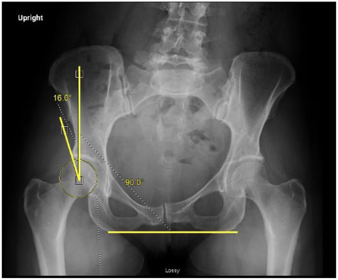
- AP pelvis - acetabular coverage, crossover sign, centre-edge angle, Tonnis grade
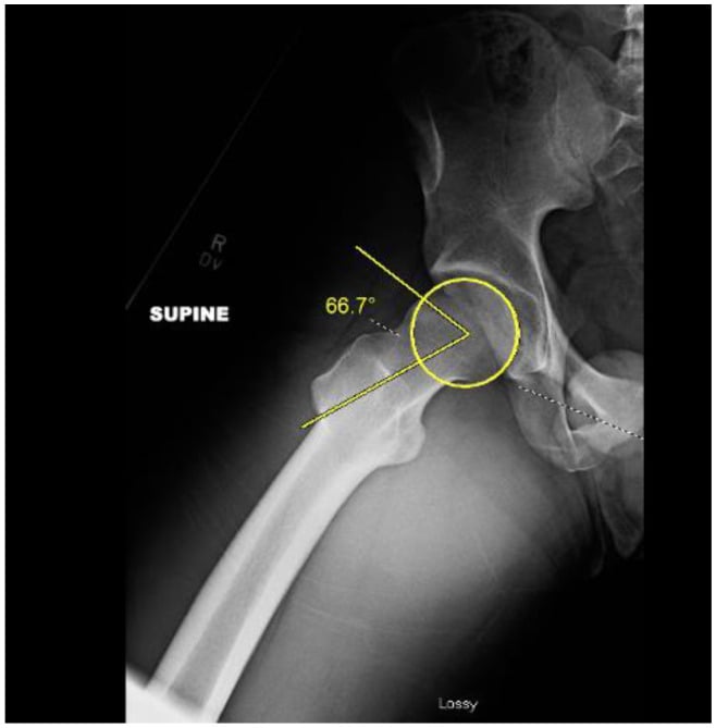
- Frog-leg lateral - alpha angle and head-neck offset
- False profile - anterior coverage
The measurements that decide management are the alpha angle (greater than 55° on the frog-leg lateral is diagnostic of cam), the centre-edge angle (greater than 40° on the AP suggests pincer overcoverage), the crossover sign of acetabular retroversion, and the Tonnis grade, which must be 0-1 for arthroscopy to be an option.
- View
- Frog-leg lateral or MRI
- Finding
- Angle between neck axis and asphericity point
- Indicates
- CAM morphology
- View
- AP pelvis
- Finding
- Anterior wall lateral to posterior wall
- Indicates
- Acetabular retroversion (pincer)
- View
- AP pelvis
- Finding
- Posterior wall medial to femoral head center
- Indicates
- Relative anterior overcoverage
- View
- AP pelvis
- Finding
- Angle from femoral head center to lateral rim
- Indicates
- Global overcoverage (pincer)
- View
- MRI or frog-leg
- Finding
- Pit at anterior femoral neck
- Indicates
- Marker of chronic FAI
- View
- AP pelvis
- Finding
- Ossicle at anterosuperior rim
- Indicates
- Chronic pincer impingement
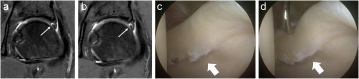
MRI arthrogram is the gold standard. It measures the alpha angle on axial oblique sequences, the most accurate method; finds labral tears with 90% sensitivity once intra-articular gadolinium is in; assesses the chondral lesions that predict outcome; maps the cam circumferentially; and shows herniation pits, synovial herniation into the femoral neck that marks FAI. The typical picture is:
- Anterosuperior labral tear (the cam pattern)
- Anterosuperior cartilage delamination or defect
- Paralabral cysts where the labrum is torn
- A herniation pit at the anterior femoral neck
CT is not routine. It is less sensitive than MRI for the labrum and cartilage, and earns its place for:
- Surgical planning in complex acetabular morphology
- 3D reconstruction to map the extent of the cam
- Bony anatomy when MRI is contraindicated
- Precise measurement of version and coverage angles
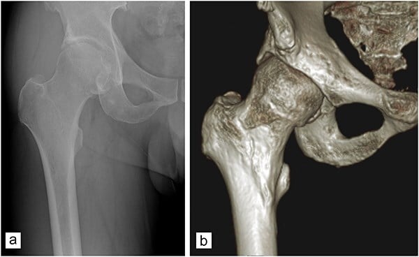
Dynamic fluoroscopy during examination under anaesthesia shows where the hip impinges as it moves, guides the extent of the osteoplasty, confirms clearance after cam resection and picks up additional sources of impingement.
The diagnostic injection. A local anaesthetic injection into the joint under fluoroscopic or ultrasound guidance can be very useful: temporary relief confirms the hip joint as the source, and persistent pain points to extra-articular pathology. It is typically combined with steroid (lidocaine or bupivacaine plus steroid) for therapeutic benefit.
Bloods are generally not needed unless an inflammatory arthropathy is in question:
- ESR and CRP if inflammatory arthritis is suspected
- Rheumatoid factor and anti-CCP if rheumatoid arthritis is suspected
- HLA-B27 if ankylosing spondylitis is suspected
Management Algorithm
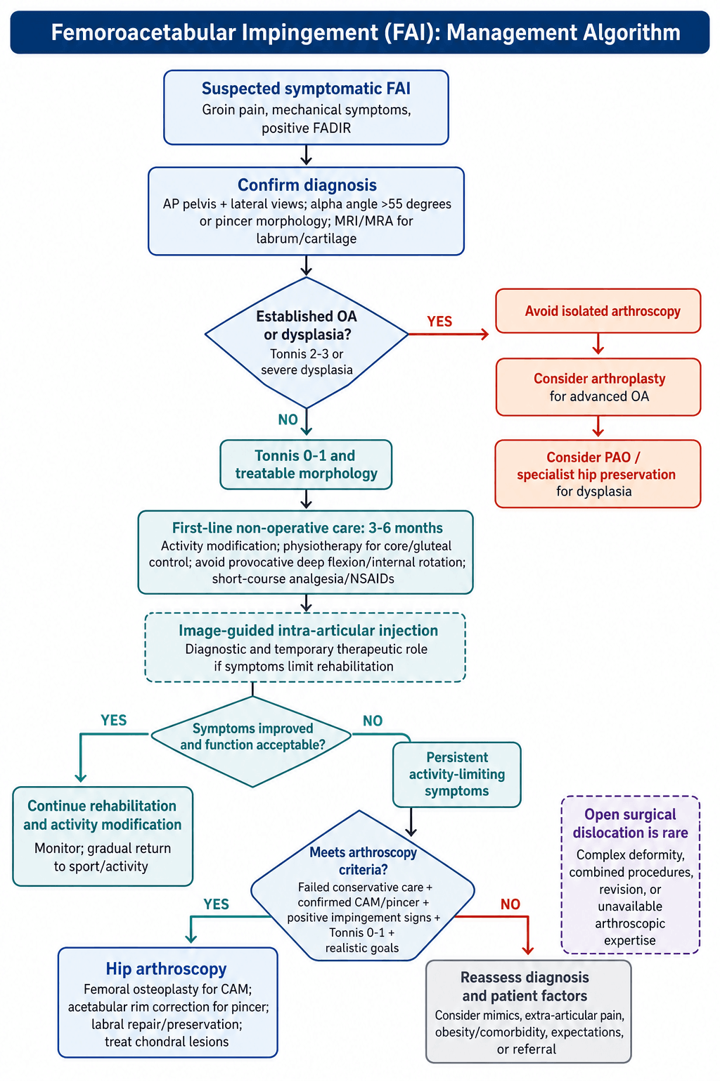
Everyone starts here. All patients should trial conservative management for 3-6 months unless the mechanical symptoms are severe.
Activity modification from the first visit:
- Limit deep flexion and internal rotation
- Reduce high-impact sport temporarily
- Avoid prolonged sitting and low chairs
- Lose weight if overweight, which reduces the forces across the hip
- Educate: what FAI is and what to expect
Physiotherapy over the 3-6 months is built around core strengthening and hip stabilisation: gluteal strengthening without aggravating the impingement, core stability, posterior chain strengthening, proprioception and balance work, and no deep hip flexion exercises. The goals are to optimise hip mechanics, reduce compensatory patterns and improve functional capacity.
Medication is for symptoms, not disease. Short courses of NSAIDs and regular paracetamol control pain during rehabilitation; long-term NSAIDs are avoided for their gastrointestinal and cardiovascular effects.
Injection when the above is failing. An image-guided steroid injection is both diagnostic and therapeutic, gives temporary relief (typically 3-6 months), may allow participation in physiotherapy, and can be repeated once if it helped. It is not a definitive treatment for FAI, and it is contraindicated in infection, severe OA and allergy.
How often it works. Conservative management succeeds in about 30-40% of symptomatic patients, more often with mild morphology (alpha angle 55-65°), no significant labral tear and good compliance. Failure means persistent symptoms limiting activities after 3-6 months of appropriate treatment.
Surgical Technique - Hip Arthroscopy
Pre-operative Planning
Consent covers the specific risks of the procedure:
- Failure to improve symptoms - 15-20%, especially with cartilage damage
- Nerve injury - pudendal and lateral femoral cutaneous, 5-8% transient
- Heterotopic ossification - 3-5%, usually asymptomatic
- Infection - under 1%
- Conversion to arthroplasty if severe cartilage damage is found at surgery
- DVT and PE at the standard orthopaedic surgical risk
Equipment is the other half of planning: a hip arthroscopy traction table with a well-padded perineal post; 30° and 70° arthroscopes; curved shavers, burrs and radiofrequency devices; a C-arm positioned for AP and lateral views; suture anchors of 2.3mm or smaller for labral repair; and a high-speed burr and arthroscopic osteotomes for the cam.
Patient Positioning
Supine on the traction table. The well-padded perineal post sits against the medial groin, both feet are secured in traction boots, the contralateral leg is abducted and secured to give the C-arm room, and the operative leg starts in neutral. Padding is where the nerve injuries are prevented: the perineal post for the pudendal nerve, no compression at the ASIS for the lateral femoral cutaneous nerve, the contralateral knee and ankle, and the arms secured across the chest or on boards.
Traction. Apply roughly 25-50 pounds and distract the joint 8-10mm, confirmed on fluoroscopy; adequate distraction is what makes portal placement safe. Monitor it through the case, release periodically, and keep total traction time under 2 hours.
Pudendal nerve injury from excessive or prolonged traction is a recognised complication of hip arthroscopy. Risk factors are traction beyond 2 hours, excessive force, inadequate perineal post padding and female gender. Release the traction whenever work moves to the peripheral compartment. The neurapraxia usually resolves within 6 months but can be permanent.
Portal Placement
Anterolateral portal first, under fluoroscopy. It lies at the intersection of a horizontal line from the superior tip of the greater trochanter and a vertical line from the ASIS. A spinal needle confirms the intra-articular position on the image, then skin incision, blunt dissection to the capsule, and the arthroscope sheath and cannula go in. This is the main viewing portal for the central compartment.
Mid-anterior portal second, under direct vision. With the scope looking at the anterolateral capsule from the first portal, a spinal needle comes in outside-in, kept a safe distance from the lateral femoral cutaneous nerve, and the working portal follows by skin incision and blunt dissection. This is the main working portal for labral repair and cam resection.
Additional portals as needed: a posterolateral portal for the posterior labrum and a distal anterolateral accessory portal for peripheral cam resection. Two or three portals are enough for most FAI cases.
Central Compartment Work (Under Traction)
Look before you cut. The central compartment is examined in a fixed order so nothing is missed:
- Labrum - the anterosuperior labrum is the usual cam tear site and the posterior labrum is checked for the contre-coup pincer lesion; classify the tear (radial flap, longitudinal, bucket-handle, degenerative) and probe it for an unstable flap
- Cartilage - map the acetabular damage by the Beck classification; the femoral head cartilage is usually better preserved; look for delamination, the carpet phenomenon of cartilage lifted off bone, and the wave sign that indicates it
- Ligamentum teres - partial or complete tear; debride if degenerate and symptomatic; not routinely reconstructed
- Loose bodies - common in FAI from cartilage delamination, in the anterior or posterior recess; remove them
Peripheral Compartment Work (Traction Released)
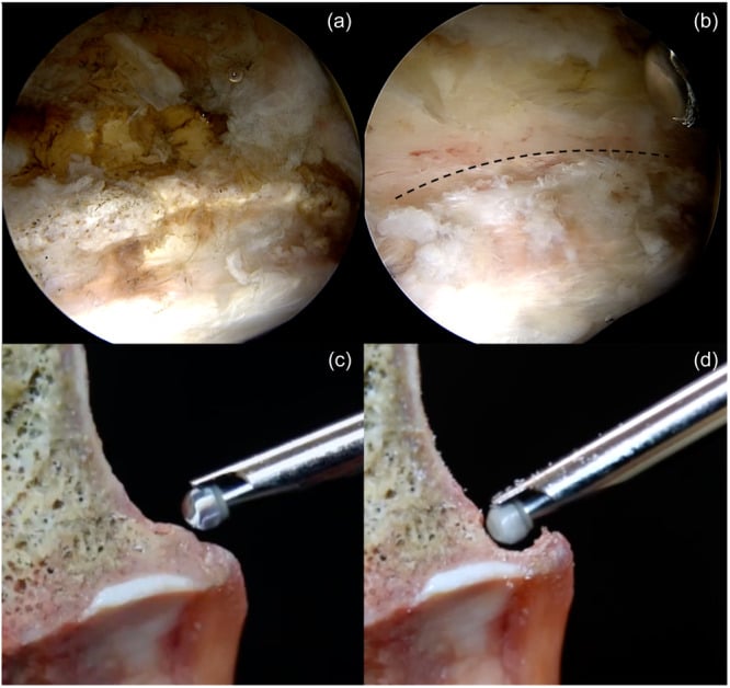
Release the traction and flex and externally rotate the hip to bring the femoral neck into view. The peripheral compartment lies between the capsule and the neck, and a limited capsulotomy may be needed for access.
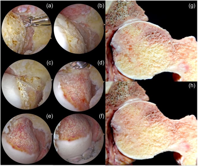
Resect the cam. The lesion sits at the anterosuperior head-neck junction. A high-speed burr takes down the aspherical portion of the head, the goal being a restored concave waist at the junction and an alpha angle under 50° on fluoroscopy. The resection typically covers the anterosuperior quadrant, and may extend from the 1 o'clock to the 4 o'clock position on a right hip, with a smooth transition from head to neck and no stress riser; resect conservatively rather than cut a notch into the neck.
Confirm clearance. On the AP view the head-neck offset should be restored; on the lateral the alpha angle should be under 50-55°; and flexing the hip under dynamic fluoroscopy should show no impingement. The arthroscope from the anterolateral portal gives a direct view as well.
Close the capsule if a capsulotomy was made. Repair with arthroscopic sutures restores capsular integrity, which may reduce postoperative instability; some surgeons leave the capsulotomy open, and the question remains controversial. The reasons for closing it are set out under Complications.
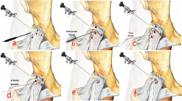
Closure
Finish as carefully as you started. Release the traction completely, make a final arthroscopic inspection, confirm no retained instruments or debris, and secure haemostasis. Remove the cannulas, close each portal with one or two simple interrupted sutures and apply sterile dressings. A compression dressing, ice and elevation, crutches with partial weight-bearing, and DVT prophylaxis as per protocol complete the case.
Complications
- Incidence
- 5-8%
- Risk Factors
- Prolonged traction, excessive force
- Management
- Usually resolves 3-6 months, observe
- Incidence
- Less than 1%
- Risk Factors
- Same as above
- Management
- May require nerve specialist referral
- Incidence
- 3-5%
- Risk Factors
- Extensive CAM resection, genetic predisposition
- Management
- Usually asymptomatic, excision if limiting motion
- Incidence
- 15-20%
- Risk Factors
- Severe cartilage damage, persistent morphology
- Management
- May require revision or conversion to THA
- Incidence
- 1-2%
- Risk Factors
- Excessive rim trimming, capsulotomy not closed
- Management
- Activity modification, may need capsular plication
- Incidence
- Less than 1%
- Risk Factors
- Excessive CAM resection creating notch
- Management
- Usually requires ORIF or arthroplasty
- Incidence
- Less than 1%
- Risk Factors
- Standard surgical risk factors
- Management
- Arthroscopic washout, antibiotics
- Incidence
- Less than 1%
- Risk Factors
- Standard orthopedic risk factors
- Management
- Anticoagulation as per protocol
- Incidence
- Less than 1%
- Risk Factors
- Technical error
- Management
- Retrieve broken instrument intraoperatively
Which nerves. The pudendal nerve suffers from perineal post pressure or excessive traction, giving numbness and sexual dysfunction; the lateral femoral cutaneous nerve from portal placement or positioning, giving lateral thigh numbness. Sciatic nerve injury from traction or positioning is rare, and femoral nerve injury very rare.
Heterotopic ossification follows 3-5% of hip arthroscopies and is usually asymptomatic. The risk rises with extensive cam resection, a history of HO, male gender and genetic predisposition, and it is more common in revision cases and after open surgery. Prophylaxis is controversial and not used by every surgeon; the options are indomethacin 75mg daily for 2-3 weeks or single-dose radiation of 7 Gy.
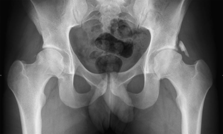
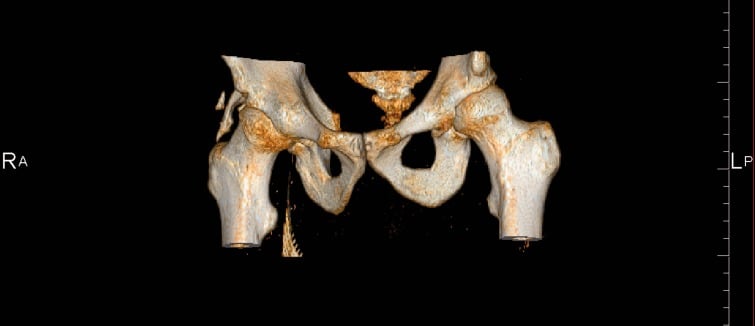
Failure to improve is the most common "complication", in 15-20%. The causes:
- Severe cartilage damage at surgery (Outerbridge grade 3-4)
- Incomplete correction of the morphology, leaving residual impingement
- Progression of osteoarthritis despite surgery
- Wrong diagnosis, the pain coming from another source
- Unrealistic expectations
- Poor rehabilitation compliance
Prevention is careful selection (Tonnis 0-1, confirmed morphology, mechanical symptoms), complete correction of both cam and pincer, and realistic counselling.
Femoral neck fracture after cam resection is rare (under 1%) but devastating. The risk factors are a resection depth greater than 30% of the neck diameter, a notch cut into the neck, and early weight-bearing before healing. Prevention is a conservative resection depth, a smooth gradual transition from head to neck with no sharp corners or notches, and protected weight-bearing for 6 weeks after surgery.
Hip Microinstability and Capsular Management
What it is. Hip microinstability is symptomatic, extra-physiological motion of the femoral head within the socket without frank dislocation. The hip resists it through three structures, each of which this operation can compromise:
- The capsule, principally the iliofemoral ligament (Y-ligament of Bigelow), the strongest anterior stabiliser limiting external rotation and extension, divided by the interportal or T-capsulotomy used for access
- The labral suction seal, which maintains negative intra-articular pressure and fluid sealing, lost if the labrum is debrided rather than repaired
- The bony architecture, which is why over-resecting the acetabular rim into iatrogenic under-coverage destabilises the joint
Do not trade impingement for instability. Capsular management is therefore part of the operation rather than an afterthought. Many surgeons now repair or plicate the capsule, and in patients with ligamentous laxity, borderline acetabular coverage or revision surgery the threshold for capsular repair or plication and labral preservation, and against aggressive rim resection, is lower; routine closure nevertheless remains debated. The aim is to relieve impingement without destabilising the joint.
Postoperative Care and Rehabilitation
Principles. Full recovery takes 6-12 months, and progression is by criteria rather than by the calendar. Weight-bearing is protected for the first 6 weeks, because early full loading stresses the femoral neck; range of motion avoids the impingement positions, because aggressive early motion risks the labral repair; strengthening centres on the gluteals, because hip control is what protects the result. Returning to sport or resuming high-impact activity too early increases the failure risk, because the hip needs full healing first. The therapist needs to know exactly what was done, and persistent symptoms are investigated rather than ignored.
Hip Arthroscopy Rehabilitation
Protection phase:
- Partial weight-bearing with crutches (20-30 pounds foot-flat)
- Hip brace optional (some surgeons, especially after capsulotomy)
- Ice and elevation
- DVT prophylaxis (aspirin or LMWH as per protocol)
- Multimodal analgesia
- Passive ROM, avoiding the extremes
- No hip flexion beyond 90° and no combined flexion-internal rotation
Progressive weight-bearing:
- Week 2: increase to 50% weight-bearing if comfortable
- Week 4: wean off crutches to full weight-bearing
- Week 6: walking normally without aids
Range of motion:
- Gentle active-assisted ROM
- Supine hip flexion to 90°, avoiding end-range flexion
- Hip abduction and extension exercises
- Still no combined flexion and internal rotation
Stationary cycling from 3-4 weeks with a high seat
Progressive loading:
- Advance ROM with a gradual increase in flexion
- Closed-chain strengthening (squats, leg press to limited depth)
- Hip abductor strengthening, gluteus medius in particular
- Core stability
- Pool work if available
- No impact activities
Goals: full ROM, normalised gait, good hip control
Sport-specific training:
- Jogging on the flat at 3 months
- Agility and cutting drills at 4-5 months
- Sport-specific training at 5 months
- Full sport at 6 months if strength testing is adequate
Criteria for return:
- Pain-free full ROM
- Hip strength 90% of the contralateral side
- Functional testing: single-leg squat, hop tests
- No effusion or mechanical symptoms
- Hip strengthening continued indefinitely
- High-impact activities avoided if possible (controversial)
- Watch for recurrence of symptoms
- Radiographs at 1 and 2 years for OA progression
Outcomes and Prognosis
What to expect. Modern hip arthroscopy for FAI gives good to excellent outcomes in about 85% of appropriately selected patients at 2 years, with high satisfaction and significant improvement in pain, function and return to sport. Selection is what moves that number.
- 2-Year Success
- Greater than 90%
- Prognostic Factors
- Age under 40, alpha angle 55-70°, Tonnis 0, minimal cartilage damage
- 2-Year Success
- 80-90%
- Prognostic Factors
- Age 40-50, mixed morphology, Tonnis 0-1, moderate cartilage damage
- 2-Year Success
- 60-70%
- Prognostic Factors
- Age over 50, Tonnis 1, significant cartilage damage (Outerbridge 3)
- 2-Year Success
- Less than 50%
- Prognostic Factors
- Tonnis 2-3, severe cartilage loss, unrealistic expectations
Return to sport. Recreational athletes return in 80-90% at 6-12 months and elite or professional athletes reach their pre-injury level in 70-85%, with contact sports possibly lower. The average time is 6 months (range 4-12 months).
Predictors of poor outcome. These patients are counselled about the higher failure risk and the possible need for arthroplasty:
- Tonnis grade 2-3 osteoarthritis, the strongest negative predictor
- Severe cartilage damage (Outerbridge grade 3-4 or Beck grade 3-4)
- Age over 50 years
- Severe joint space narrowing, under 2mm
- A worker's compensation claim
- Isolated pincer morphology, which does worse than cam or mixed
- Previous hip surgery, revision cases doing worse
Does surgery prevent osteoarthritis? Satisfaction and function are high in the short term (2-5 years) and good outcomes are maintained in most at 5-10 years, but the long-term data are only emerging: at 10 years and beyond, 20-30% may develop OA despite surgery. What decides progression is the severity of cartilage damage at surgery above all, then the completeness of the morphological correction, the age at surgery (younger is better for prevention), and genetic factors and joint loading. Arthroscopy improves symptoms and function and may delay OA; it may not prevent it in every patient, especially with significant cartilage damage at the time of surgery, and studies out to 15-20 years are still lacking.
Revision. Around 10-15% need revision within 5 years, for incomplete initial correction, progression of cartilage damage or adhesions, and revision succeeds less often than primary surgery, at 60-70%.
Conversion to arthroplasty. In the figures usually quoted, 5-10% progress to total hip arthroplasty within 10 years, on average 5-7 years after the failed arthroscopy, with severe cartilage damage at the index operation, older age and incomplete correction the risk factors. Set that against the Menge cohort in the Evidence Base, where 34% converted within 10 years, with older age and a joint space of 2mm or less the independent predictors: the same factors that mark the marginal and poor candidates in the table above.
Guidelines, Registries & Global Practice
Global epidemiology:
- Finding
- Common; markedly higher in male athletes performing high-impact sports during skeletal maturation
- Source population
- Imaging cohorts and athlete screening studies
- Finding
- Moderate cam (alpha over 60°) adjusted OR 3.67; severe cam (alpha over 83°) adjusted OR 9.66 within 5 years
- Source population
- CHECK cohort, Ann Rheum Dis 2013 (PMID 22730371)
- Finding
- Most people with cam/pincer morphology are asymptomatic - morphology alone is NOT FAI syndrome
- Source population
- Warwick Agreement, Br J Sports Med 2016 (PMID 27629403)
- Finding
- Cam-predominant FAI more common in young men; pincer-predominant pattern described more in middle-aged women
- Source population
- Clinical cohorts (descriptive)
The defining, registry- and cohort-level numbers for FAI are the CHECK cohort odds ratios for cam-driven OA and the FASHIoN / FAIT treatment-effect sizes. Quote these with their sources. Precise single-figure "prevalence", "satisfaction" and "return-to-sport" percentages vary widely between studies and populations - present them as ranges or attribute them, never as universal facts.
Major guidelines and consensus, side by side:
- Position on diagnosis
- FAI syndrome = symptoms + clinical signs + imaging (cam/pincer); incidental morphology is not a disease
- Position on treatment
- Conservative care/rehabilitation AND arthroscopic or open surgery are all legitimate options
- Evidence basis
- Expert consensus (Level V), PMID 27629403
- Position on diagnosis
- FAI is a recognised cause of young-adult hip pain warranting structured assessment
- Position on treatment
- Open and arthroscopic FAI surgery supported for use with standard arrangements for governance and audit
- Evidence basis
- Procedure-specific review
- Position on diagnosis
- Diagnosis as per Warwick; exclude established OA (KL grade 2 or more) and dysplasia
- Position on treatment
- Arthroscopy gives a clinically meaningful benefit over physiotherapy in referred secondary/tertiary patients
- Evidence basis
- Level 1, PMID 29893223 and 30733197
- Position on diagnosis
- Confirm intra-articular source (e.g. diagnostic injection) before surgery
- Position on treatment
- Trial structured physiotherapy first; reserve surgery for confirmed, refractory, Tonnis 0-1 disease
- Evidence basis
- Cohort and consensus
No major national body publishes a prescriptive step-by-step FAI algorithm. The Warwick Agreement is the closest thing to a global standard and is endorsed by societies across the UK, USA, Europe, Australasia and beyond. Examiners worldwide expect the symptom-sign-imaging triad and the principle that imaging morphology without symptoms is not treated.
Registry evidence:
Unlike arthroplasty, FAI/hip-preservation surgery is tracked mainly through dedicated hip arthroscopy registries rather than the national joint replacement registries. The most informative is the Danish Hip Arthroscopy Registry (DHAR), which reported significant improvements in HAGOS, EQ-5D and pain scores across 2054 procedures (PMID 28630739). National joint replacement registries (NJR, AJRR, AOANJRR, SHAR, NZJR) become relevant only once FAI has progressed to end-stage OA requiring total hip arthroplasty, where they inform implant and bearing selection.
Global practice variation:
- Hip arthroscopy concentrated in specialist hip-preservation and sports units with traction tables and intra-operative fluoroscopy
- MRI arthrogram and 3D CT readily available for planning
- Strong emphasis on labral preservation/repair and capsular management
- Access still varies by funding model and surgeon volume
- Diagnosis may rest on history, examination and plain radiographs (alpha angle, crossover sign, Tonnis grade)
- Structured physiotherapy and activity modification are the mainstay first-line treatment everywhere
- Arthroscopy availability limited by equipment and trained surgeons; open surgical dislocation may be used where expertise exists
- End-stage disease managed with arthroplasty as elsewhere
Regardless of health system, consent for hip arthroscopy should cover: transient nerve injury (commonly quoted around 5-8%, usually traction-related and resolving), permanent nerve injury (under 1%), heterotopic ossification (around 3-5%), infection (under 1%), failure to improve (commonly 15-20%, higher with cartilage damage), iatrogenic instability or femoral neck fracture from over-resection, and possible future conversion to arthroplasty. Document the conservative trial, patient-selection criteria (Tonnis grade, alpha angle, symptoms) and realistic expectations.
MCQ Practice Points
Q: What are the two main morphological types of femoroacetabular impingement? A: CAM impingement (aspherical femoral head with decreased head-neck offset) and Pincer impingement (acetabular overcoverage, focal or global). Mixed morphology with both CAM and pincer components is present in 85% of symptomatic FAI cases.
Q: What is the diagnostic threshold for CAM morphology based on alpha angle measurement? A: Alpha angle greater than 55 degrees is diagnostic for CAM morphology. The alpha angle is measured on frog-leg lateral X-ray or MRI axial oblique view. It represents the angle between the femoral neck axis and the point where the femoral head becomes aspherical. Normal is under 50-55 degrees.
Q: Which clinical test has the highest sensitivity for diagnosing FAI and how is it performed? A: The FADIR test (Flexion-Adduction-Internal Rotation) has 94-99% sensitivity. Patient supine, flex hip to 90 degrees, then adduct across the body and internally rotate. Positive test = reproduction of anterior groin pain. This position maximizes impingement at the anterosuperior acetabulum.
Q: What is the crossover sign and what does it indicate? A: The crossover sign is when the anterior acetabular wall projects lateral to (crosses over) the posterior wall on AP pelvis X-ray. It indicates acetabular retroversion (focal anterior overcoverage), a type of pincer morphology. Normal acetabulum has anterior wall medial to posterior wall throughout.
Q: What is the contraindication to hip arthroscopy for FAI based on osteoarthritis status? A: Tonnis grade 2-3 osteoarthritis is a contraindication to hip arthroscopy. Only Tonnis grade 0-1 (no OA or mild OA) should be considered for arthroscopy. Patients with established OA (grade 2-3) have poor outcomes with arthroscopy and should be offered total hip arthroplasty instead.
Q: In hip arthroscopy for FAI, is labral repair or labral debridement preferred, and why? A: Labral repair is preferred over labral debridement where the tissue is repairable. The labrum functions as a suction seal maintaining negative intra-articular pressure and reducing microinstability. Meta-analysis data show repair trends toward better patient-reported outcomes, pain and satisfaction than simple debridement, with comparable complication rates. Preserve the labral seal whenever possible; debridement remains acceptable for irreparable or degenerate tissue.
Exam Viva Scenarios
Practise clinical reasoning and management decisions out loud
“A 28-year-old semi-professional soccer player presents with 12 months of progressive right groin pain. Pain is worse with running, changing direction, and getting out of his car. He has tried rest and physiotherapy without improvement. On examination, he has a positive FADIR test reproducing his groin pain. X-rays show an alpha angle of 68 degrees on frog-leg lateral view. What is your assessment and management?”
“You are planning hip arthroscopy for a 32-year-old female with mixed CAM-pincer FAI and anterosuperior labral tear on MRI. Alpha angle is 62 degrees, and she has crossover sign indicating acetabular retroversion. Tonnis grade 0. Walk me through your surgical approach and decision-making.”
“A 45-year-old patient had hip arthroscopy for FAI 18 months ago with CAM osteoplasty and labral debridement. She initially improved for 6 months but now has recurrent groin pain, mechanical symptoms, and limited hip flexion. X-rays show alpha angle now 52 degrees (was 70 degrees pre-op), but joint space has narrowed from 4mm to 2mm with Tonnis grade 2 changes. What is your assessment and management?”
Key Anatomy and Biomechanics
- Normal head-neck offset = concave waist at anterosuperior junction allows clearance
- CAM = aspherical femoral head, decreased offset, shear forces cause outside-in damage
- Pincer = acetabular overcoverage, rim contact causes inside-out damage via levering
- Labrum = suction seal maintaining negative intra-articular pressure and load distribution
- Impingement zone: CAM damages anterosuperior labrum/cartilage, pincer damages posteroinferior
Classification and Diagnosis
- CAM (25%) = alpha angle greater than 55°, male predominance 3:1
- Pincer (10%) = crossover sign, coxa profunda, center-edge angle greater than 40°
- Mixed (85%) = both CAM and pincer components - most common
- FADIR test = 94-99% sensitivity (flex 90°, adduct, internal rotate)
- Tonnis 0-1 = arthroscopy candidate, Tonnis 2-3 = contraindication (need THA)
Imaging and Measurements
- X-rays: AP pelvis + frog-leg lateral (alpha angle, crossover sign, Tonnis grade)
- Alpha angle greater than 55° = CAM morphology (measure on frog-leg or MRI)
- MRI arthrogram = gold standard (labral tears 90% sensitivity, cartilage assessment)
- Crossover sign = anterior wall crosses posterior wall (acetabular retroversion)
- Center-edge angle greater than 40° = pincer overcoverage
Treatment Algorithm
- Conservative first: 3-6 months physio, activity modification, NSAIDs (30-40% success)
- Surgery if: failed conservative, confirmed morphology, Tonnis 0-1, positive FADIR
- Hip arthroscopy (95% of cases): CAM osteoplasty + labral repair + pincer trimming if needed
- Target: alpha angle under 50°, center-edge angle 25-35° if pincer resection
- Contraindications: Tonnis 2-3 OA, active infection, severe dysplasia, unrealistic expectations
Surgical Pearls and Complications
- Labral repair preferred to debridement where repairable (better pain/function/satisfaction on meta-analysis; restores suction seal)
- Conservative resection: avoid femoral notch (fracture risk) or over-trimmed rim (instability)
- Limit traction under 2 hours (pudendal nerve injury risk 5-8% transient, less than 1% permanent)
- Post-op: partial weight-bearing 6 weeks, return to sport 6 months if criteria met
- Complications: nerve injury 5-8%, HO 3-5%, failure to improve 15-20%, revision 10-15% at 5 years
Key Evidence and Outcomes
- FASHIoN (Lancet 2018) and FAIT (BMJ 2019): arthroscopy superior to physiotherapy (Level 1)
- Warwick Agreement: diagnose FAI syndrome on symptoms + signs + imaging triad (not morphology alone)
- Poor predictors: Tonnis grade 2-3, severe cartilage damage, age over 50, worker's comp
- Long-term: 20-30% may develop OA by 10 years despite surgery
- Conversion to THA: 5-10% within 10 years, higher if severe cartilage damage at index surgery
Evidence Base and Key Trials
CHECK Cohort - Cam Morphology and Incident Hip Osteoarthritis
- Nationwide prospective cohort of 1002 patients with early symptomatic OA (CHECK), with standardised radiographs at baseline, 2 and 5 years
- Moderate cam deformity (alpha angle over 60 degrees) gave an adjusted OR of 3.67 (95% CI 1.68-8.01) for end-stage OA within 5 years
- Severe cam deformity (alpha angle over 83 degrees) gave an adjusted OR of 9.66 (95% CI 4.72-19.78) for end-stage OA
- Severe cam plus reduced internal rotation (20 degrees or less) gave a 52.6% positive predictive value for end-stage OA
- Only 2.76% of hips developed end-stage OA overall, so most cam morphology does NOT progress quickly
UK FASHIoN Trial - Hip Arthroscopy vs Personalised Hip Therapy
- Pragmatic multicentre RCT across 23 UK NHS hospitals: 348 participants randomised (171 hip arthroscopy, 177 personalised hip therapy)
- Primary outcome: hip-related quality of life (iHOT-33) at 12 months
- iHOT-33 improved 39.2 to 58.8 (arthroscopy) versus 35.6 to 49.7 (physiotherapy)
- Adjusted mean difference 6.8 points (95% CI 1.7-12.0) against a 6.1-point minimum clinically important difference - the POINT ESTIMATE clears the MCID but the confidence interval does not, so the data are equally compatible with a 1.7-point benefit that no patient would notice
- Both treatments improved hip-related quality of life; one post-surgical hip joint infection reported
FAIT Trial - Arthroscopic Surgery vs Physiotherapy and Activity Modification
- Two-group assessor-blinded pragmatic RCT across seven NHS England sites: 222 participants aged 18-60 (112 arthroscopy, 110 physiotherapy and activity modification)
- Primary outcome: Hip Outcome Score Activities of Daily Living (HOS-ADL) subscale at 8 months
- Adjusted HOS-ADL 10.0 points higher (95% CI 6.4-13.6) against a 9-point minimum clinically important difference - the same pattern as UK FASHIoN: the point estimate clears the MCID, the lower confidence bound (6.4) does not
- Patients with established osteoarthritis (Kellgren-Lawrence grade 2 or more) and dysplasia (centre-edge angle under 20 degrees) were excluded
- No serious adverse events reported in either group
Labral Debridement vs Repair - 10-Year Survivorship
- Single-surgeon prospective cohort with minimum 10-year follow-up: 79 labral repair and 75 labral debridement hips (94% followed)
- 50 patients (34%) underwent total hip arthroplasty within 10 years
- Independent predictors of conversion to THA: increased age, joint space 2 mm or less (HR 4.26), and acetabular microfracture
- After adjustment, no significant difference in THA hazard or outcome scores between repair and debridement
- Both groups had large, durable improvements in HOS, modified Harris Hip Score and SF-12 in hips that survived
Labral Repair vs Debridement - Meta-analysis
- Systematic review and meta-analysis of five studies (four observational, one RCT) comparing arthroscopic labral repair and debridement
- Modified Harris Hip Score, VAS pain and satisfaction favoured labral repair (statistically significant)
- Non-Arthritic Hip Score, failure rate and complication rate did not differ significantly between the two techniques
- Repair is proposed to better re-create the suction-seal effect and reduce microinstability
- Low to moderate risk of bias across included studies
Danish Hip Arthroscopy Registry (DHAR) - National Outcomes
- National web-based prospective registry: 2054 FAI procedures in 1835 patients (219 bilateral), 2012-2015
- Radiographic definitions used: cam = alpha angle over 55 degrees, pincer = lateral centre-edge angle over 35 degrees
- All Copenhagen Hip and Groin Outcome Score (HAGOS) subscales improved significantly at 1 and 2 years
- EQ-5D improved 0.66 to 0.78 and Hip Sports Activity Scale 2.5 to 3.3 by 2 years
- Pain scores fell (NRS-rest 39 to 17, NRS-walk 49 to 22)
Warwick Agreement - International Consensus on FAI Syndrome
- International multidisciplinary consensus: 22 panellists and 1 patient from 9 countries and 5 specialties, endorsed by 25 clinical societies
- Introduced the term 'FAI syndrome' to emphasise that symptoms - not morphology alone - define the disorder
- Diagnosis requires a triad: appropriate symptoms, positive clinical signs, and imaging findings of cam or pincer morphology
- Endorsed treatment options of conservative care/rehabilitation and arthroscopic or open surgery
- Imaging morphology in an asymptomatic person is NOT FAI syndrome and should not be treated