Jones Fracture | Zone Classification | High Non-Union Risk
- Zone 2 (Jones fracture) has watershed blood supply - high non-union risk
- Intramedullary screw fixation is treatment of choice in athletes
- Zone 1 (tuberosity) avulsions heal reliably with conservative treatment
- Delayed union common if conservative treatment of Zone 2
- Return to sport faster with surgical fixation (6-8 vs 12-20 weeks)
- “Jones fracture is Zone 2 (NOT Zone 1 tuberosity avulsion)
- “Blood supply enters distally - Zone 2/3 is watershed area
- “Athletes with Zone 2 fractures should be offered surgery
- “Zone 1 avulsions rarely need surgery - peroneus brevis attachment
Overview and Epidemiology
Fifth metatarsal fractures are common foot injuries, and the proximal fifth metatarsal is the most commonly fractured metatarsal. Prognosis varies significantly with the anatomical zone: a fracture a few millimetres more distal has a different blood supply and therefore a different natural history.
Who. Peak incidence is in the 20-40 year age group, with a male predominance that is strongest in athletes. The injury is common in basketball, football, soccer and running sports.
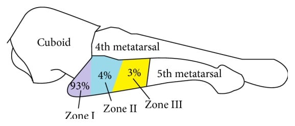
Where. Zone I tuberosity avulsions account for the overwhelming majority, about 93%, with zone II Jones fractures near 4% and zone III proximal diaphyseal stress fractures around 3%. The rarest zones are the ones that fail to unite, which is why the zone must be named before treatment is chosen.
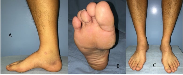
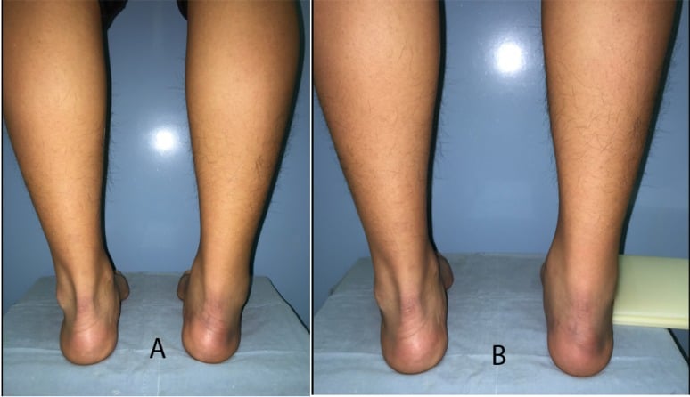
Foot morphology matters, but "pes cavus" is only half the story. The unifying mechanism is lateral column overload, and two quite different feet produce it. A cavovarus foot with a varus hindfoot and a tight gastrocnemius drives load laterally in the classic recreational pattern. But in O'Malley's series of elite basketball players, cited in the Evidence Base below, most players were pes planus, with a curved, prominently based fifth metatarsal and forefoot metatarsus adductus. Do not exclude the diagnosis, or the morphological risk, because the arch looks normal or flat.
Metatarsus adductus is the measurement that changes management, because it was the feature distinguishing those who refractured. Assess forefoot adduction, fourth-to-fifth intermetatarsal angle and fifth metatarsal lateral deviation in any athlete you are fixing, and consider prophylactic open bone grafting when they are high.
The eponym. Sir Robert Jones described the fracture in 1902 from his own injury, sustained while dancing; it was at the proximal diaphysis, not the tuberosity. The original Jones fracture was a Zone 2 fracture, and the term is often incorrectly applied to the Zone 1 tuberosity avulsion. Torg's classification of 1984 refined the understanding of healing potential.
Pathophysiology and Mechanisms
The bone. The fifth metatarsal is the most lateral of the five and articulates with the cuboid proximally. Its styloid process, the tuberosity, projects proximally and laterally and receives the insertion of peroneus brevis; peroneus tertius inserts on the dorsal shaft.
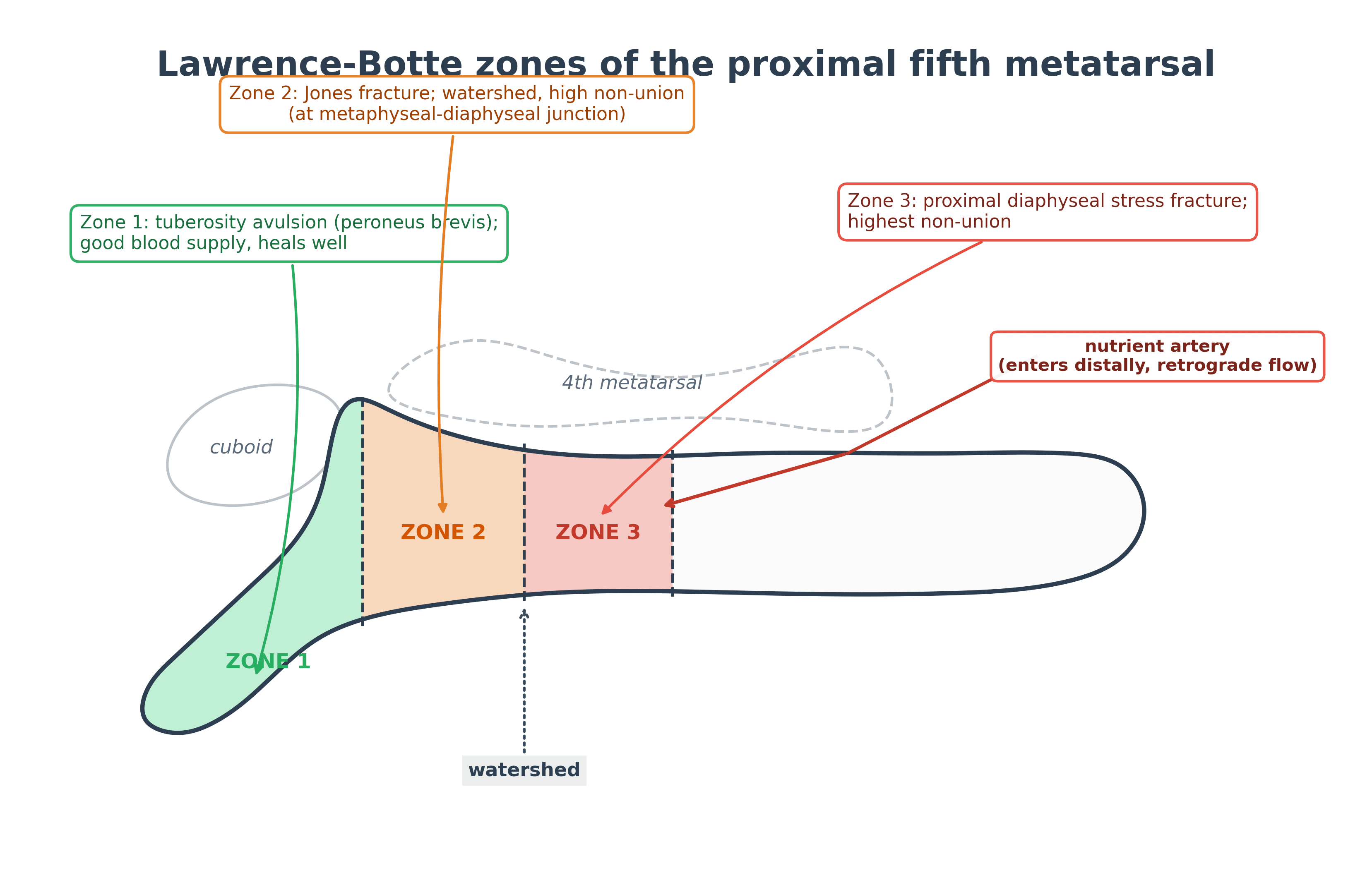
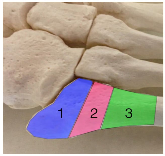
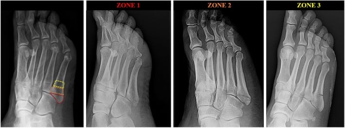
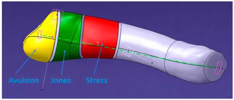
Zone 1, the tuberosity. The proximal tip of the styloid process, where peroneus brevis inserts. It is cancellous bone, well vascularised from metaphyseal vessels, and it heals readily. The mechanism is an inversion injury in which peroneus brevis avulses the fragment.
Zone 2, the metaphyseal-diaphyseal junction. The junction between metaphysis and diaphysis, extending into the fourth-fifth intermetatarsal articulation. It is cortical bone with a limited cancellous component and sits in the watershed between the metaphyseal and diaphyseal blood supplies, which is why its non-union risk is high.
Zone 3, the proximal diaphysis. The proximal 1.5cm of diaphysis distal to Zone 2, pure cortical bone, and the site of the highest mechanical stress concentration. A chronic stress reaction may precede the complete fracture, and the radiograph often shows medullary sclerosis. This is the zone with the highest non-union risk, and it often requires bone grafting.
The blood supply. The nutrient artery enters the distal third of the shaft and its blood flows retrograde, from distal to proximal. Separate metaphyseal vessels supply the proximal tuberosity, and the two territories meet at Zone 2, the watershed. The proximal diaphysis has only a limited periosteal supply to fall back on.
Why it matters. A fracture through the watershed has a compromised blood supply, so Zone 2 and 3 fractures have far higher non-union rates than Zone 1, and they fail to unite even when the reduction is adequate.
The nutrient artery enters distally and flows retrograde; Zone 2 is the watershed where its territory meets the metaphyseal vessels.
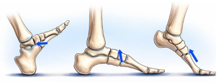
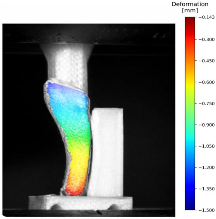
Where the load comes from. The fifth metatarsal takes a rising lateral bending moment through midstance into push-off, concentrated at the proximal metaphyseal-diaphyseal junction. Repetition of that bending, not a single event, produces the stress fracture. Under simulated gait loading the displacement concentrates at the proximal metaphyseal-diaphyseal region, the same watershed where the nutrient artery supply is poorest; mechanical concentration and vascular poverty coinciding is the reason Zone 2 and 3 fractures fail to unite.
Classification Systems
Lawrence and Botte's three zones are the shared language and the standard for the exam, and the most clinically useful of the systems: the zone is read on the oblique view, and getting it right is what predicts union. Torg grades the chronicity of a Zone 2 or 3 fracture, and Stewart subdivides Zone 1.
Lawrence and Botte Zone Classification (1993)
- Location
- Tuberosity
- Characteristic
- Avulsion fracture
- Treatment
- Conservative
- Prognosis
- Excellent
- Location
- Metaphyseal-diaphyseal junction
- Characteristic
- Jones fracture
- Treatment
- Surgical for athletes
- Prognosis
- Guarded
- Location
- Proximal diaphysis
- Characteristic
- Stress fracture
- Treatment
- Surgical
- Prognosis
- Poor if delayed
The dancer's fracture is not a Jones fracture. The Lawrence-Botte zones cover the proximal fifth metatarsal, but the commonest fifth metatarsal fracture overall is the dancer's fracture, a spiral or oblique fracture of the distal shaft or neck caused by twisting on a plantarflexed, externally rotated foot: classically rolling off a demi-pointe in dance, but also any twisting-plant sport. Unlike the proximal Jones fracture, the distal shaft is well vascularised, so the dancer's fracture heals reliably with non-operative functional treatment, a stiff-soled shoe or walking boot with weight-bearing as tolerated and early mobilisation, even when moderately displaced or angulated. Surgery is reserved for marked displacement or shortening, intra-articular extension into the MTP joint, or symptomatic non-union.
The exam contrast is the whole point. A proximal meta-diaphyseal fracture (Jones) is a watershed, high-non-union, often-operative lesion; a distal fifth metatarsal (dancer's) fracture is a benign, conservatively managed injury: location dictates prognosis.
History
Acute or insidious. The first question is whether this was a single event or an insidious onset, since a thorough history is what distinguishes the acute injury from the chronic stress reaction. Ask about the mechanism (inversion, direct blow, repetitive stress), previous foot injuries or fractures, training changes in mileage, intensity or surface, shoe wear and orthotics, and the sport and activity level.
Risk factors for stress fracture
- Rapid training increases
- Change in running surface
- Improper footwear
- Foot morphology loading the lateral column: cavovarus with a varus hindfoot, or equally metatarsus adductus with a curved, prominent-based fifth metatarsal
- Previous fifth metatarsal fracture
- Female athlete triad
- Vitamin D deficiency
Examination
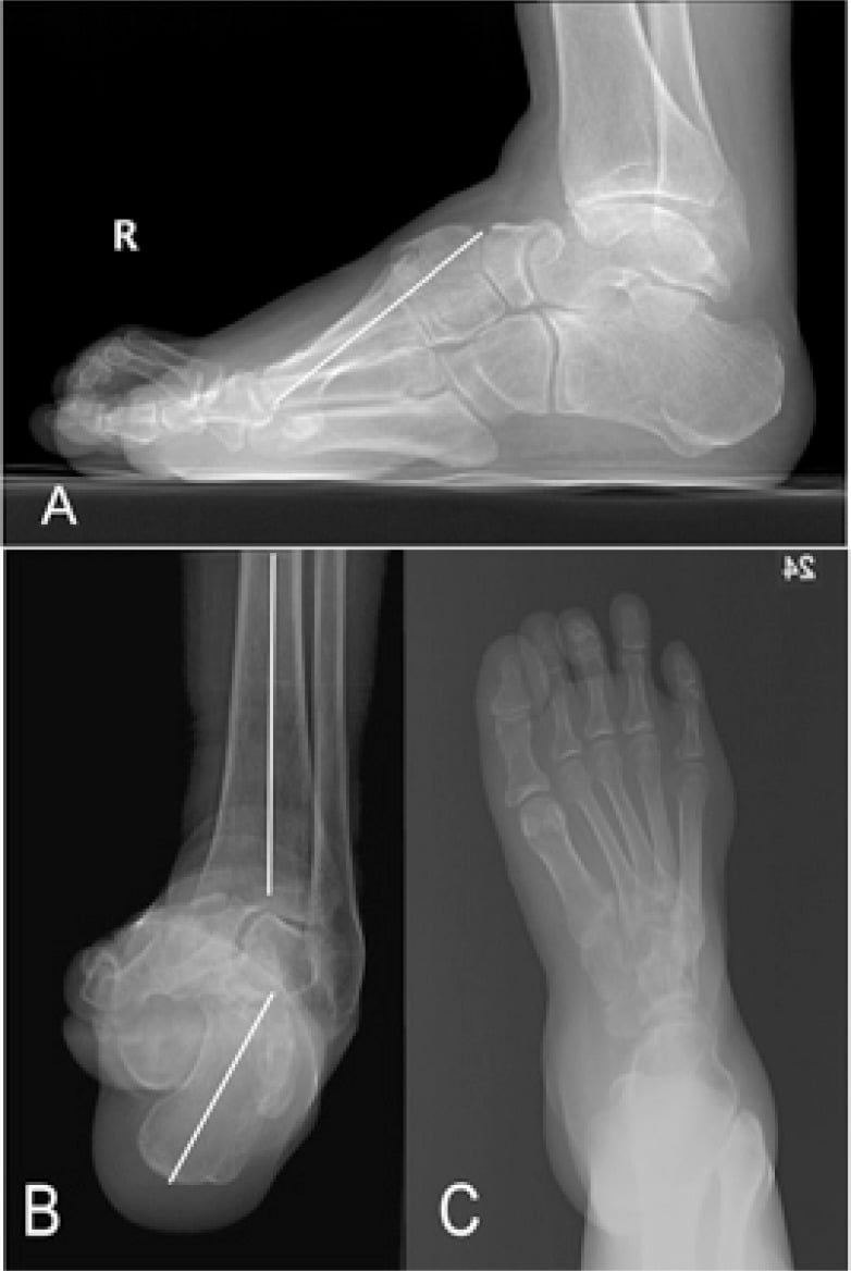
Inspection. Look for swelling over the lateral midfoot and for ecchymosis, then at the alignment of the foot (pes cavus, hindfoot varus) and the weight-bearing posture. The radiographic counterpart of the clinical cavovarus foot is a plantarflexed first ray, a high navicular and a depressed fifth metatarsal base sitting directly under load.
Palpation. Point tenderness over the base of the fifth metatarsal, localised as precisely as possible to Zone 1, 2 or 3 and compared with the other side. Precise localisation of tenderness helps determine the zone involved.
Special tests. Weight-bearing tolerance and the single-leg hop test screen for a stress fracture; gastrocnemius-soleus flexibility and ankle range of motion and stability complete the assessment.
Differential diagnosis. Lateral midfoot pain has a short list of mimics. The os vesalianum is an accessory ossicle at the base of the fifth metatarsal that can pass for a Zone 1 avulsion; it has smooth corticated margins, is bilateral in 90%, and sits separate from the tuberosity.
- Key feature
- Accessory ossicle at 5th MT base
- Imaging clue
- Smooth corticated margins, bilateral in most
- Distinguishes from fracture
- Rounded, separate from tuberosity (not a sharp lucency)
- Key feature
- Ossicle within peroneus longus tendon
- Imaging clue
- Lies near cuboid, not at MT base
- Distinguishes from fracture
- Located plantar/lateral to cuboid
- Key feature
- Traction apophysitis in adolescents
- Imaging clue
- Apophysis oriented parallel to shaft
- Distinguishes from fracture
- Open apophysis, age 9 to 14, no true fracture line
- Key feature
- Tenderness over cuboid
- Imaging clue
- Marrow oedema in cuboid on MRI
- Distinguishes from fracture
- Pain proximal and dorsal to MT base
- Key feature
- Tenderness along tendon
- Imaging clue
- Tendon thickening / split on MRI
- Distinguishes from fracture
- Bone is non-tender; pain reproduced on resisted eversion
- Key feature
- Midfoot instability
- Imaging clue
- Tarsometatarsal malalignment, fleck sign
- Distinguishes from fracture
- Tenderness more medial at TMT joints
Iselin disease, the adolescent's pseudo-avulsion. In the skeletally immature athlete, typically 9-14 years, the base of the fifth metatarsal has a secondary ossification centre, an apophysis, that appears around age 10 and fuses by the mid-teens. Repetitive traction from peroneus brevis causes a traction apophysitis presenting as activity-related lateral foot pain, tenderness and swelling over the tuberosity in a young runner, dancer or footballer.
The radiographic trap is that the normal apophysis is a flake-like ossicle oriented obliquely, roughly parallel to the shaft, and can be mistaken for a Zone 1 avulsion, whereas a true fracture line runs transverse to the shaft. The oblique orientation, smooth corticated margins and symmetry on the contralateral foot mark it as the apophysis; image the other side if unsure. Iselin disease is self-limiting, managed with rest from sport, activity modification and a stiff-soled shoe or boot, and heals with skeletal maturity; surgery is essentially never needed.
Investigations
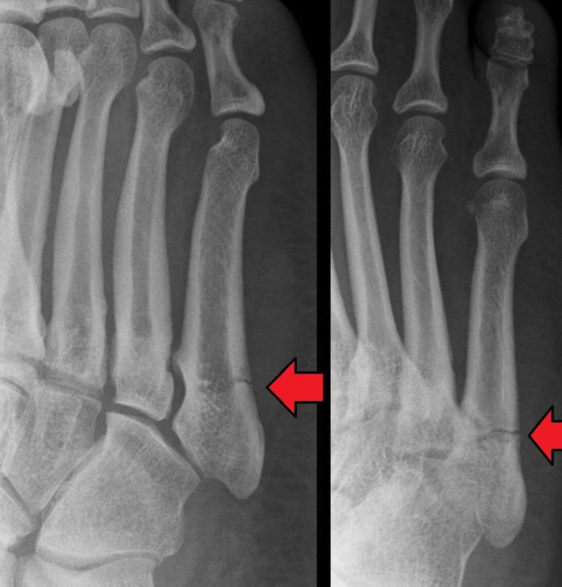
Radiographs. AP, lateral and oblique views of the foot are first-line for the acute injury. An acute fracture shows a visible fracture line; a stress reaction shows periosteal reaction and cortical thickening; a chronic fracture shows medullary sclerosis and a widened fracture line. Compare with the contralateral foot for subtle findings, and grade any Zone 2 or 3 fracture by the Torg signs of chronicity (Classification Systems).
MRI is the most sensitive test for the occult fracture and detects the early stress reaction, as bone marrow oedema, before there is any cortical break. It also assesses the soft tissues and any associated injury.
CT assesses fracture healing and the degree of medullary canal sclerosis, plans the screw size before surgery, and can detect an occult stress fracture.
Bone health. Consider a bone health work-up for any stress fracture; optimising bone health is essential for fracture healing and for preventing recurrence.
- Vitamin D (25-OH)
- Calcium
- TSH
- Female athlete triad screening
- DEXA if recurrent stress fractures
Management Algorithm
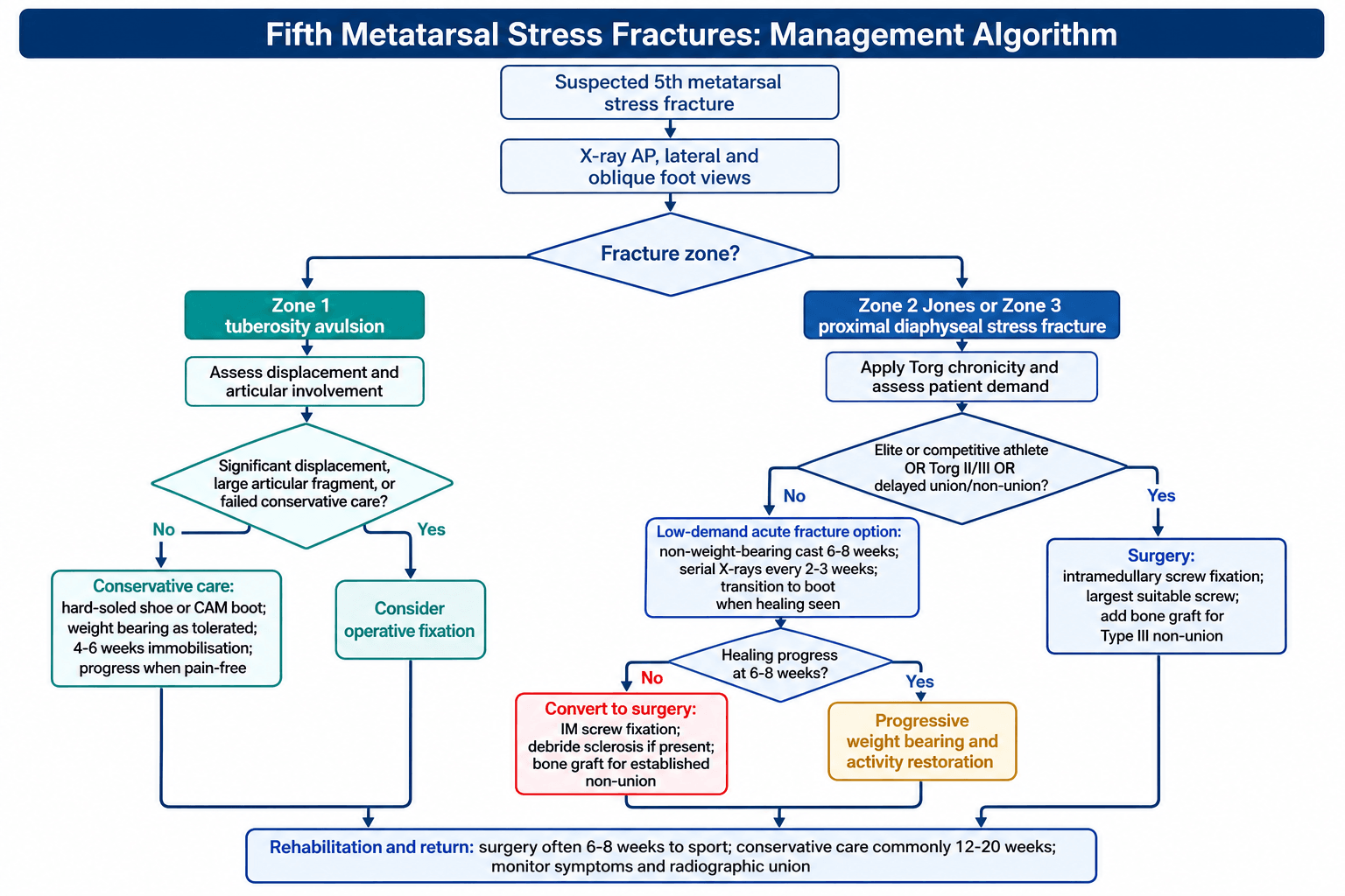
The decision. Three radiographs place the fracture in its zone, and the zone makes the first decision. A Zone 1 avulsion is treated conservatively for most patients, with the Stewart grade and displacement deciding the exceptions. A Zone 2 or 3 fracture is stratified by Torg chronicity and by patient demand: the competitive athlete and the Torg II or III fracture go to intramedullary screw fixation, while the low-demand acute fracture may be trialled in a non-weight-bearing cast and converted to surgery if it fails to progress.
- Location
- Tuberosity
- Mechanism
- Avulsion (inversion)
- Non-Union Risk
- Less than 5%
- Treatment
- Conservative - NWB cast 4-6 weeks
- Location
- Metaphyseal-diaphyseal junction
- Mechanism
- Acute stress/trauma
- Non-Union Risk
- 25-50%
- Treatment
- Surgery for athletes; conservative option for non-athletes
- Location
- Proximal diaphysis
- Mechanism
- Repetitive stress
- Non-Union Risk
- 30-50%
- Treatment
- Surgery strongly recommended - bone graft may be needed
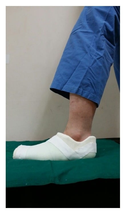
Zone 1. Conservative treatment is the standard, because the tuberosity has an excellent blood supply and these avulsions heal reliably. The non-displaced fracture, which is the common one, goes into a hard-soled shoe or short leg walking boot with weight-bearing as tolerated for 4-6 weeks, progressing to activity when pain-free. The displaced fracture is treated in a short leg non-weight-bearing cast for 4-6 weeks followed by a walking boot for 2-4 weeks.
Surgery for a Zone 1 fracture is rare. The indications:
- Displacement greater than 2mm
- Large fragment with significant articular involvement
- Failed conservative treatment
Zone 2 and 3 in the athlete. Surgery is recommended, because fixation offers faster and more reliable healing. The indications for fixation:
Absolute
- Competitive athletes with Zone 2/3 fractures
- Torg Type III non-unions
- Failed conservative management, including delayed union and non-union
Relative
- Torg Type II delayed unions
- High-demand recreational athletes
- Patient preference for faster return, or a need for expedited return to sport
Contraindications
- Active infection
- Severe peripheral vascular disease
- Medical comorbidities precluding surgery
A displaced or complete Zone 2/3 fracture, a recurrent fracture, and a widened fracture line signalling chronic stress reaction all push the same way. The operation is intramedullary screw fixation with the largest diameter screw that fits, 4.5-6.5mm, a headless compression screw preferred, with bone graft for a Type III non-union. Postoperatively the athlete is non-weight-bearing for 2-3 weeks, progresses through weight-bearing over weeks 3-6, and returns to sport at 6-8 weeks, against 12-20 weeks with conservative treatment; that difference is what drives the recommendation for surgery in athletes.
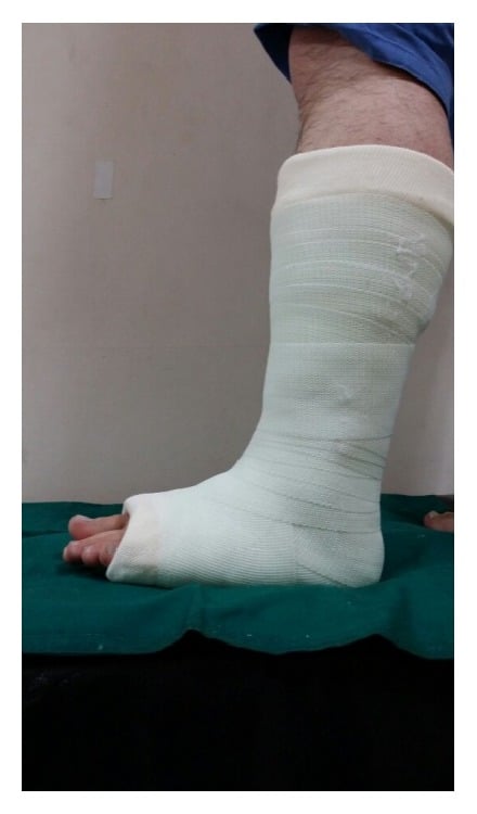
Zone 2 and 3 in the low-demand patient. Conservative management is an option, and it is a demanding one: it requires strict compliance and close monitoring. Partial loading through the cast is what converts a slow union into a non-union.
- Short leg non-weight-bearing cast for 6-8 weeks
- Serial radiographs every 2-3 weeks
- Transition to a walking boot when healing is evident, then progressive weight-bearing
Expect a healing rate of 70-75% for acute fractures, a time to union of 12-20 weeks, and a refracture risk of 15-25%. Convert to surgery when there is no healing progress at 6-8 weeks, when medullary sclerosis develops, or when symptoms worsen.
The established non-union is treated surgically, with debridement, bone grafting and screw fixation; the technique and its results are in the next section.
Screw size and the evidence. The instruction to use the largest screw that fits should be read against the trials. Porter's comparison of 5.5mm and 4.5mm cannulated screws could not show that the larger screw was more effective, so size superiority is unproven. In Glasgow and Torg's analysis of surgical failures, using other than a 4.5mm ASIF malleolar screw correlated with failure of screw fixation, and undersized grafts and incomplete medullary reaming with failure of bone grafting. The reconciliation is to fill the canal without over-reaming.
Surgical Technique
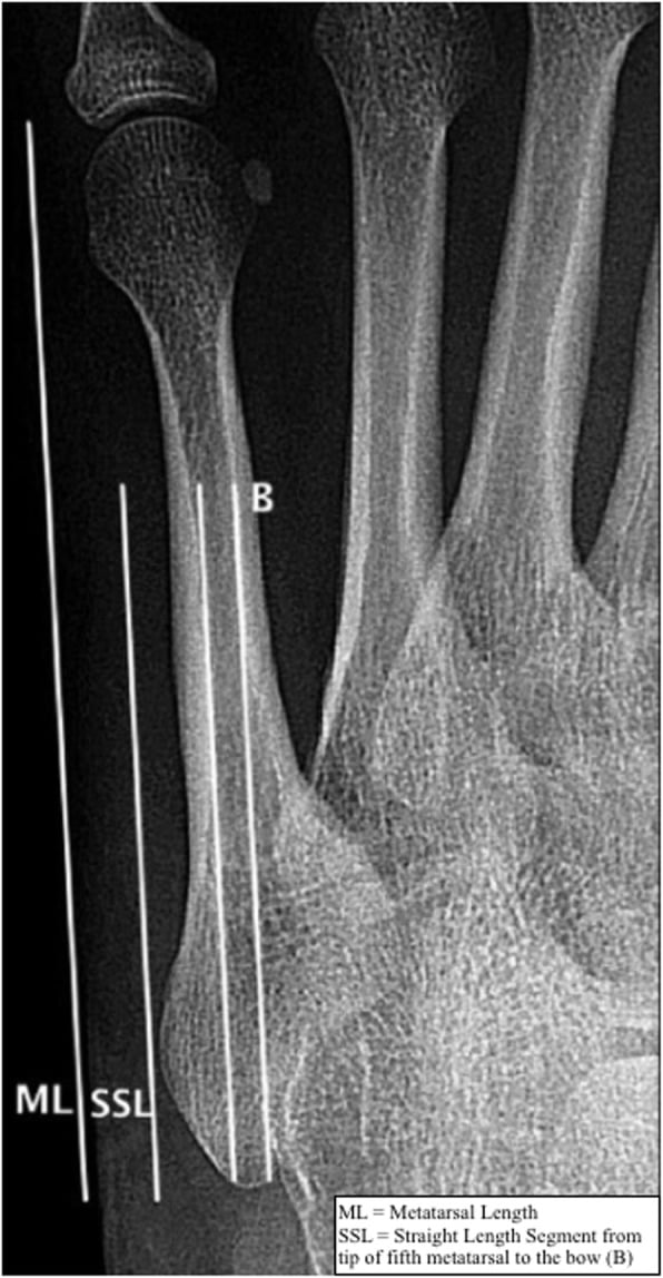
Planning the screw. Measure the medullary canal diameter at the bow before ordering an implant: the screw must fill the canal to control rotation, and the curvature of the bone limits how long a straight screw can be. An undersized screw is a recognised cause of refracture. Screw length must respect the lateral bow, so a shorter, wider screw is safer than a longer, narrower one; a screw that is too long creates a new stress riser, and a fresh stress fracture at the screw tip with reactive dorsal cortical thickening is the result.
Surgical Steps
Supine with bump under ipsilateral hip. Foot at end of table for fluoroscopy access. Tourniquet optional.
Longitudinal incision over lateral tuberosity. Protect sural nerve branches. Identify peroneus brevis tendon.
Start point at tip of tuberosity (high and lateral). Use 2.0mm K-wire for guide placement. Confirm position with fluoroscopy.
Drill and tap medullary canal. Insert largest screw that fits (4.5-6.5mm). Advance to distal metaphysis. Confirm compression.
Irrigate wound. Layered closure. Bulky dressing and posterior splint.
The entry point decides the operation. Start high and lateral, at the most proximal and lateral aspect of the tuberosity, and at the very base so the screw does not penetrate the lateral cortex at the bow. Use the largest diameter that fits, aiming for greater than 5mm, prefer a headless compression screw, make sure it crosses the fracture into the distal metaphysis, and confirm compression on the C-arm. Proper placement is what gives compression and stability.
Entry point too medial is the most common technical error, leading to varus angulation and poor screw purchase. Always start at the most proximal and lateral aspect of the tuberosity.
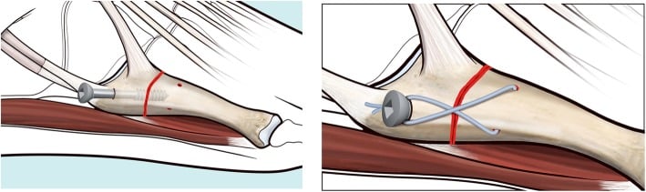
Pitfalls. An entry point too medial, an undersized screw with inadequate compression, sclerotic bone left undebrided in a non-union, and concomitant soft-tissue pathology that goes unrecognised.
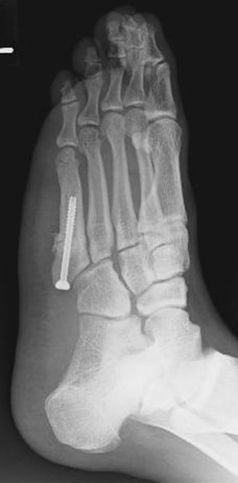
Complications
Non-union is the most common complication, and it is common with Zone 2 and 3 fractures. The risk rises with:
- Zone 2/3 location
- Conservative treatment of a high-risk fracture
- Torg Type II or III at presentation
- Non-compliance with weight-bearing restrictions
- Smoking
Treatment is the revision surgery described above, debridement and autograft with a larger diameter screw, and low-intensity pulsed ultrasound (LIPUS) can be considered. Return to sport is typically possible, though it may need extended rehabilitation.
Refracture. The rate is 15-25% with conservative treatment against less than 5% with surgery. The risk factors are premature return to sport, a previous non-union, undersized screw fixation and persistent biomechanical abnormalities; treatment is revision fixation with a larger screw, bone grafting and extended rehabilitation. Prevention:
- Confirm radiographic union before return to sport
- Gradual activity progression
- Address the underlying biomechanics with orthotics and flexibility work
- Consider screw removal with caution
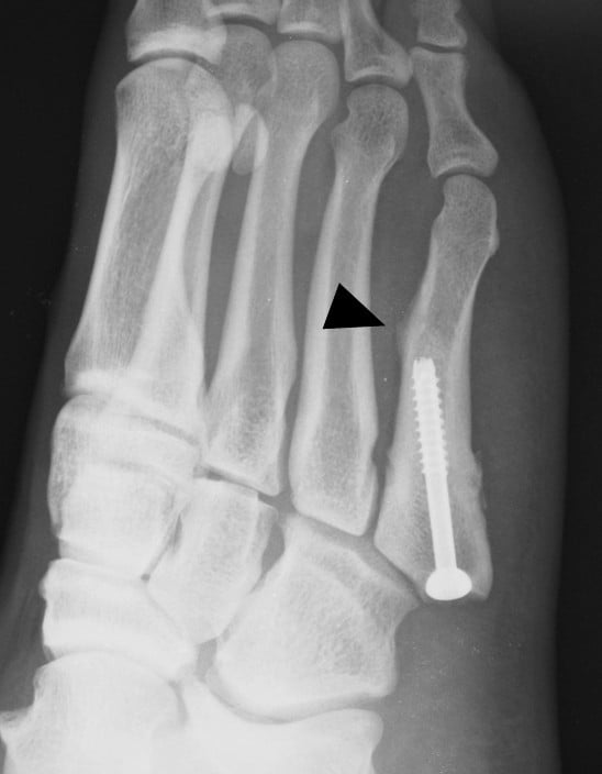
Hardware. Complications of the screw are uncommon with proper technique. Prominence may cause pain in shoes, and a symptomatic screw can be removed after union. Breakage is rare with proper sizing and may need removal and revision fixation. A loose screw usually indicates a non-union, and the non-union is what is treated.
Other. Wound infection in less than 1%, sural nerve injury from the approach (numbness of the lateral foot), peroneus brevis tendon injury, chronic pain and stiffness. Prevention is meticulous soft-tissue handling, protection of the sural nerve branches, a closure that is not over-tightened and early range-of-motion exercises; most complications are minor and resolve with conservative measures.
Postoperative Care
After intramedullary screw fixation, initial protection allows healing to begin and gradual progression prevents stress on the healing bone; the athlete typically returns to sport at 8-10 weeks.
- Posterior splint or CAM boot, non-weight-bearing
- Elevation and ice for swelling; pain management
- Wound care at 10-14 days
- Upper body and core exercises; ankle range of motion in the boot
- Transition to walking boot
- Progressive partial weight-bearing
- Pool exercises if wound healed; stationary cycling with the non-operative leg
- Full weight-bearing in the boot
- Radiograph to assess healing
- Closed-chain strengthening and gait training
- Regular footwear
- Progressive walking and light jogging; sport-specific conditioning
- Confirm radiographic healing
- Gradual return to sport: agility and cutting drills, full training with the team
- Competition when the functional goals are met
Return-to-sport criteria. Meeting all the criteria minimises refracture risk:
- Radiographic union: bridging callus visible on the radiograph
- Pain-free weight-bearing: no tenderness at the fracture site
- Full range of motion of the ankle and foot
- Strength restored: single-leg hop test equal to the opposite side
- Sport-specific function: passes agility and performance tests
Protect the return with an orthotic for pes cavus, correction of flexibility deficits, a gradual training progression, and watchfulness for symptoms of recurrence.
Outcomes and Prognosis
- Treatment
- Conservative
- Union Rate
- 95%+
- Time to Union
- 6-8 weeks
- Return to Sport
- 8-10 weeks
- Treatment
- Conservative
- Union Rate
- 70-75%
- Time to Union
- 12-20 weeks
- Return to Sport
- 15-25 weeks
- Treatment
- Surgical (screw)
- Union Rate
- 90-95%
- Time to Union
- 6-8 weeks
- Return to Sport
- 8-10 weeks
- Treatment
- Surgery + graft
- Union Rate
- 90-95%
- Time to Union
- 10-14 weeks
- Return to Sport
- 12-16 weeks
Reading the table against the headline figures. The headline comparison, 6-8 weeks with surgery against 12-20 weeks conservatively, is quoted on this page for healing and for return to sport alike. The table separates the two: time to union 6-8 weeks after fixation and 12-20 weeks in a cast, return to sport 8-10 weeks and 15-25 weeks respectively, which is the 8-10 weeks the rehabilitation protocol works to. The return-to-play figures in the Evidence Base after fixation are a median of 8.0 weeks (Mologne), 9.3 weeks (Porter) and 9.8 weeks (O'Malley).
What decides the outcome. The zone (Zone 1 best, Zone 3 worst), the chronicity at presentation (acute better than chronic), the treatment chosen (surgery better for athletes), compliance with rehabilitation, and the underlying biomechanics. In the long term the majority return to their pre-injury level of sport; recurrence is more common after conservative treatment, hardware symptoms may need screw removal, and addressing the biomechanics reduces recurrence.
Guidelines, Registries & Global Practice
Global epidemiology:
- Figure
- 61% to 78%
- Source
- Buskova/Rammelt, JBJS Rev 2021 (PMID 34673663)
- Figure
- Fifth (proximal)
- Source
- Cheung & Lui, Arch Trauma Res 2016 (PMID 28144601)
- Figure
- Zone 2/3 (meta-diaphyseal)
- Source
- Polzer, Injury 2012 (PMID 22465516)
There is no dedicated international registry for fifth metatarsal fractures (unlike arthroplasty registries such as AOANJRR or NJR). The evidence base is therefore built on a single Level I RCT (Mologne, PMID 15888715) plus systematic reviews and athlete case series, which drives the practice variation below.
Side-by-side guidance and consensus (no single global guideline exists):
- Position on Zone 1
- Functional treatment for all tuberosity avulsions
- Position on Zone 2/3 in athletes
- Early IM screw fixation for meta-diaphyseal fractures
- Evidence level
- Systematic review (Level II)
- Position on Zone 1
- Non-operative; functional
- Position on Zone 2/3 in athletes
- IM fixation to reduce healing time / expedite RTS
- Evidence level
- Review (Level III)
- Position on Zone 1
- Symptomatic, weight-bear as tolerated
- Position on Zone 2/3 in athletes
- IM screw fixation, often with biologic augmentation
- Evidence level
- Expert consensus + RCT
- Position on Zone 1
- Functional bracing, early mobilisation
- Position on Zone 2/3 in athletes
- Surgery for elite/high-demand; non-op acceptable in low-demand
- Evidence level
- Expert consensus
- North American sports practice favours early fixation plus bone marrow aspirate concentrate in elite athletes (O'Malley, PMID 26781131).
- European practice (Polzer) emphasises functional treatment for metaphyseal fractures and reserves fixation for true Zone 3 meta-diaphyseal fractures.
- Screw size remains debated: Porter (PMID 19176182) found no proven advantage of 5.5 mm over 4.5 mm, while failure analyses (Glasgow/Torg, PMID 8863022) link undersized fixation to failure.
- High-risk sports share cutting, pivoting and rapid-acceleration demands — including rugby codes, soccer, basketball and other field or court sports — with injury peaks during pre-season loading.
- In elite and professional settings, sports physicians and orthopaedic surgeons co-manage athletes; professional team medical units use structured early-detection and return-to-sport protocols and frequently proceed to in-season surgical fixation.
- Where MRI access is available it supports early stress-reaction detection; bone-health screening, training-load monitoring and orthotic assessment are used for prevention.
Fifth metatarsal stress fractures are common in sports with cutting, pivoting and rapid-acceleration demands — such as rugby codes, soccer and basketball. Professional team medical units use structured protocols for early detection and often proceed directly to surgical fixation for in-season fractures.
MCQ Practice Points
Q: Which nerve is most commonly injured during the approach for 5th metatarsal screw fixation? A: Dorsolateral Branch of the Sural Nerve. It runs along the lateral aspect of the foot and is at risk with lateral dissection or drill guide placement.
Q: Which arterial supply is deficient at the Zone 2/3 junction? A: The area is a watershed vascular zone between the intramedullary nutrient artery (diaphyseal) and the metaphyseal/periosteal vessels.
Q: What is the optimal starting point for an intramedullary screw in the 5th metatarsal? A: High and Inside (Dorsal and Medial) on the base. This aligns the screw with the straight distal segment of the canal.
Q: What defines a Torg Type II fracture? A: Widened fracture line with intramedullary sclerosis. Type I has no sclerosis. Type III has complete canal obliteration.
Q: How long does conservative treatment typically take for union in a Zone 3 stress fracture? A: 12-20 weeks (3-5 months). This prolonged time is why surgical fixation (6-8 weeks) is preferred for athletes.
Exam Viva Scenarios
Practise clinical reasoning and management decisions out loud
“A 25-year-old professional footballer presents with an acute Jones fracture (Zone 2) after a twisting injury. X-ray shows a sharp fracture line without sclerosis. He has an important match in 8 weeks. How would you manage this patient?”
“A 40-year-old recreational tennis player presents after an inversion injury with pain at the base of the fifth metatarsal. X-ray shows a 4mm displaced avulsion of the tuberosity (Zone 1). How would you manage this?”
“A 28-year-old amateur footballer was treated conservatively for a Jones fracture 4 months ago. He continues to have pain and X-ray shows a widened fracture line with medullary sclerosis. How would you manage this non-union?”
Zone Classification
- Zone 1: Tuberosity avulsion - excellent prognosis
- Zone 2: Jones fracture (metaphyseal-diaphyseal junction) - high non-union
- Zone 3: Proximal diaphyseal stress fracture - highest non-union risk
- True Jones = Zone 2, NOT Zone 1
Blood Supply
- Nutrient artery enters distally - retrograde flow
- Zone 2 is watershed zone
- Poor blood supply = high non-union rate
- Metaphyseal vessels supply tuberosity (Zone 1)
Torg Classification
- Type I (acute): Sharp margins, no sclerosis
- Type II (delayed): Widened line, some sclerosis
- Type III (non-union): Wide gap, complete sclerosis
- Sclerosis indicates need for surgery
Treatment by Zone
- Zone 1: Conservative - NWB cast 4-6 weeks
- Zone 2/3 athletes: Surgery - IM screw fixation
- Zone 2/3 non-athletes: Conservative option with monitoring
- Non-union: Surgery + bone graft
Surgical Technique
- Entry point: most proximal/lateral tuberosity
- Largest diameter screw (5mm or greater preferred)
- Headless compression screw
- Pitfall: entry too medial = varus angulation
Outcomes
- Zone 1 conservative: 95% union, 8-10 weeks RTS
- Zone 2/3 surgical: 90-95% union, 6-8 weeks RTS
- Zone 2/3 conservative: 70-75% union, 12-20 weeks RTS
- Surgery offers faster, more reliable healing
Evidence Base
Mologne RCT: Early Screw Fixation vs Casting for Acute Jones Fractures
- Cast group: 8 of 18 (44%) treatment failures (5 non-unions, 1 delayed union, 2 refractures)
- Screw group: 1 of 19 a treatment failure
- Median union and return to sport: 7.5 and 8.0 weeks (screw) vs 14.5 and 15.0 weeks (cast)
- Difference statistically significant for both endpoints (P less than 0.01)
Porter: 5.5 mm vs 4.5 mm Cannulated Screw for Jones Fracture
- 5.5 mm group: 96.7% average radiographic healing, 100% clinical healing
- Mean return to sport 9.3 weeks; three re-injuries managed in a boot
- No significant difference demonstrated between 5.5 mm and 4.5 mm screws
- Authors could not conclude a larger screw is more effective with the numbers available
Glasgow & Torg: Why Surgical Jones Fracture Treatment Fails
- Failures split between intramedullary screw fixation (6) and inlaid corticocancellous graft (5)
- For screw fixation, using other than a 4.5 mm ASIF malleolar screw correlated with failure
- For bone graft, undersized grafts and incomplete medullary reaming correlated with failure
- Early return to vigorous activity contributed to delayed union and refracture
Torg: Original Classification of Base of Fifth Metatarsal Fractures
- Three types defined: acute (narrow line, no sclerosis), delayed union, and non-union (medullary obliteration)
- Acute fractures in a non-weight-bearing toe-to-knee cast: 14 of 15 healed (mean 7 weeks)
- Acute fractures treated with weight-bearing methods: only 4 of 10 united
- Non-unions treated with medullary curettage and bone graft: 8 of 9 healed (mean 3 months)
Wright: Refracture After Intramedullary Screw Fixation in Athletes
- Athletes released to full activity at a mean of 8.5 weeks (range 5.5-12)
- Three footballers refractured within one day of returning to full activity
- Two footballers re-fixed with larger screws returned to play the same season
- Authors advise larger-diameter screws for high-body-mass athletes plus bracing or orthoses for return to play
O'Malley: Operative Treatment of Jones Fractures in the NBA
- Percutaneous fixation augmented with bone marrow aspirate concentrate; 3 had primary open bone grafting
- Average radiographic healing 7.5 weeks; return to play 9.8 weeks
- Three athletes refractured; the refracture group had the highest metatarsus adductus angles
- Most players were pes planus with a curved, prominent fifth metatarsal base
Buskova & Rammelt: Critical Analysis Review of Base of Fifth Metatarsal Fractures
- Proximal fifth metatarsal fractures account for 61% to 78% of all foot fractures
- Lawrence and Botte three-zone scheme is the most widely accepted classification
- Zone 1 avulsions generally heal with non-operative functional treatment
- Zone 3 diaphyseal stress fractures are preferentially treated operatively, especially with delayed union
Polzer: Systematic Review and Refined Treatment Recommendations
- All Zone 1 tuberosity avulsions healed well with functional treatment, even when displaced or intra-articular
- A short leg cast significantly delayed return to pre-injury level versus functional treatment
- Zone 3 meta-diaphyseal fractures had significantly higher non-operative failure than Zone 1/2
- Early intramedullary screw fixation shortened time to union and return to sport