Loss of lumbar lordosis causing forward trunk lean with inability to stand upright
- Flatback = loss of lumbar lordosis causing sagittal imbalance with forward trunk lean
- SVA (sagittal vertical axis): distance from C7 plumb line to S1; normal under 5cm
- PI-LL mismatch: difference between pelvic incidence and lumbar lordosis; normal under 10 degrees
- Osteotomies by correction power: SPO (posterior column, 10 degrees), PSO (three-column, 30 degrees), VCR (40 degrees or more)
- Iatrogenic most common cause: distraction instrumentation (Harrington rods)
- “Patients cannot stand upright without compensation (knee flexion, hip extension)
- “ODI and SF-36 correlate with SVA - most important predictor of disability
- “Always assess pelvic compensation (pelvic tilt, pelvic incidence-lumbar lordosis)
- “Rod fracture risk high if SVA not corrected adequately
Flatback Syndrome
Overview and Epidemiology
Flatback syndrome is loss of the normal lumbar lordosis, producing positive sagittal balance: the C7 plumb line falls anterior to the posterosuperior corner of S1 and the patient cannot hold an upright posture without significant compensation. A normal spine carries the head centred over the pelvis with minimal energy expenditure. In flatback the head sits anterior to the pelvis and the posture is held by constant muscular work.
Who gets it. Flatback followed 20-40% of Harrington rod instrumentations; after modern spinal fusion the prevalence is 5-10%. Iatrogenic cases peak at 50-70 years, degenerative cases at a variable age, and women outnumber men about 2:1 because more of them have had fusions for scoliosis.
Risk factors. Four groups:
- Iatrogenic: Harrington rods, long fusion to the sacrum, flat rod contouring
- Degenerative: disc collapse, compression fractures, ankylosing spondylitis
- Post-surgical: pseudarthrosis, junctional kyphosis, hardware failure
- Patient factors: osteoporosis, smoking, obesity, prior laminectomy
The Harrington era. From the 1960s to the 1990s scoliosis was corrected with distraction instrumentation, which straightened the spine in the coronal plane and eliminated lumbar lordosis in the sagittal plane. The result was an epidemic of flatback syndrome. Modern instrumentation preserves lordosis with contoured rods and pedicle screw fixation.
Pathophysiology
Normal sagittal alignment. The spine balances through reciprocal curves: cervical lordosis 20-40 degrees, thoracic kyphosis 20-40 degrees and lumbar lordosis 40-60 degrees from L1 to S1. When the C7 plumb line falls within 2-5cm of the posterosuperior corner of S1, standing needs minimal muscular effort. That is the cone of economy: the small region in which the trunk can be held with little muscular work, and the region a flat lumbar segment pushes the patient out of.
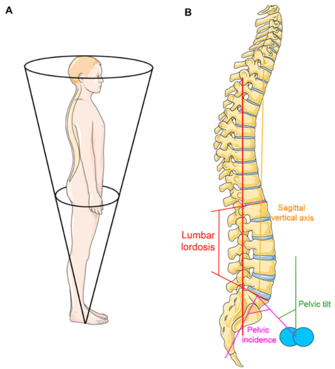
Pelvic parameters. Three angles describe the pelvis, and only one of them is fixed:
- Definition
- Angle between the perpendicular to the S1 endplate at its midpoint and the line to the hip axis
- Normal
- 35-80 degrees
- Behaviour
- Fixed anatomical parameter; cannot change
- Definition
- Angle between the vertical and the line from the S1 midpoint to the hip axis
- Normal
- 10-25 degrees
- Behaviour
- Variable; increases with compensation
- Definition
- Angle between the S1 endplate and the horizontal
- Normal
- 30-50 degrees
- Behaviour
- Variable; decreases in flatback
PI = PT + SS, always, which is a check on the measurement. Lumbar lordosis should approximately equal pelvic incidence, with a mismatch (PI-LL) under 10 degrees. PI cannot change, so once lordosis falls below it the pelvis is the first thing to move.
How imbalance develops. The sequence is the same whatever the cause.
- Stage 1, loss of lordosis. Flat rod contouring or distraction forces in the iatrogenic case, disc collapse or compression fractures in the degenerative one. Lumbar lordosis falls below PI.
- Stage 2, pelvic compensation. The pelvis rotates posteriorly: PT rises and SS falls, bringing the C7 plumb line back over the sacrum at a moderate increase in energy cost.
- Stage 3, hip and knee compensation. Once the pelvis has nothing more to give, the hips extend (limited by the hip flexors) and the knees flex to shift the centre of mass posteriorly. The energy cost is now severe and fatigue comes early.
- Stage 4, decompensation. Every mechanism is exhausted. The trunk lean becomes fixed, the patient cannot stand upright without support, and disability is severe.
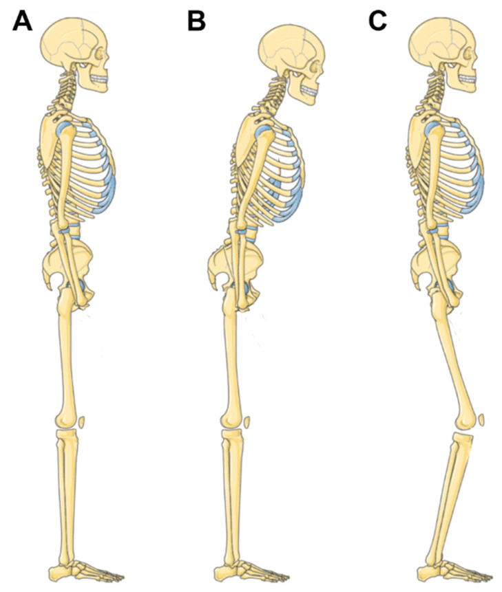
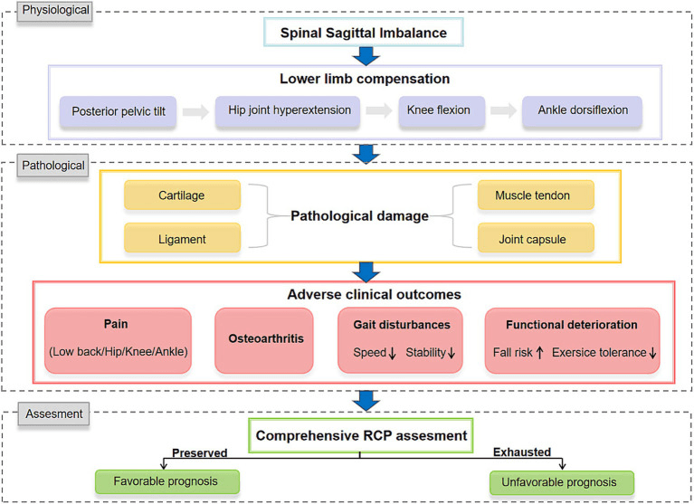
Compensation exhaustion. A pelvic tilt over 25 degrees means pelvic compensation is used up. These patients have severe disability and limited reserve for further degeneration; surgical correction should restore balance to stop the progression.
Iatrogenic flatback, the most common cause. The Harrington construct combined distraction without lordosis preservation, long fusions often to L5 or the sacrum, and flat rods with no sagittal profile, and it removed the lumbar lordosis entirely. Modern instrumentation can produce the same deformity:
- Undercorrection of lordosis in long fusions
- Flat rod placement in degenerative fusions
- Pseudarthrosis with rod fracture and kyphosis
- Adjacent segment degeneration above the fusion
Prevention. Contour the rods to restore a lordosis matched to the pelvic incidence, control it with pedicle screw fixation, use interbody cages with lordotic angles of 20-30 degrees, and avoid long fusions unless they are necessary.
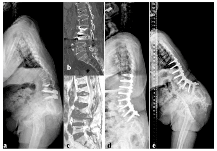
Degenerative flatback. Lordosis is lost progressively over time. Multiple-level disc collapse loses height anteriorly and with it the disc lordosis, and facet arthropathy prevents extension. Osteoporotic compression fractures, thoracolumbar Scheuermann kyphosis and the fixed kyphosis of ankylosing spondylitis do the same from the vertebrae. After extensive laminectomy the facets become incompetent and progressive kyphosis follows, especially at the thoracolumbar junction.
Post-traumatic flatback. Alignment changes through bony collapse or malunion: burst fractures with anterior column collapse, compression fractures at several levels, or a fracture-dislocation that has malunited. Pelvic injuries act through the pelvic parameters, a sacral fracture changing the sacral slope and an acetabular fracture altering hip mechanics.
Lordosis distribution matters as much as its magnitude. The Roussouly classification describes four normal sagittal lumbar morphotypes defined by the sacral slope, each with a characteristic lordosis magnitude, apex and number of vertebrae in the curve:
- Type 1 - low sacral slope (under approximately 35 degrees): a short, sharply-angled lower-lumbar hyperlordosis with a low apex (near L5) and a thoracolumbar kyphosis above. Low pelvic incidence.
- Type 2 - low sacral slope but a flat, low-magnitude lordosis ("flat back" morphotype). Low pelvic incidence. This shape is the most prone to degenerative disc disease and to degenerative flatback - the spine has little lordotic reserve to lose.
- Type 3 - average sacral slope (approximately 35 to 45 degrees): a well-balanced, harmonious lordosis with the apex at L4. The most common and most forgiving morphotype.
- Type 4 - high sacral slope (over approximately 45 degrees) and high pelvic incidence: a long, curved hyperlordosis with a high apex (L3 or above) and many vertebrae recruited into the curve.
Why it matters for flatback correction: restoring the correct total lumbar lordosis is not enough - the lordosis must be distributed correctly (the right apex and the right proportion in the lower arc, where roughly two-thirds of lordosis normally sits below L4). A correction that gives an adequate number of degrees but the wrong shape (for example a uniform, low-apex lordosis in a patient whose pelvic incidence demands a Type 3 or 4 distribution) leaves the construct mismatched to the patient's ideal Roussouly type and is a recognised driver of proximal junctional kyphosis and mechanical failure. Plan to recreate the patient's predicted ideal morphotype, not simply a target number.
Clinical Presentation
The cardinal symptom is forward trunk inclination. Patients say they cannot stand upright, that they are "looking at the ground" when they walk, that they need to lean on a shopping trolley or walker, and that standing or walking exhausts them. Standing tolerance is often under 15-30 minutes and walking distance is limited by fatigue, not neurogenic claudication. The social cost is real: they cannot make eye contact, and cannot stand long enough to cook, shop or socialise.
Pain. It follows the compensation, so it is felt where the work is being done:
- Mechanism
- Constant isometric contraction
- Character
- Burning, aching, fatigue
- Mechanism
- Hip extension compensation
- Character
- Cramping, tightness
- Mechanism
- Flexion compensation
- Character
- Aching, early arthritis
- Mechanism
- Gluteal fatigue
- Character
- Weakness, pain on standing
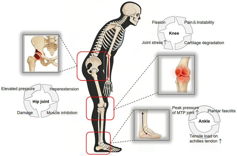
Inspection. The trunk leans forward and the patient cannot stand upright without support. The knees are flexed in compensation, hip extension is limited by tight hip flexors, and the gait is shuffling, antalgic and requires an assistive device.
Palpation and movement. The paraspinal muscles are in spasm and tender, the lumbar lordotic curve is gone, there is a step-off if a spondylolisthesis is present, and hardware is palpable after prior surgery. Lumbar extension is severely limited or absent while flexion is often preserved, and hip extension is limited, with a positive Thomas test.
Neurological examination. Usually normal: this is not a neurological condition. Look for radiculopathy if there is foraminal stenosis, and for myelopathy if the cervical spine is compensating.
- Technique
- Patient flexes forward, attempts to stand upright
- Positive Finding
- Cannot achieve upright position without knee flexion
- Technique
- Patient stands with back to wall, attempts to touch wall with back of head
- Positive Finding
- Head far anterior to wall (over 15cm abnormal)
- Technique
- Patient bends forward from waist
- Positive Finding
- Assess coronal deformity if scoliosis present
Quality of life. The Oswestry Disability Index correlates strongly with SVA, SF-36 physical function is severely impaired, and depression and anxiety are common with the chronic pain and disability.
Investigations
The standing full-length lateral radiograph. Everything in this topic is measured on one film, and the film is only valid if it is taken properly:
- True standing position, no leaning, no support
- Arms positioned with the fists on the clavicles or grasping horizontal bars, to clear the thoracic spine
- Field from the skull to the femoral heads
- Cassette at least 36 inches long
Radiographs must be taken standing. Supine films cannot assess sagittal balance and underestimate the deformity.
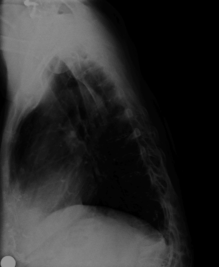
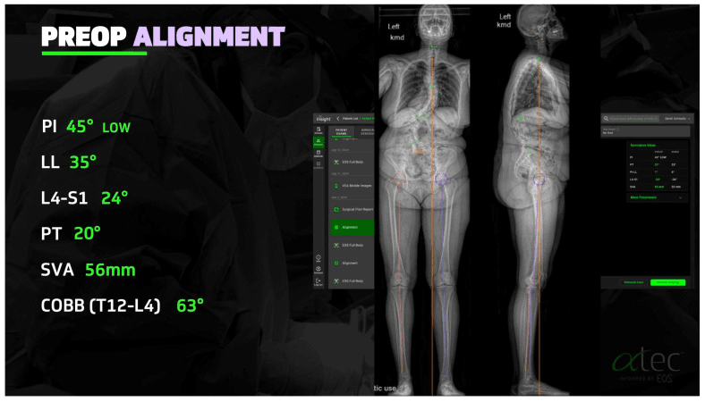
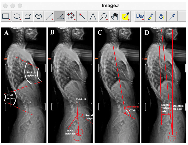
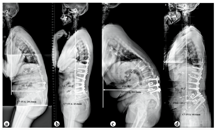
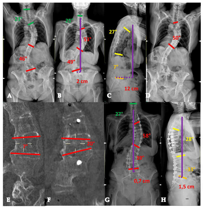
Sagittal vertical axis. Drop a plumb line from the centre of the C7 vertebral body and measure its horizontal distance from the posterosuperior corner of S1. A positive value means C7 sits anterior to S1, the flatback direction. Under 5cm is normal and goes with minimal disability; 5-10cm is abnormal, with moderate disability; over 10cm is severe.
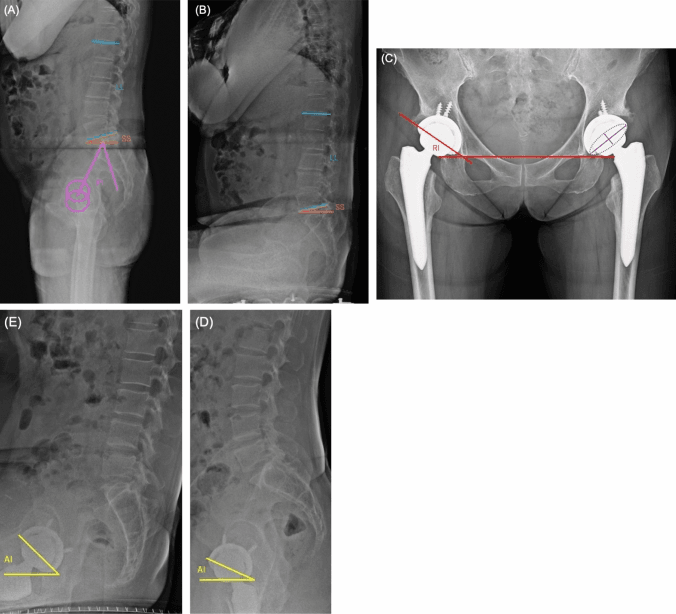
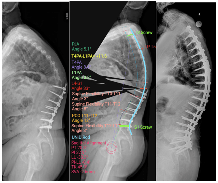
The pelvis and the lordosis on the flatback film. The parameters are defined in Pathophysiology; on the film of an established flatback PT has risen above 25 degrees as compensation is spent, SS has fallen below 30 degrees, and lumbar lordosis, measured as the Cobb angle from the superior endplate of L1 to the superior endplate of S1, is under 30 degrees. Subtract that lordosis from the pelvic incidence: a PI-LL under 10 degrees is normal, over 10 degrees is symptomatic, and over 20 degrees is severe.
PI-LL is the key surgical parameter. The goal of surgery is to restore PI-LL to under 10 degrees. If PI is 60 degrees, the target lumbar lordosis is 50-70 degrees.
CT is the planning study: bony anatomy and pedicle size, the prior fusion mass, the hardware (loosening, fracture) and the choice of osteotomy level. MRI answers the neurological and soft-tissue questions: neural compression from stenosis or foraminal narrowing, disc degeneration and how much disc health remains, fluid signal at a fusion site suggesting pseudarthrosis, and cord integrity if myelopathy is suspected. Flexion-extension radiographs show whether the adjacent segments move, pick up motion through a pseudarthrosis, and tell you which segments are mobile and which are fused when the osteotomy is being planned.
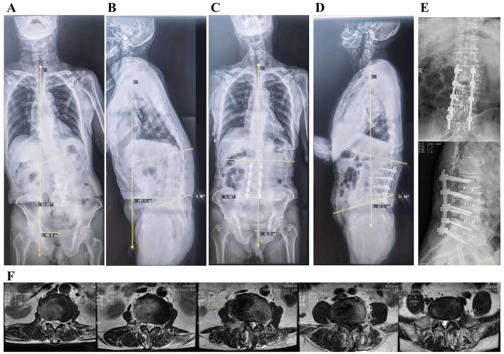
Ames-ISSG classification. The most used grading of flatback deformity, built from SVA and PT, and it maps onto management:
- SVA
- Under 5cm
- PT
- Under 25 degrees
- Interpretation
- Compensated, minimal symptoms
- Management
- Conservative
- SVA
- 5-10cm
- PT
- Under 25 degrees
- Interpretation
- Mild imbalance
- Management
- Consider surgery if symptomatic
- SVA
- Over 10cm
- PT
- Under 25 degrees
- Interpretation
- Severe imbalance, compensation failing
- Management
- Surgery usually indicated
- SVA
- Any
- PT
- Over 25 degrees
- Interpretation
- Severe, pelvic compensation exhausted
- Management
- Surgery usually indicated
SVA is the headline parameter, but two newer measures overcome its weaknesses.
T1 Pelvic Angle (TPA) is the angle subtended at the femoral head axis between a line to the centroid of the T1 vertebral body and a line to the centre of the S1 endplate. It effectively combines trunk inclination and pelvic tilt into one number. Its key advantage over SVA is that it is position-independent: SVA can be falsely "normalised" by a patient recruiting compensation (pelvic retroversion, knee flexion), so a measured SVA may underestimate the true deformity, whereas TPA captures both the trunk lean and the pelvic compensation and is not erased by that recruitment. A TPA of over approximately 20 degrees indicates severe deformity; the realignment target is roughly under 14 degrees.
The GAP score (Global Alignment and Proportion) is a proportional, pelvic-incidence-based score rather than an absolute threshold. It combines relative pelvic version, relative lumbar lordosis, the lordosis distribution index (echoing the Roussouly principle that the apex and lower-arc proportion matter), relative spinopelvic alignment, and an age factor, and grades the reconstruction as proportioned, moderately disproportioned, or severely disproportioned. Its value is predictive: a disproportioned construct carries a substantially higher rate of mechanical complications (rod fracture, proximal junctional kyphosis, pseudarthrosis), so aiming for a proportioned GAP result - not merely an SVA under 5cm - is the modern planning goal. Both measures reinforce the same lesson as age-adjusted targets: alignment must be individualised to the patient's pelvic incidence and age, not forced to a single universal number.
Management Algorithm
What conservative treatment can do. Its efficacy in established flatback is limited: it may relieve symptoms, and it does not correct the deformity or restore lost lordosis. It is still the first step.
Physiotherapy. The goals are to strengthen the core, lengthen the hip flexors and keep whatever lumbar motion remains:
- Examples
- Planks, bridges, dead bugs
- Benefit
- Paraspinal endurance
- Examples
- Thomas stretch, kneeling lunge
- Benefit
- Reduces compensatory hip extension
- Examples
- Seated hamstring stretch
- Benefit
- Allows pelvic rotation
- Examples
- Wall slides, chin tucks
- Benefit
- Awareness and positioning
Medication. Nothing here treats the mechanics:
- Examples
- Ibuprofen, naproxen
- Use
- Muscle inflammation, pain
- Limitation
- GI side effects, limited efficacy
- Examples
- Cyclobenzaprine
- Use
- Paraspinal spasm
- Limitation
- Sedation; not for long-term use
- Examples
- Gabapentin, pregabalin
- Use
- If there is a radicular component
- Limitation
- Minimal benefit for mechanical pain
Injections. An epidural steroid injection is for radiculopathy from foraminal stenosis and gives temporary relief, weeks to months; it does nothing for the mechanical back pain of imbalance. Facet injections are for facet-mediated pain, with variable and temporary effect, and are not a treatment for sagittal imbalance.
Assistive devices. A walker with forearm supports unloads the spine and permits the forward lean, at the cost of dependence and social stigma. A cane gives minimal support and is insufficient for severe flatback. A corset or brace may reduce pain and does not correct the deformity.
Natural history if untreated. SVA and PT worsen progressively, pain and disability increase, the segment above a prior fusion degenerates, and the compensating hips and knees develop arthritis.
Indications for surgery. Any of:
- Failed conservative management after a 3-6 month trial
- Severe disability, ODI over 40
- SVA over 5cm with symptoms
- PI-LL mismatch over 10 degrees with symptoms
- Progressive deformity
Surgical Management
Goals. Radiographically the operation aims for an SVA under 5cm, ideally 0-3cm, a PI-LL mismatch under 10 degrees, a lumbar lordosis restored to match the pelvic incidence, and coronal balance maintained. The SRS-Schwab targets quoted in the guidelines section are tighter, SVA under 4cm and PT under 20-25 degrees, and age-adjusted targets deliberately leave older patients in mild positive balance. Clinically the aims are an upright posture without compensation, less pain and disability, better walking tolerance and daily function, and the fewest complications.
Choosing the osteotomy. Three operations, in ascending order of power and risk. The choice turns on how much correction is needed and whether the anterior column is mobile or fused.
What it is. A posterior column osteotomy (Schwab grade I). The inferior and superior facets and the ligamentum flavum are removed to leave a V-shaped gap in the posterior elements, and closing the gap creates lordosis. It needs a mobile anterior column, a disc or a pseudarthrosis, to hinge through; forcing closure against a stiff anterior column risks an anterior column fracture.
Correction. Approximately 10 degrees per level, cumulative across multiple levels.
Indications.
- Mild sagittal imbalance, SVA under 10cm
- PI-LL mismatch under 20 degrees
- Mobile anterior column present
- Multiple-level correction planned
For and against. It has the lowest complication rate of the three, preserves the vertebral body and can be repeated at several levels. Against it: the correction per level is limited, it requires a mobile disc, and inadequate correction is common if it is used alone.
Complications. Neurological injury 2-5%; vertebral body fracture 5-10%.
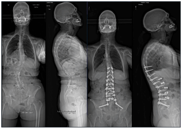
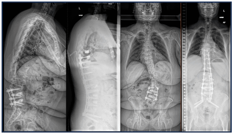
Planning the correction. The radiographic parameters set the size of the operation, and the patient's bone sets its limits:
- Finding
- 5-10cm
- Surgical plan
- Single-level PSO may suffice
- Finding
- Over 10cm
- Surgical plan
- May need a two-level PSO or PSO plus SPO
- Finding
- 20-30 degrees
- Surgical plan
- Single-level PSO (30 degrees of correction)
- Finding
- Over 40 degrees
- Surgical plan
- Consider VCR or a two-level PSO
- Finding
- Solid fusion mass
- Surgical plan
- Osteotomy through the fusion or adjacent to it
- Finding
- DEXA T-score under -2.5
- Surgical plan
- Optimise bone health; consider cement augmentation
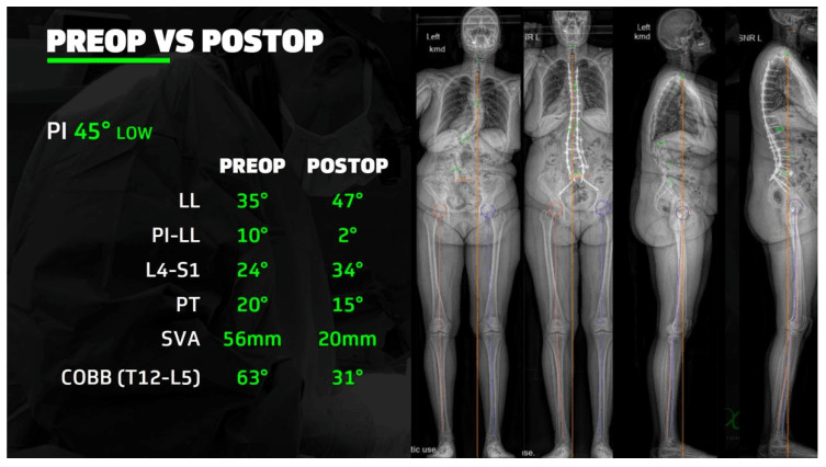
Choosing the level. L3 is the most common PSO level: a large body, safer than L4-5, and the best balance of safety and correction:
- Advantages
- Large vertebral body, high lordosis potential
- Disadvantages
- High in the lumbar spine; junctional stress
- Advantages
- Most common PSO level; large body; safer
- Disadvantages
- Moderate lordosis contribution
- Advantages
- Good lordosis; mid-lumbar
- Disadvantages
- Smaller body; L5 root at risk
- Advantages
- Maximum lordosis potential
- Disadvantages
- Small body; sacral roots at risk; not recommended
The distal foundation. Osteotomy level and correction magnitude get the attention, but the construct fails at its ends, and the distal end of a long lumbar reconstruction is the harder problem. A long fusion stopping at S1 alone loads two S1 screws with the entire cantilever of a corrected lumbar spine, and the predictable results are S1 screw loosening or pull-out and lumbosacral pseudarthrosis. Pelvic fixation exists to protect the S1 screws and the lumbosacral fusion by moving the distal anchor beyond the sacrum, and S2-alar-iliac screws are generally preferred over classic iliac screws because their medial, in-line starting point reduces implant prominence and avoids a separate offset connector.
- When it is adequate
- Short constructs with a healthy, non-degenerate L5/S1 disc
- Problem
- Leaves a mobile segment under a stiff construct; L5/S1 commonly degenerates and re-imbalances
- When it is adequate
- Rarely adequate for a long deformity fusion
- Problem
- S1 screws take the full lever arm; loosening and lumbosacral pseudarthrosis
- When it is adequate
- Standard for long fusions to the sacrum, and effectively mandatory alongside a lumbar PSO
- Problem
- Adds operative time; iliac screws need a connector and can be prominent, which S2AI largely solves by starting more medially and in line with the S1 screws
Pedicle subtraction osteotomy, step by step. The patient is prone on a Jackson table or radiolucent frame with the hips extended to allow lordosis, the arms abducted to 90 degrees and the neuromonitoring leads placed. A single-level PSO typically takes 4-6 hours.
- Exposure. Midline incision and subperiosteal dissection
- Screws. Pedicle screws 3-4 levels above and below the osteotomy
- Temporary rods. Placed to hold the spine stable while the osteotomy is made
- Laminectomy. Complete removal of the lamina and spinous process at the osteotomy level
- Facetectomy. Inferior and superior facets bilaterally
- Pedicle resection. Both pedicles, identifying the nerve roots
- Posterior body resection. The posterior third to half of the vertebral body, as a 30-40 degree wedge, tapered laterally so the cavity is egg-shaped
- Nerve root decompression. Mobilise the roots and protect them through closure
- Closure. Remove the temporary rods, place the final rods pre-contoured with lordosis, and close the osteotomy by cantilever slowly, 1-2mm at a time, checking the neuromonitoring after each increment
- Compress the instrumentation, decorticate the fusion bed and apply bone graft and BMP
- Haemostasis and closure. Large drains, subfascial, with layered closure over them
Complications
Neurological injury. The rate of a new or worsening deficit after PSO is 10-30%, and most recover: 50% have resolved by 1 year and 5-10% are permanent, with motor deficit the most concerning. Prevention is slow closure under continuous neuromonitoring, adequate decompression and no overstretching of the neural elements.
- Incidence
- 5-10%
- Mechanism
- Direct injury, ischaemia, haematoma
- Management
- Stop closure, release the compression, image
- Incidence
- 15-25%
- Mechanism
- Nerve root traction
- Management
- Often transient; monitor
- Incidence
- 1-2%
- Mechanism
- Severe canal compromise
- Management
- Immediate decompression
Vascular injury. Prevention is careful anterior dissection, staying midline and palpating the aorta.
- Incidence
- 0.5-1%
- Risk factors
- PSO at L4-5; osteoporotic collapse
- Management
- Vascular surgery consult; open repair
- Incidence
- 0.5-1%
- Risk factors
- Anterior perforation
- Management
- Direct repair; difficult access
- Incidence
- 5-10%
- Risk factors
- Lateral dissection
- Management
- Usually self-limiting; electrocautery
Haemorrhage. Average blood loss is 1-3 litres for a PSO and 3-5 litres for a VCR, and 70-90% of patients are transfused. Consider cell salvage and antifibrinolytics (tranexamic acid).
The first six weeks. Epidural haematoma occurs in 2-5% and may need evacuation; nerve root injury in 5-10%, usually transient. Wound problems are superficial infection 5-10%, deep infection 3-5%, seroma or haematoma 5-10% and CSF leak 3-5%; prophylactic antibiotics, meticulous haemostasis and vancomycin powder are the defence against infection. The medical complications have their own prevention:
- Incidence
- 5-10%
- Risk factors
- Long surgery, immobility
- Prevention
- Chemoprophylaxis, sequential compression devices
- Incidence
- 5-8%
- Risk factors
- Prolonged intubation, pain
- Prevention
- Early mobilisation, incentive spirometry
- Incidence
- 10-20%
- Risk factors
- Bowel retraction, opioids
- Prevention
- Early feeding, bowel regimen
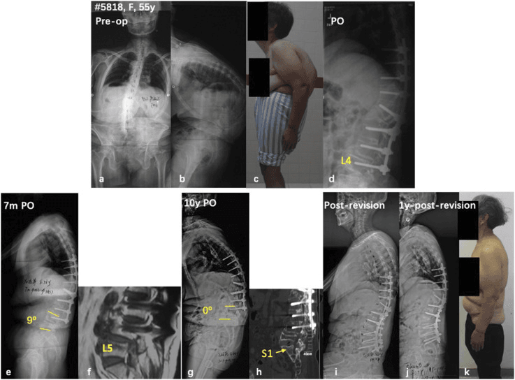
Proximal junctional kyphosis. Kyphosis over 10 degrees at the upper instrumented vertebra, seen in 20-40% after PSO; 10-15% need revision. The risk factors are stopping the construct at the thoracolumbar junction, osteoporosis and overcorrection, so prevention is to extend the construct to the upper thoracic spine (T9-T10), avoid overcorrection and add prophylactic vertebroplasty at the upper instrumented vertebra. Treatment is extension of the fusion if it is symptomatic or progressive.
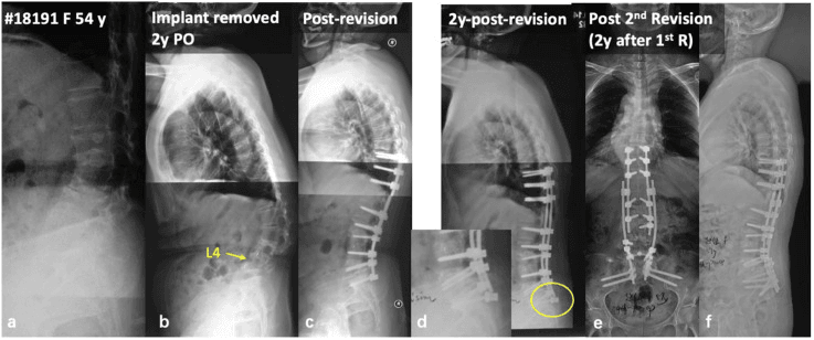
Rod fracture. 10-20% overall, higher when the postoperative SVA is over 5cm, and it presents between 6 months and 2 years. Undercorrection, pseudarthrosis and single rods are the risk factors; adequate SVA correction, dual rods and a solid fusion are the prevention, and revision is for symptomatic or progressive deformity.
Pseudarthrosis. 10-20% at the osteotomy site, with smoking, osteoporosis, infection and rod fracture as its risk factors. It is diagnosed on CT, motion or lucency at the site, or declared by a rod fracture, and treatment is revision fusion with BMP and anterior support, needed in 5-10%. BMP and bone graft at the index operation, stopping smoking and optimising nutrition are the prevention.
Implant failure and reoperation. Screw pull-out occurs in 5-10%, from osteoporosis or the forces of overcorrection, screw fracture in 2-5% and cross-link fracture in 5-10%. Reoperation for any reason runs at 20-30% at 2-5 years, driven by rod fracture, PJK and pseudarthrosis.
Postoperative Care
The first two days. The patient is monitored in intensive care: haemodynamic stability, because the blood loss is often significant; a neurological examination every 2 hours; drain output; and pain control with a PCA or epidural. Mobilisation starts early, out of bed to a chair on day 1 if stable and walking with the physiotherapist on days 2-3.
Days 3 to 7. Daily neurological examination, drains out when output is under 30mL per 8 hours, early AP and lateral radiographs to check the alignment, and a transition to oral analgesia. Physiotherapy progresses ambulation, isometric core work, daily-living tasks and stairs before discharge. The patient goes home when haemodynamically stable, neurologically stable or improving, controlled on oral analgesia, ambulating independently or with a walker, and drain-free.
Follow-up. At each visit the standing films are read for the same five things: SVA (goal under 5cm), PI-LL (goal under 10 degrees), hardware integrity, bridging bone across the fusion, and any proximal junctional kyphosis.
- Assessment
- Wound check, suture removal
- Imaging
- None unless there is a concern
- Assessment
- Clinical examination, pain
- Imaging
- Standing AP and lateral
- Assessment
- Neurological examination, function
- Imaging
- Standing AP and lateral
- Assessment
- Function, return to activities
- Imaging
- AP and lateral
- Assessment
- Final assessment
- Imaging
- AP and lateral; CT if fusion is in doubt
- Assessment
- Long-term outcome
- Imaging
- As needed
Activity.
- Restriction
- Nothing over 10 lbs for 3 months
- Rationale
- Protect the fusion, prevent hardware failure
- Restriction
- None for 6 weeks (opioids)
- Rationale
- Safety, pain control
- Restriction
- 3 months
- Rationale
- Depends on pain and function
- Restriction
- 6-12 months
- Rationale
- Heavy labour risks the fusion
- Restriction
- Light at 6 months, full at 1 year
- Rationale
- Fusion maturation
Bracing. Not usually needed: rigid pedicle screw fixation provides the stability and bracing does not improve fusion rates with modern instrumentation, though a soft lumbar corset may be worn for comfort. The exceptions are osteoporotic bone where screw pull-out is a concern, an extended laminectomy where instability is a concern, and patient preference.
Guidelines, Registries & Global Practice
Global Epidemiology
Flatback historically affected 20-40% of patients after Harrington distraction instrumentation; with lordosis-preserving pedicle screw constructs the rate of clinically significant iatrogenic flatback has fallen to roughly 5-10% after long fusion. Adult spinal deformity overall affects up to 60% of people over 60 years on radiographic survey, with a female predominance reflecting the higher rate of scoliosis fusion.
Side-by-Side Society and Group Guidance
- Position on Sagittal Realignment
- Classification grading by SVA, PT and PI-LL; targets SVA under 4cm, PT under 20-25 degrees, PI-LL under 10 degrees
- Position on Sagittal Realignment
- Endorses age-adjusted alignment targets (older patients tolerate higher SVA/PI-LL); three-column osteotomy for fixed deformity
- Position on Sagittal Realignment
- Maintains adult deformity outcome registry; emphasises sagittal over coronal correction (Glassman 2005)
- Position on Sagittal Realignment
- Specialist commissioning only; deformity correction confined to tertiary units with neuromonitoring
Registry and Outcome Notes
- Multicentre deformity databases (e.g. ISSG, European Spine Study Group) report symptomatic rod fracture of 6-8% overall and approximately 16% after PSO, concentrated at the osteotomy site.
- Reoperation rates of 20-30% at 5 years are consistent across large series, driven by rod fracture, PJK and pseudarthrosis.
- Age-adjusted alignment is now standard practice: older patients are deliberately left in mild positive sagittal balance to reduce proximal junctional failure.
High- vs Limited-Resource Variation
- High-resource settings: Routine standing full-length EOS or stitched radiography, intraoperative neuromonitoring (SSEP/MEP/EMG), cell salvage, tranexamic acid, and dedicated spinal rehabilitation units.
- Limited-resource settings: Reliance on long-cassette plain films, selective neuromonitoring, and staged or lower-risk osteotomy choices; PSO/VCR may be deferred or referred to deformity centres given blood-loss and monitoring requirements.
Perioperative Optimisation (Globally Applicable)
- Bone health: DEXA, treat osteoporosis (e.g. teriparatide, antiresorptives) before major instrumented correction.
- Antifibrinolytics (tranexamic acid) and cell salvage to manage 1-3 litre blood loss in PSO.
- Mandatory intraoperative neuromonitoring for all three-column osteotomies.
- Nutritional and cardiopulmonary optimisation; smoking cessation to reduce pseudarthrosis.
Perioperative Protocols (Global Standard)
Infection Prophylaxis
- Cefazolin 2g IV at induction (3g if over 120kg), re-dosed every 3-4 hours intraoperatively; vancomycin 15mg/kg if beta-lactam allergy.
- Intrawound vancomycin powder is widely used to reduce deep surgical site infection in instrumented deformity surgery.
- Continue systemic prophylaxis for no more than 24 hours postoperatively.
Venous Thromboembolism Prophylaxis
- Mechanical prophylaxis (sequential compression devices) intraoperatively and until mobile.
- Chemical prophylaxis (e.g. low-molecular-weight heparin) typically started 24-48 hours postoperatively once epidural haematoma risk is acceptable, balancing bleeding against thromboembolic risk.
- Early mobilisation is the single most effective measure.
Blood and Bone Management
- Tranexamic acid and cell salvage to manage 1-3 litre blood loss in PSO (3-5 litres in VCR).
- Bone morphogenetic protein and autograft/allograft at the osteotomy and fusion bed; optimise osteoporosis preoperatively.
This topic provides comprehensive coverage of flatback syndrome pathophysiology, radiographic assessment (SVA, PI-LL mismatch), conservative management, and surgical correction with Smith-Petersen, PSO, and VCR osteotomies for global fellowship examination preparation.
MCQ Practice Points
Q: What is flatback syndrome and what are its causes?
A: Flatback syndrome: Loss of normal lumbar lordosis causing positive sagittal balance (sagittal vertical axis greater than 5cm anterior to S1). Patients lean forward and cannot stand upright without hip/knee flexion. Causes: (1) Iatrogenic - distraction instrumentation (Harrington rods), hypolordotic fusion constructs; (2) Degenerative disc disease with disc height loss; (3) Vertebral fractures; (4) Ankylosing spondylitis; (5) Adjacent segment degeneration after fusion.
Q: What are the clinical features and compensatory mechanisms in flatback syndrome?
A: Symptoms: Back pain (fatigue), inability to stand erect, forward stooped posture, need to lean on objects. Compensatory mechanisms (from spine distally): Thoracic hyperkyphosis; Hip hyperextension; Knee flexion; Ankle dorsiflexion. With exhaustion, compensatory mechanisms fail and patient leans progressively forward. Physical exam: Forward trunk inclination; Positive sagittal balance; Hip flexion contractures may develop; Diminished lumbar lordosis or frank kyphosis.
Q: How do you assess sagittal balance radiographically?
A: Standing full-spine radiographs essential. Key measurements: SVA (sagittal vertical axis): C7 plumb line to posterior S1 - normal less than 5cm, positive values indicate anterior shift. Pelvic incidence (PI): Fixed anatomic value. Lumbar lordosis (LL): Should approximately equal PI ± 10°. Pelvic tilt (PT): Increases with compensation (pelvis retroversion). T1 pelvic angle: Global sagittal alignment measure. Goal is to restore PI-LL match and normalize SVA.
Q: What are the surgical options for correcting flatback syndrome?
A: Osteotomy techniques (increasing correction): Smith-Petersen osteotomy (SPO): Posterior column shortening through facets, 10° per level. Pedicle subtraction osteotomy (PSO): Wedge resection through all three columns, 30-35° correction per level. Vertebral column resection (VCR): Complete removal of vertebral segment, greatest correction but highest risk. Extension of fusion: Address adjacent segment disease. Selection based on magnitude of deformity and prior fusion status. Often multiple osteotomies required.
Q: What are the complications specific to flatback correction surgery?
A: Neurological injury: Especially with PSO/VCR - cord monitoring essential; Root injury from nerve stretch or direct trauma. Pseudarthrosis: High mechanical load at osteotomy site. Hardware failure: Rod fracture at osteotomy site (stress riser). Proximal junctional kyphosis: Failure above fusion construct. Adjacent segment disease: Increased stress at adjacent levels. Medical complications: High blood loss, prolonged surgery, age-related comorbidities. Mortality rates 1-5% in revision deformity surgery.
Exam Viva Scenarios
Practise clinical reasoning and management decisions out loud
“A 62-year-old woman presents with severe back pain and forward trunk lean. She had scoliosis surgery with Harrington rods 30 years ago. She can only stand upright for 10 minutes and requires a walker. Full-length standing lateral radiograph shows SVA of 14cm, PI of 58 degrees, LL of 25 degrees, and PT of 32 degrees.”
“A 68-year-old man with no prior spine surgery presents with progressive forward lean over 5 years. He has severe back and leg fatigue with standing and walking. Standing lateral shows SVA 8cm, PI 62 degrees, LL 38 degrees, PT 28 degrees. Multiple disc collapse L2-5. How would you manage this?”
“You are performing a PSO at L3 for flatback correction. After closing the osteotomy, the neurophysiologist reports loss of motor evoked potentials in bilateral lower extremities. What do you do?”
Key Definitions
- Flatback = loss of lumbar lordosis causing sagittal imbalance (forward trunk lean)
- SVA = C7 plumb to posterosuperior S1; normal under 5cm, severe over 10cm
- PI-LL mismatch = Pelvic Incidence minus Lumbar Lordosis; normal under 10 degrees
- Pelvic compensation = posterior pelvic tilt to restore balance (PT normal 10-25 degrees)
Radiographic Parameters
- SVA (Sagittal Vertical Axis): Normal under 5cm, symptoms over 5cm, severe over 10cm
- PI (Pelvic Incidence): FIXED anatomic (35-80 degrees), does NOT change
- LL (Lumbar Lordosis): VARIABLE (40-60 degrees L1-S1), should approximate PI
- PT (Pelvic Tilt): VARIABLE compensation (10-25 degrees), over 25 = exhausted
- PI = PT + SS (sacral slope) - use to check measurement accuracy
Etiologies
- Iatrogenic (most common): Harrington rods, flat fusion, long fusion to sacrum
- Degenerative: Disc collapse, compression fractures, ankylosing spondylitis
- Post-laminectomy: Extensive posterior element removal causing kyphosis
- Traumatic: Vertebral compression fractures, pelvic malunion
Clinical Presentation
- Forward trunk lean with inability to stand upright (cardinal finding)
- Compensatory mechanisms: knee flexion, hip extension, pelvic retroversion
- Severe back and leg fatigue (paraspinals, hip flexors, quadriceps)
- ODI and SF-36 correlate with SVA (worse disability with higher SVA)
- Standing tolerance often under 15-30 minutes
Imaging Requirements
- Standing full-length lateral spine (skull to femoral heads) - MUST be standing
- Arms positioned: fists on clavicles or grasping horizontal bars
- Measure SVA, PI, PT, SS, LL (all on same standing lateral film)
- CT for surgical planning (bony anatomy, pedicle size, prior fusion)
- MRI if radiculopathy or stenosis (neural compression assessment)
Surgical Goals
- Radiographic: SVA under 5cm (ideally 0-3cm), PI-LL under 10 degrees
- Clinical: Upright posture without compensation, reduce pain/disability
- Restore lumbar lordosis to approximately match pelvic incidence
- Avoid complications: neurologic injury, rod fracture, PJK
Osteotomy Options
- SPO (Smith-Petersen): Posterior column only, approximately 10 degrees/level, needs mobile disc
- PSO (Pedicle Subtraction): 3-column, 30 degrees/level, workhorse for flatback
- VCR (Vertebral Column Resection): Complete vertebrectomy, 40 degrees/level, severe cases
- PSO at L3 most common (large body, safer than L4-5, good lordosis contribution)
PSO Technique Key Points
- Instrumentation 3-4 levels above/below, temporary rods for stability
- Laminectomy, pedicle removal, posterior body wedge (30-40 degrees)
- Close osteotomy SLOWLY (1-2mm increments, check neuromonitoring)
- Cantilever closure technique, neural elements shorten (accordion)
- BMP at osteotomy site, decorticate fusion bed, rigid fixation
Complications and Rates
- Neurologic injury: 10-30% (motor 5-10%, sensory 15-25%), 50% resolve by 1 year
- Rod fracture: 10-20% (higher if SVA over 5cm postop, undercorrection)
- PJK (Proximal Junctional Kyphosis): 20-40% (extend to upper thoracic to prevent)
- Pseudarthrosis: 10-20% at osteotomy site (smoking, infection, rod fracture)
- Vascular injury: 1-3% (aorta, vena cava - L4-5 PSO higher risk)
- Infection: 5-10% (deep 3-5%), bleeding: 1-3 liters average PSO
Outcomes
- Patient satisfaction: 70-80% satisfied at 2 years if adequate correction
- ODI improvement: 40-60% reduction (from 50 preop to 25 postop average)
- SVA correction: Average 8-10cm improvement (from 14cm to 4cm)
- Revision rate: 20-30% at 5 years (rod fracture, PJK, pseudarthrosis)
- Correlation: SVA under 5cm postop = best outcomes, over 5cm = rod fracture risk
Exam Pearls
- SVA is KING: Most important outcome predictor (correlates with ODI, SF-36)
- PI-LL target: Under 10 degrees mismatch (predicts rod fracture if over 10)
- PSO at L3: Most common level (large body, safer, good lordosis)
- Neurologic loss during closure: STOP, open osteotomy, optimize perfusion
- Rod fracture = undercorrection: Usually means SVA over 5cm or pseudarthrosis
Outcomes and Evidence
Patient-reported outcomes. At 2 years the improvements are large:
- Preoperative
- 40-60 (severe disability)
- 2-year postoperative
- 20-30 (moderate)
- Improvement
- 40-60% improvement
- Preoperative
- 25-35
- 2-year postoperative
- 45-55
- Improvement
- Significant improvement
- Preoperative
- 7-8/10
- 2-year postoperative
- 3-4/10
- Improvement
- 50% reduction
- Preoperative
- 5-6/10
- 2-year postoperative
- 2-3/10
- Improvement
- 50% reduction
Satisfaction. 70-80% of patients are satisfied or very satisfied, 10-20% satisfied with reservations and 5-10% dissatisfied.
Radiographic outcomes. The average SVA falls from 12-15cm to 3-5cm, an 8-10cm gain; the target of under 5cm is achieved in 70-80% and correlates with satisfaction. The average PI-LL falls from 25-35 degrees to 5-15 degrees, a 20-25 degree improvement; the target of under 10 degrees is achieved in 60-70% and correlates with rod fracture risk.
What predicts the result. A postoperative SVA under 5cm, a PI-LL under 10 degrees, a solid fusion at 1 year and no neurological complication predict a good outcome. Undercorrection with an SVA over 5cm, rod fracture, proximal junctional kyphosis, neurological injury and infection predict a poor one.
Schwab et al. (2010) - Spinopelvic Realignment Targets
- Current concepts review establishing surgical realignment objectives for adult spinal deformity
- Restoring low sagittal vertical axis and pelvic tilt identified as critical surgical goals
- Lumbar lordosis must be proportional to pelvic incidence (PI-LL match)
- Emphasises global alignment and individualised, proportional targets over single thresholds
Smith et al. (2012) - Symptomatic Rod Fracture After Deformity Fusion
- Multicentre retrospective review of 442 adult deformity patients with long posterior fusion
- Symptomatic rod fracture in 6.8% overall but 15.8% after pedicle subtraction osteotomy (PSO)
- Among PSO rod fractures, 89% occurred at or adjacent to the PSO site
- Postoperative sagittal malalignment (SVA over 50mm) increased rod fracture risk; cobalt chromium rods fractured less than titanium or stainless steel
Bridwell et al. (2009) - Operative vs Nonoperative Treatment of Adult Deformity
- Prospective multicentre study of 160 adult symptomatic lumbar scoliosis patients (ages 40-80)
- Operative cohort improved significantly across all QOL measures (SRS, ODI, pain scores) at 2 years
- Nonoperative cohort showed no improvement, with frequent nonsignificant decline
- Limited by only 45% follow-up in the nonoperative group versus 95% operative
Lagrone et al. (1988) - Landmark Description of Flatback After Fusion
- 55 patients with symptomatic loss of lumbar lordosis after spinal fusion treated with corrective osteotomy
- 95% could not stand erect and 89% had back pain at presentation
- Distraction instrumentation with a hook in the lower lumbar spine or sacrum was the most frequent contributing factor
- Failure to restore sagittal balance led to higher pseudarthrosis and recurrent deformity; complications in 60%
- The outcomes are the sobering part, and are what justify the paper's emphasis on prevention: at a mean of 6 years, 47% STILL leaned forward and 36% STILL had moderate or severe back pain despite osteotomy
Bridwell et al. (2003) - Pedicle Subtraction Osteotomy for Fixed Sagittal Imbalance
- 27 consecutive patients with fixed sagittal imbalance treated by lumbar pedicle subtraction osteotomy
- Average lordosis increase of 34.1 degrees achieved through a single posterior osteotomy
- Average C7 sagittal plumb line improvement of 13.5cm
- Significant improvement in Oswestry and pain scores; pseudarthrosis and breakdown caudal to fusion were the main failure modes
Glassman et al. (2005) - Sagittal Balance Predicts Symptoms
- 298 adult deformity patients (172 unoperated, 126 with prior fusion) studied on standing radiographs
- Positive sagittal balance was the most reliable radiographic predictor of pain and function in both groups
- Magnitude of coronal deformity and coronal correction were less critical parameters
- Restoration of normal sagittal balance is the key goal of reconstructive spine surgery
References
-
Schwab F, Patel A, Ungar B, et al. Adult spinal deformity-postoperative standing imbalance: how much can you tolerate? An overview of key parameters in assessing alignment and planning corrective surgery. Spine. 2010;35(25):2224-31.
-
Smith JS, Shaffrey CI, Ames CP, et al. Assessment of symptomatic rod fracture after posterior instrumented fusion for adult spinal deformity. Neurosurgery. 2012;71(4):862-7.
-
Bridwell KH, Glassman S, Horton W, et al. Does treatment (nonoperative and operative) improve the two-year quality of life in patients with adult symptomatic lumbar scoliosis: a prospective multicenter evidence-based medicine study. Spine. 2009;34(20):2171-8.
-
Glassman SD, Berven S, Bridwell K, et al. Correlation of radiographic parameters and clinical symptoms in adult scoliosis. Spine. 2005;30(6):682-8.
-
Bridwell KH, Lewis SJ, Lenke LG, et al. Pedicle subtraction osteotomy for the treatment of fixed sagittal imbalance. J Bone Joint Surg Am. 2003;85(3):454-63.
-
Kim YJ, Bridwell KH, Lenke LG, et al. Proximal junctional kyphosis in adult spinal deformity after segmental posterior spinal instrumentation and fusion: minimum five-year follow-up. Spine. 2008;33(20):2179-84.
-
Yagi M, Akilah KB, Boachie-Adjei O. Incidence, risk factors and classification of proximal junctional kyphosis: surgical outcomes review of adult idiopathic scoliosis. Spine. 2011;36(1):E60-8.
-
Smith JS, Klineberg E, Schwab F, et al. Change in classification grade by the SRS-Schwab Adult Spinal Deformity Classification predicts impact on health-related quality of life measures: prospective analysis of operative and nonoperative treatment. Spine. 2013;38(19):1663-71.
-
Schwab FJ, Blondel B, Bess S, et al. Radiographical spinopelvic parameters and disability in the setting of adult spinal deformity: a prospective multicenter analysis. Spine. 2013;38(13):E803-12.
-
Kim HJ, Bridwell KH, Lenke LG, et al. Patients with proximal junctional kyphosis requiring revision surgery have higher postoperative lumbar lordosis and larger sagittal balance corrections. Spine. 2014;39(9):E576-80.
-
Cho KJ, Bridwell KH, Lenke LG, et al. Comparison of Smith-Petersen versus pedicle subtraction osteotomy for the correction of fixed sagittal imbalance. Spine. 2005;30(18):2030-7.
-
Yang BP, Ondra SL, Chen LA, et al. Clinical and radiographic outcomes of thoracic and lumbar pedicle subtraction osteotomy for fixed sagittal imbalance. J Neurosurg Spine. 2006;5(1):9-17.
-
Auerbach JD, Lenke LG, Bridwell KH, et al. Major complications and comparison between 3-column osteotomy techniques in 105 consecutive spinal deformity procedures. Spine. 2012;37(14):1198-210.
-
Ames CP, Smith JS, Scheer JK, et al. Impact of spinopelvic alignment on decision making in deformity surgery in adults: A review. J Neurosurg Spine. 2012;16(6):547-64.
-
Lafage V, Schwab F, Patel A, et al. Pelvic tilt and truncal inclination: two key radiographic parameters in the setting of adults with spinal deformity. Spine. 2009;34(17):E599-606.