Biceps Femoris Most Common | Grading I-III | Proximal Repair if Greater Than 2cm Retraction
- Biceps femoris long head (BFlh) most commonly injured - eccentric loading at late swing phase
- Proximal hamstring avulsion requires surgical repair if greater than 2cm retraction or more than 2 tendons
- MRI essential for surgical planning - measure retraction distance and number of tendons involved
- Nordic hamstring exercises are evidence-based for prevention and rehabilitation
- Return to sport requires hamstring:quadriceps ratio greater than 0.8 and Askling H-test negative
- “Hamstring injuries peak at late swing phase (eccentric contraction during deceleration)
- “Proximal avulsion off ischial tuberosity more common in water-skiing, splits injuries
- “Mid-substance tears (myotendinous junction) more common in sprinting, kicking sports
- “Bent-knee stretch test distinguishes proximal (positive) from mid-substance (negative) tears
Overview and Epidemiology
Hamstring injuries are among the most common muscle injuries in sport. They affect sprinters, footballers of every code (AFL, soccer, rugby) and track athletes, and they run from a mild strain to a complete proximal avulsion that needs surgical repair.
Who. Hamstring injuries make up 12-16% of all sports injuries in running-based sports, and approximately 15-20% of all injuries in elite running-and-kicking sport (AFL, soccer, rugby), where the average time loss for a Grade II injury is 3-4 weeks of missed playing time. The peak age is 20-40 years, the years of peak athletic participation, and men outnumber women about 2:1, reflecting higher male participation in sprinting sports.
Which muscle. The long head of biceps femoris takes 80% of injuries, semimembranosus 15% and semitendinosus 5%. Why the long head is the one that tears is set out in the anatomy section.
Mechanism. Four patterns account for the spectrum:
- Eccentric overload in the late swing phase of sprinting, the muscle lengthening under load
- Sudden acceleration or deceleration: change of direction, explosive sprinting
- Extreme hip flexion with the knee extended (the splits, water-skiing): the mechanism of proximal avulsion
- Chronic overuse: accumulated microtears in high-volume training
Sprint-type or stretch-type. The single most useful thing to establish from the history is which of two injuries this is, because they differ in the muscle involved, the site within it, and above all in how long the athlete will be out. Askling's work, which underpins the rehabilitation protocols in this topic, rests on this split.
- Sprint-type. Terminal swing phase at high speed, sudden onset, usually the biceps femoris long head at its proximal myotendinous junction. The athlete typically knows the exact stride. These are the commoner injuries and, grade for grade, the quicker to return.
- Stretch-type. A slow, high-amplitude stretch rather than speed: the sliding tackle, the high kick, dancing, a hurdle, doing the splits. These characteristically involve the proximal semimembranosus close to its free tendon, often with less dramatic initial symptoms, and they are substantially slower to recover, sometimes several times longer than a sprint-type injury that looked worse on day one.
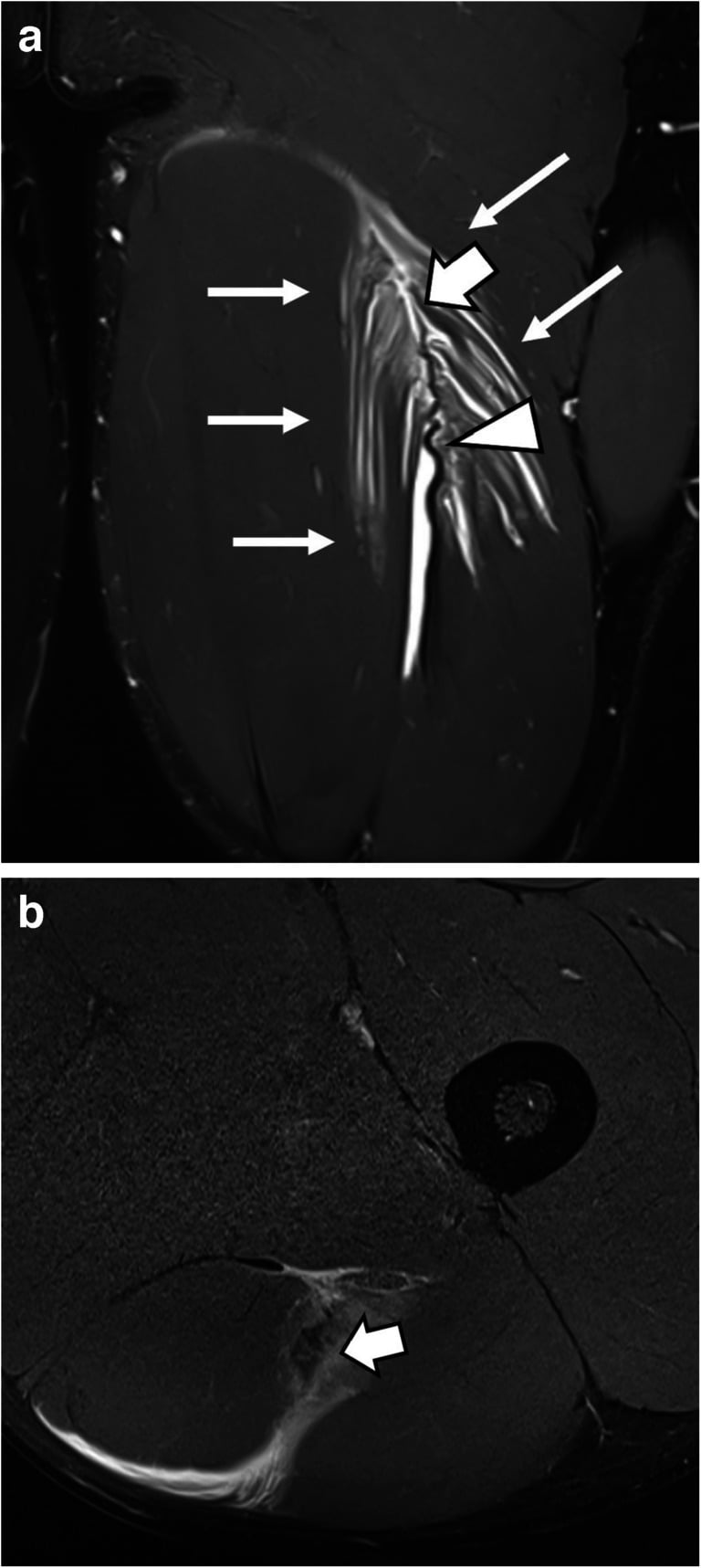
A stretch-type injury with modest pain and a nearly full range of movement can be badly underestimated at first assessment, and an optimistic early prognosis will be wrong. Ask specifically what the leg was doing at the moment of pain, and let the answer, not the initial severity, anchor the timeline you give. The same history decides where to look on MRI: a stretch-type mechanism should send you to the proximal free tendon and its aponeurosis rather than the mid-substance myotendinous junction, which is exactly the free-tendon and intratendinous involvement the BAMIC "c" suffix is designed to capture, and which carries the longer return and higher reinjury risk.
Pathophysiology and Mechanisms
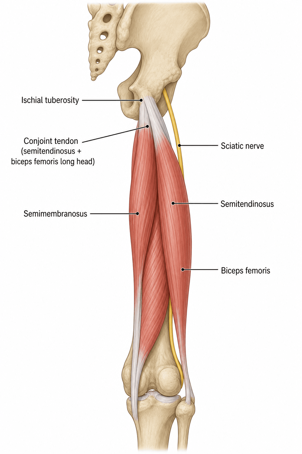
The three muscles. Semimembranosus, semitendinosus and biceps femoris make up the posterior thigh. Semitendinosus and the long head of biceps femoris arise from the ischial tuberosity as a shared conjoint tendon; semimembranosus arises just lateral to it. All three flex the knee and extend the hip, and semitendinosus also internally rotates the tibia.
- Origin
- Ischial tuberosity (medial facet)
- Insertion
- Fibular head
- Innervation
- Tibial division of sciatic nerve
- Function
- Knee flexion, hip extension
- Origin
- Ischial tuberosity (medial facet)
- Insertion
- Proximal medial tibia (pes anserinus)
- Innervation
- Tibial nerve
- Function
- Knee flexion, hip extension, tibial internal rotation
- Origin
- Ischial tuberosity (lateral facet)
- Insertion
- Posterior medial tibial condyle
- Innervation
- Tibial nerve
- Function
- Knee flexion, hip extension
The innervation, stated correctly. Biceps femoris as a whole has two nerve supplies because it has two heads: the long head from the tibial division of the sciatic nerve (L5, S1, S2) and the short head from the common peroneal division (L4, L5, S1). There is no medial-to-lateral split within the long head, and "the long head has dual innervation" is routinely written in exam answers and is an anatomical error. The proposed consequence, asynchronous activation between the two heads during high-speed running, remains a hypothesis rather than a demonstrated cause. The short head is the odd one out twice over: a different nerve, and the only hamstring that does not arise from the ischial tuberosity, taking origin from the linea aspera instead.
Why the long head tears. The better-established reasons are mechanical. It is the most eccentrically loaded hamstring in terminal swing, it has the longest fascicles and greatest musculotendinous excursion, and it has a long, thin proximal aponeurosis over which strain concentrates, which is where the tear usually starts.
Two nerves because two heads: long head tibial division, short head common peroneal division. Never "the long head is dually innervated".
The sciatic nerve. It lies immediately lateral to the hamstring origin and descends through the posterior thigh. That proximity is why a proximal avulsion can produce sciatic symptoms, and why protecting the nerve is the central step of surgical repair.
Blood supply. Perforating branches of the profunda femoris artery supply the muscles, with the inferior gluteal artery supplying the proximal portion. The myotendinous junction, the most common site of Grade I-II tears, is a watershed zone with relatively poor vascularity and the highest stress concentration during eccentric loading, and that poor blood supply is why it is slow to heal.
The running cycle. The hamstrings fail in late swing (70-80% of the gait cycle), when the muscle lengthens while contracting to decelerate the leg. Peak force occurs just before foot strike, and this eccentric load, up to 2.5 times body weight, exceeds the muscle's capacity in fatigue.
- Hamstring activity
- Minimal activity
- Risk
- Low
- Hamstring activity
- Concentric contraction (hip extension)
- Risk
- Low
- Hamstring activity
- Eccentric contraction (deceleration)
- Risk
- High: the peak injury phase
- Hamstring activity
- Isometric contraction
- Risk
- Low
Proximal avulsion is a different injury from the strain: extreme hip flexion with the knee extended (the splits, water-skiing, martial arts kicks) separates one or more tendons completely from the ischial tuberosity, and it is the Grade III injury that may need surgical repair.
Risk factors. Previous hamstring injury is the greatest risk factor.
- Modifiable: inadequate warm-up, hamstring weakness (H:Q ratio less than 0.6), fatigue, limited flexibility
- Non-modifiable: age over 30, previous injury, the anatomy of the long head (long fascicles)
Classification Systems
The traditional three-grade model. The grade is set by how much of the muscle's cross-sectional area is disrupted, and each grade maps onto a return time.
- MRI Findings
- Normal MRI, no oedema
- Clinical Features
- Hamstring tightness, no structural injury
- Return Time
- 1-3 days
- MRI Findings
- Less than 10% cross-sectional area, feathery oedema
- Clinical Features
- Mild pain, minimal strength loss, able to continue activity
- Return Time
- 2-3 weeks
- MRI Findings
- 10-50% cross-sectional area, visible fibre disruption
- Clinical Features
- Moderate pain, palpable defect, significant strength loss, limp
- Return Time
- 4-8 weeks
- MRI Findings
- Greater than 50% or complete tear, gap visible
- Clinical Features
- Severe pain, visible deformity, complete loss of function
- Return Time
- 12+ weeks or surgery
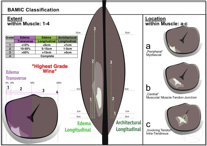
BAMIC (Pollock 2014). The British Athletics Muscle Injury Classification, in this topic's evidence base, is the contemporary MRI-based system examiners now expect, because it adds prognostic resolution the three-grade model lacks. It grades injuries 0-4 by extent and appends a site suffix (a/b/c) to grades 1-4 for where in the muscle-tendon unit the injury sits.
- MRI extent
- 0a: MRI normal / 0b: oedema only, no architectural disruption
- Site suffix (grades 1-4)
- No suffix
- MRI extent
- Small injury
- Site suffix (grades 1-4)
- a / b / c
- MRI extent
- Moderate injury
- Site suffix (grades 1-4)
- a / b / c
- MRI extent
- Extensive injury
- Site suffix (grades 1-4)
- a / b / c
- MRI extent
- Complete tear (muscle or tendon)
- Site suffix (grades 1-4)
- a / b / c
The suffix is the prognostic refinement:
- a (myofascial): at the muscle periphery or fascia; generally the shortest recovery
- b (musculotendinous): at the myotendinous junction; intermediate
- c (intratendinous): involves the central, intramuscular tendon; the longest recovery and a higher re-injury risk, even at the same grade
For a given grade, an intratendinous injury takes substantially longer to return and carries a higher re-injury risk than a myofascial injury of the same grade, so a central-tendon lesion is not equivalent to a peripheral myofascial strain even when both are labelled grade 3 or 4. Reporting the suffix, not just the grade, is the examinable refinement over the Grade I-III model.
Clinical Presentation and Assessment
History. The mechanism names the injury. Sprinting or acceleration produces a mid-substance or proximal myotendinous-junction tear; sudden deceleration or a change of direction is eccentric overload; extreme hip flexion with the knee extended (the splits, water-skiing) is the mechanism of proximal avulsion. An acute pop or tearing sensation suggests a Grade II-III injury. Then establish:
- Where the pain is: the posterior thigh points to the mid-substance, the ischium or buttock to the proximal tendon
- How severe it was: mild enough to continue playing, or severe enough to stop immediately
- What the athlete can no longer do: sprint, kick or accelerate
- Any previous hamstring injury, the most significant risk factor for re-injury
Examination. The findings scale with grade, and the discriminators are gait, a palpable defect and strength.
- Grade I
- Normal or minimal limp
- Grade II
- Antalgic gait, avoids heel strike
- Grade III/Avulsion
- Severe limp, unable to bear weight
- Grade I
- Minimal swelling
- Grade II
- Swelling, bruising appears 24-48h
- Grade III/Avulsion
- Visible deformity, loss of hamstring contour, extensive bruising
- Grade I
- Tender but no defect
- Grade II
- Palpable defect, tender area
- Grade III/Avulsion
- Large palpable gap, ischial tenderness
- Grade I
- Minimal limitation, pain at end range
- Grade II
- Limited active knee flexion, painful passive stretch
- Grade III/Avulsion
- Severe limitation, unable to flex knee against gravity
- Grade I
- 4 out of 5 (mild weakness)
- Grade II
- 3 out of 5 (moderate weakness)
- Grade III/Avulsion
- 1-2 out of 5 or complete loss of function
Special tests. Three tests, each with a different job: one clears the athlete for sport, one separates a proximal from a mid-substance injury, and one is specific for a proximal avulsion.
The Askling H-test is the functional test for return-to-sport readiness: the athlete must pass it, pain-free, before returning to sprinting or sport. Its reported sensitivity is 80% and specificity 85% for predicting safe return, and it is combined with an H:Q strength ratio greater than 0.8 for the decision.
Technique:
- Patient standing, hip neutral
- Actively flex the knee maximally (heel to buttock)
- Extend the hip while maintaining knee flexion
- Hold the position for 5 seconds
The test is positive if there is pain or discomfort, the athlete cannot complete the full range, or shows apprehension or fear of injury. A negative, pain-free test means the athlete is safe to return to sport; a positive test means rehabilitation continues and the test is repeated weekly.
Chronic sitting pain (ischial discomfort) is pathognomonic for proximal hamstring pathology. If a patient reports pain sitting on hard surfaces, especially weeks after injury, think proximal avulsion or incomplete healing. This indicates surgical consultation is warranted.
Differential diagnosis. Posterior thigh and buttock pain has a wider differential than the hamstring, and one entry on it must not be missed.
- Distinguishing Features
- Acute sprinting/eccentric mechanism, mid-posterior thigh pain, palpable defect, pain on resisted knee flexion
- Key Investigation
- MRI (feathery oedema, cross-sectional area)
- Distinguishing Features
- Pop with hip flexion/knee extension, ischial bruising, weakness, chronic sitting pain, bowstring sign
- Key Investigation
- MRI (retraction distance, number of tendons)
- Distinguishing Features
- Insidious deep buttock pain at the ischium, worse with sitting and sprinting, no acute tear; overuse
- Key Investigation
- MRI/ultrasound (tendinosis, no discontinuity)
- Distinguishing Features
- Skeletally immature athlete, sudden pain with sprint/kick, bony fragment off ischium
- Key Investigation
- Plain radiograph (displaced apophysis)
- Distinguishing Features
- Back pain, dermatomal radiation below knee, positive straight-leg raise, neurological signs, no localised tenderness
- Key Investigation
- MRI lumbar spine
- Distinguishing Features
- Buttock pain with neuropathic quality, no true weakness on isolated knee flexion, provocative sciatic tests
- Key Investigation
- MRI, dynamic ultrasound, diagnostic injection
- Distinguishing Features
- Posterior hip/buttock pain in adduction/extension, narrowed ischiofemoral space, quadratus femoris oedema
- Key Investigation
- MRI (ischiofemoral and quadratus femoris space)
- Distinguishing Features
- Calf/thigh swelling, no clear injury, risk factors; must not be missed
- Key Investigation
- Doppler ultrasound
Investigations
Who needs imaging. The diagnosis is clinical first: severity is determined from the history and examination, and imaging is not required for a Grade 0-I injury when the diagnosis is clear. MRI is indicated for a Grade II or suspected Grade III injury, when surgical planning is needed, and in the elite athlete.
MRI is the gold standard and is mandatory for accurate grading and surgical planning, obtained at 24-72 hours. T2-weighted sequences show muscle oedema as bright signal and T1-weighted sequences show the anatomy, in coronal and axial views. Three measurements predict the return-to-sport timeline: the percentage of cross-sectional area involved, the longitudinal length of the injury and, for a Grade III injury, the retraction distance.
- T2 Signal
- Feathery high signal, less than 10% area
- Muscle Architecture
- Muscle fibres intact
- Measurements Required
- Note location, no measurement needed
- T2 Signal
- High signal, 10-50% area, partial fibre disruption
- Muscle Architecture
- Partial gap, some fibres torn
- Measurements Required
- Measure cross-sectional area percentage, length
- T2 Signal
- Complete gap, retracted tendon, haematoma
- Muscle Architecture
- Complete disruption, visible retraction
- Measurements Required
- Measure retraction distance from ischial tuberosity, count tendons involved
For a proximal avulsion, MRI must document four things, because they decide the surgical approach and the prognosis:
- The number of tendons avulsed (semitendinosus, semimembranosus, long head of biceps femoris), each reported separately
- The retraction distance, measured from the ischial tuberosity to the proximal tendon edge on the coronal view, in the position used for surgical planning
- Muscle quality: fatty infiltration suggests a chronic injury
- Haematoma size
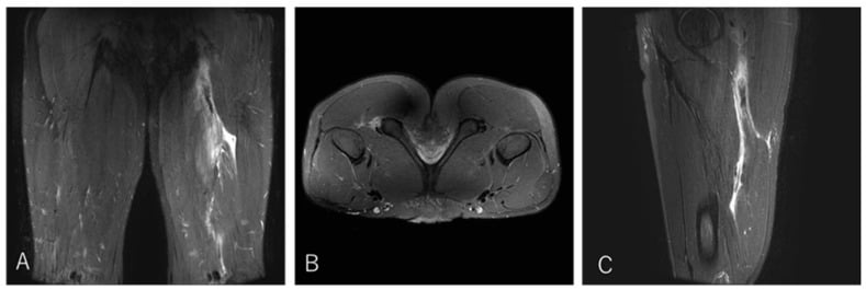
Localising the lesion by tendon and zone says more than a generic "hamstring tear", and it separates an avulsion suitable for repair from a muscle-belly or distal injury.
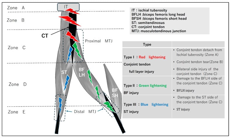
Ultrasound is the alternative: real-time and dynamic, but operator-dependent. It can show tendon retraction and haematoma, it is less sensitive than MRI for muscle oedema, and it is useful for serial monitoring of healing. Correct identification of the compartments prevents mislabelling the injured muscle and guides safe intervention, and a systematic scan sequence is more reproducible than scanning the painful point alone.
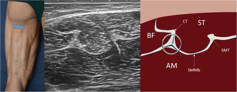
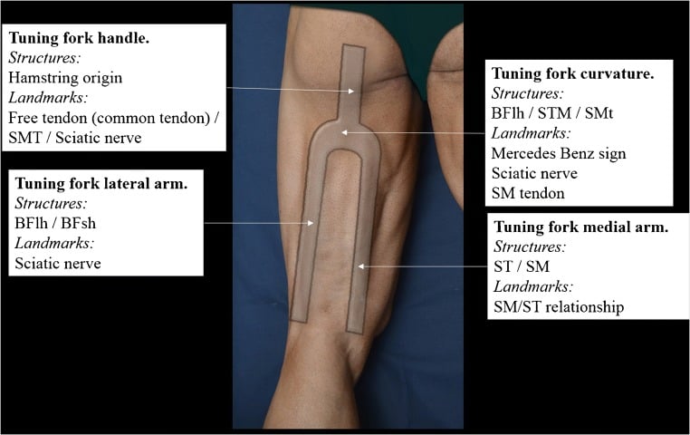
Repeat imaging. If the athlete is not progressing at 4-6 weeks, reassess with MRI: persistent oedema or an enlarging gap suggests the need for intervention. Serial ultrasound tracks rehabilitation progress.
Other investigations. A plain radiograph only if there is concern for an ischial tuberosity avulsion fracture (rare, adolescents). CT is not routinely indicated.
Management Algorithm
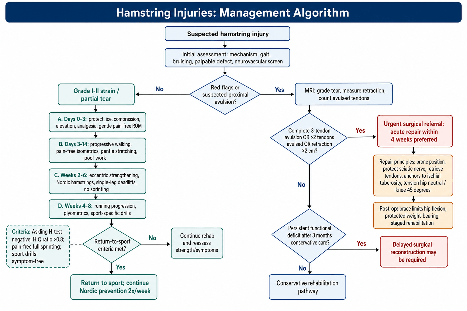
The decision. Grade decides treatment. Grade I and II strains are treated conservatively with progressive loading, and return is gated on functional criteria rather than time alone. A proximal avulsion is decided by MRI: a complete three-tendon avulsion, more than two avulsed tendons, or retraction over 2cm goes to urgent surgical referral, with acute repair within 4 weeks preferred. A lesser avulsion has a conservative trial with close monitoring, and a persistent functional deficit after 3 months of conservative care may need delayed reconstruction.
- Clinical Features
- Mild pain, minimal loss of motion, able to walk normally
- Treatment
- RICE, gentle stretching, progressive loading
- Return Timeline
- 2-3 weeks (when Askling test negative)
- Clinical Features
- Moderate pain, palpable defect, limp, reduced ROM
- Treatment
- Conservative: progressive eccentric exercises, Nordic hamstrings
- Return Timeline
- 4-8 weeks (functional criteria essential)
- Clinical Features
- Severe pain, bruising, weakness, positive bent-knee test
- Treatment
- Conservative trial (non-operative) with close monitoring
- Return Timeline
- 12-16 weeks (may need surgery if fails)
- Clinical Features
- Complete loss of hamstring contour, palpable gap, severe weakness
- Treatment
- URGENT surgical repair (within 4 weeks optimal)
- Return Timeline
- 12 weeks minimum post-op (functional testing required)
- Clinical Features
- Persistent weakness, sitting pain (ischial), functional limitation
- Treatment
- Delayed surgical reconstruction (may need graft augmentation)
- Return Timeline
- 16-24 weeks post-op (lower success than acute)
The goal is progressive loading that restores strength and prevents re-injury. The phases below are gated by criteria, not the calendar, and the evidence-based exercise is the eccentric one.
Rehabilitation Timeline
Protect the muscle: rest from aggravating activities, ice for 15-20 minutes every 2-3 hours, a compression bandage and elevation. NSAIDs (ibuprofen 400mg three times daily) for 3-5 days. Pain-free knee flexion and extension, avoiding stretching. Crutches for a Grade II injury with a significant limp.
Pain-free isometric hamstring contractions (seated knee flexion against resistance), gentle stretching within the pain-free range (aggressive stretching delays healing), walking that increases in distance and speed as tolerated, and aqua jogging in the pool for unloaded work.
Nordic hamstring curls, 3 sets of 5-8 repetitions three times a week, are the evidence-based core of rehabilitation. Add single-leg deadlifts for controlled eccentric loading and increase the resistance as strength improves. No sprinting or kicking yet.
Running progresses from light jogging to straight-line sprinting to change of direction. Hopping and bounding plyometrics begin once the Askling test is negative, followed by kicking and acceleration drills specific to the sport. Strength testing: the H:Q ratio must be greater than 0.8.
Clearance requires all four criteria: (1) Askling H-test negative, (2) H:Q ratio greater than 0.8, (3) pain-free full sprinting, (4) sport-specific drills completed without symptoms. Minimum time is 2-3 weeks for Grade I and 4-8 weeks for Grade II. Nordic hamstring curls continue twice a week indefinitely.
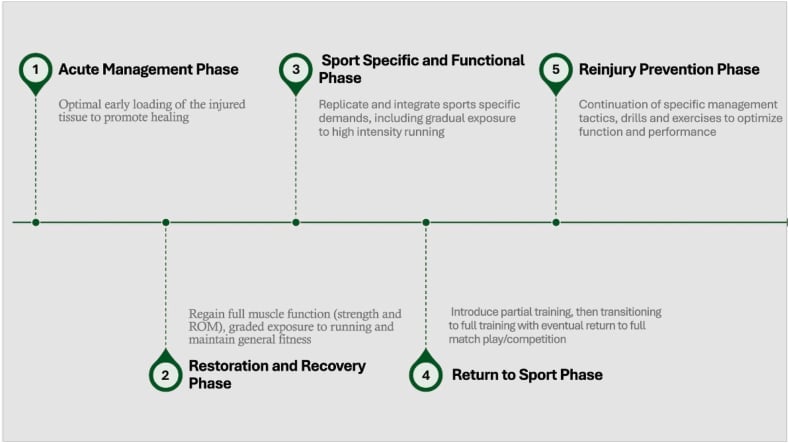
Premature return, within 3 weeks of a Grade II injury, carries a 34% re-injury rate. Athletes who complete the full rehabilitation, including eccentric strengthening, re-injure at 12%, the same figure seen in Grade II athletes who return after 6 weeks, and every additional week of rehabilitation reduces re-injury risk by approximately 5%. Take a minimum of 6 weeks for a Grade II injury, and 12 weeks after surgery for Grade III, and rest clearance on functional criteria (strength, the Askling test, pain-free sprinting), not time alone.
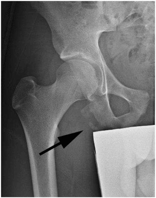
Surgical Technique - Proximal Repair Detail
Consent. The figures the patient hears:
- Sciatic nerve injury 1-3% (numbness, weakness, a permanent risk)
- Infection 2-5%, superficial wound infection most common
- Re-rupture 5-10%, if non-compliant with the post-operative protocol
- Persistent weakness 10-20%, especially after chronic repairs
- Sitting discomfort, which may persist for 6-12 months
- Failure to return to sport 10-20%
Equipment. Suture anchors, 5.5mm, 3-5 of them; No.2 non-absorbable suture (Fiberwire or Ethibond); deep retractors for gluteus maximus and the sciatic nerve; a C-arm for anchor placement, the lateral view confirming depth; and a post-operative hip brace limiting flexion to 60 degrees.
Position. Prone on a radiolucent table so the C-arm can reach, with chest padding to reduce pressure on the chest and abdomen, pelvic bolsters that support the pelvis and allow hip mobility, the knees padded to protect the patellae, and the arms on arm boards or alongside the body. Slight hip and knee flexion reduces tension on the retracted stump and the sciatic nerve. The set-up must still allow fluoroscopy, and distal access if a second incision is needed; confirm on the C-arm lateral view that the ischial tuberosity can be seen.
Preparation. Wide, from the lumbar spine to mid-thigh bilaterally, including the perineum in the field for ischial access, under a transparent adhesive drape.
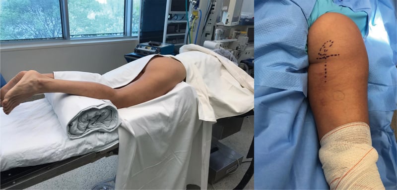
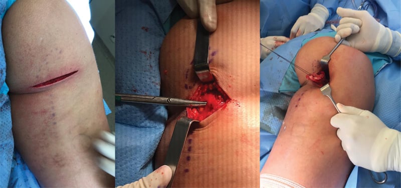
Incision. Palpate the ischial tuberosity, the bony prominence inferior to the gluteal fold. A transverse incision in the gluteal crease is preferred, or a longitudinal one, centred over the tuberosity and 8-12cm long, with a longitudinal extension available for chronic retraction. Deepen through the subcutaneous fat, identifying and protecting the posterior femoral cutaneous nerve.
Deep dissection. Split gluteus maximus longitudinally along its fibres, by blunt dissection to avoid bleeding. Identify the sciatic nerve at the lateral border of the incision, deep to gluteus maximus, and place a deep retractor to hold it laterally and protect it throughout. The nerve lies lateral and deep to the ischial tuberosity and is vulnerable during lateral retraction and anchor placement; where scar tethers it and obscures the plane, it must be identified and released.
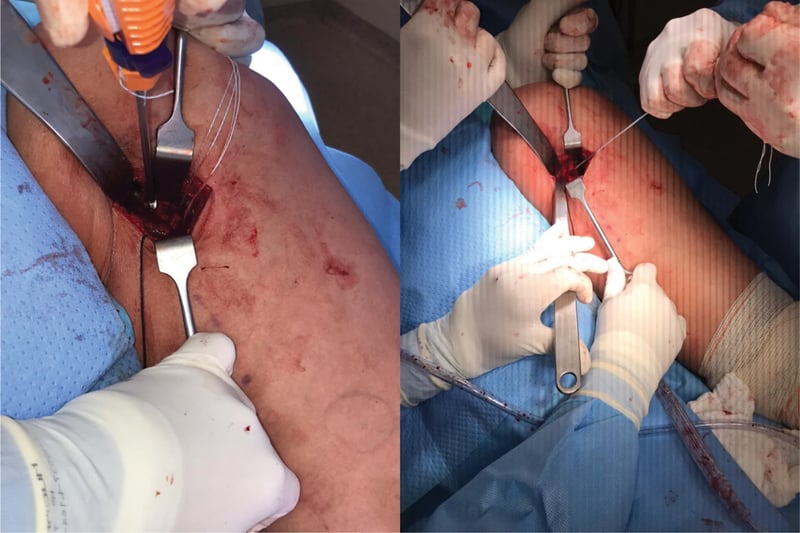
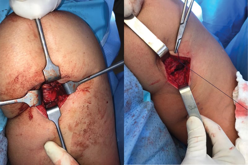
Intraoperative troubleshooting. Four problems recur, and each has a standard answer:
- Cause
- Extensive retraction (greater than 10cm)
- Solution
- Extend incision distally, palpate along posterior thigh
- Cause
- Chronic retraction, scarring
- Solution
- Consider allograft augmentation or accept tension
- Cause
- Nerve adherent to scar or stretched
- Solution
- Mobilise nerve carefully, may need external neurolysis
- Cause
- Poor bone quality or incorrect angle
- Solution
- Re-drill, use larger anchor, or add additional anchor
Complications
- Incidence
- 12-34%
- Risk Factors
- Premature return, inadequate rehab, previous injury
- Management
- Prevention: Nordic hamstrings, functional criteria, minimum 6 weeks
- Incidence
- 5-15%
- Risk Factors
- Incomplete healing, inadequate rehab
- Management
- Extended physiotherapy, consider PRP injection, surgical consultation
- Incidence
- 1-3%
- Risk Factors
- Intraoperative traction, anchor malposition
- Management
- Observation (most resolve), EMG at 6 weeks, neurosurgery if persistent
- Incidence
- 2-5%
- Risk Factors
- Deep dissection, haematoma
- Management
- Antibiotics, drainage if collection, debridement if deep
- Incidence
- 5-10%
- Risk Factors
- Non-compliance with brace, premature loading
- Management
- Prevention: strict brace protocol 6 weeks, controlled rehab
- Incidence
- 10-15%
- Risk Factors
- Incomplete haemostasis
- Management
- Observation if small, aspiration if large, drain if recurrent
- Incidence
- 20-40% (6-12 months)
- Risk Factors
- Ischial tuberosity sensitivity, hardware prominence
- Management
- Cushioned seating, time, hardware removal if persistent
Re-injury is the complication that defines this injury, and the AFL data put numbers on it. Risk remains elevated for approximately 15 weeks after return to play (Orchard 2020), and a recent prior hamstring injury is by far the strongest risk factor (adjusted OR 13.1). Prevention rests on three things: Nordic hamstring exercises (approximately 70% reduction in overall injury and approximately 85% reduction in recurrence, Petersen 2011), functional testing before clearance (the Askling test and the H:Q ratio), and continued eccentric loading and load monitoring well beyond clearance (4-8 weeks for Grade II, 12 or more weeks after surgery).
Platelet-rich plasma. PRP appears in this topic as an "option", in the complications table and the recurrent-injury scenario, so the evidence deserves to be stated plainly. For acute hamstring muscle injury, placebo-controlled randomised trials and subsequent meta-analyses have not shown that PRP shortens return-to-play time or reduces re-injury compared with a structured rehabilitation programme, with or without a blinded placebo injection; the early enthusiasm was not borne out once PRP was tested against blinded saline controls. PRP is therefore not recommended as routine treatment for acute hamstring strain. The intervention with robust (Level 1) evidence remains progressive eccentric loading, the Nordic programme, supported by criterion-based return to play. PRP and other biologics may still be discussed with elite athletes under shared decision-making, but without an evidence-based expectation of faster or safer return.
Postoperative Care and Rehabilitation
The principles are strict brace compliance for 6 weeks to prevent re-rupture, Nordic hamstring curls from week 6 onwards, loading that increases gradually with symptoms monitored, and objective functional testing before return. Pain during rehabilitation means the load is excessive. No aggressive stretching for the first 12 weeks, because it delays healing; no running before 12 weeks; and no return to sport before 4 months for athletes.
Post-Surgical Protocol (Proximal Hamstring Repair)
The hip brace limits flexion to 60 degrees and is worn at all times except for exercises. Toe-touch weight-bearing with crutches for 2 weeks. Passive knee flexion and extension, with no hip flexion beyond 60 degrees. Keep the wound dry; staples out at 14 days. Analgesia is paracetamol and tramadol, avoiding NSAIDs, which may impair healing. DVT prophylaxis: enoxaparin 40mg daily for 14 days, extended if high risk.
The brace continues with its 60-degree flexion limit while weight-bearing progresses to full as tolerated. Hip flexion is increased gradually, targeting 10 degrees a week. Isometric hamstring contractions (seated) and quadriceps strengthening; aqua walking from week 4 for unloaded range of motion. No aggressive hamstring stretching, which delays healing.
Wean off the brace at 6 weeks if the range is adequate, aiming for full hip flexion by 8 weeks. Begin Nordic hamstring curls three times a week, progressing resistance with single-leg deadlifts and hamstring curls, and a stationary bike at low resistance for endurance. No running, jumping or explosive movements.
Running starts as light treadmill jogging and progresses outdoors. Nordic hamstrings continue as the eccentric focus, the key to success. The H:Q ratio should be greater than 0.7 by 16 weeks. Hopping and bounding plyometrics if the Askling test is negative, then sport-specific movements.
Return requires the same four criteria as after a strain: Askling H-test negative (pain-free), H:Q ratio greater than 0.8 on isokinetic testing, pain-free full sprinting, and sport-specific drills completed without symptoms. The minimum is 4-6 months after surgery for high-demand athletes, and Nordic hamstring curls continue twice a week indefinitely.
Outcomes and Prognosis
Conservative management (Grade I-II). Almost every Grade I injury and most Grade II injuries return, and the number that matters is the re-injury column:
- Return to Sport
- 95-100% at 2-3 weeks
- Re-injury Risk
- 10-15%
- Key Prognostic Factor
- Compliance with Nordic exercises
- Return to Sport
- 85-95% at 6-8 weeks
- Re-injury Risk
- 20-34%
- Key Prognostic Factor
- Time to return (longer is better)
Surgical repair (proximal avulsion). Every measure falls as the delay to repair lengthens:
- Return to Sport
- 90-95%
- Strength Recovery
- 90-100% (isokinetic testing)
- Patient Satisfaction
- Excellent (90%)
- Return to Sport
- 80-90%
- Strength Recovery
- 80-90%
- Patient Satisfaction
- Good (80%)
- Return to Sport
- 65-80%
- Strength Recovery
- 70-80%
- Patient Satisfaction
- Fair (70%)
Prognostic factors. The good and the poor are mirror images of each other.
- Good: acute repair within 4 weeks; 2 or fewer tendons involved; no muscle atrophy or fatty infiltration on MRI; a young patient (under 40); compliance with the post-operative protocol (brace, restricted range); a maintained Nordic hamstring programme
- Poor: delayed repair beyond 12 weeks; complete 3-tendon avulsion with extensive retraction; chronic muscle changes (atrophy, fatty infiltration); an older patient (over 50); non-compliance with rehabilitation; premature return to high-demand activities
Long term. Sitting discomfort resolves in 80% by 12 months, and 20% have persistent mild discomfort. Strength recovers to 90-95% in acute repairs and 70-80% in chronic ones, and after an acute repair 85-90% return to their pre-injury level.
Guidelines, Registries & Global Practice
Global epidemiology (PubMed-backed):
- Figure
- Most frequent and most prevalent injury
- Source population / evidence
- AFL surveillance 1992-2012 (Orchard 2013, PMID 23460329)
- Figure
- Approximately 26% (historically up to 25-26% before modern prevention)
- Source population / evidence
- AFL surveillance (Orchard 2013)
- Figure
- Elevated for approximately 15 weeks; approximately 9% in first match back
- Source population / evidence
- AFL cohort 1992-2014 (Orchard 2020, PMID 32024646)
- Figure
- Recent (8 weeks or less) prior hamstring injury, adjusted OR 13.1
- Source population / evidence
- Orchard 2020
- Figure
- Approximately 70% reduction overall, approximately 85% reduction in recurrence
- Source population / evidence
- Petersen 2011 RCT (PMID 21825112)
Hamstring strain injury is consistently the single most common time-loss injury across football codes worldwide (soccer, rugby, AFL, American football, Gaelic football), so the principles below are examinable on any board.
Major guidance and consensus, side by side:
- Position
- Use MRI grade 0-4 with myofascial/musculotendinous/intratendinous suffix for prognosis; intratendinous injuries take longest
- Evidence level
- Expert/classification (Level 5), validation ongoing
- Position
- Criterion-based return to play (clinical, strength symmetry, Askling H-test); MRI normalisation NOT required before return
- Evidence level
- Level 2 (systematic reviews, Reurink 2014 / Hickey 2017)
- Position
- Eccentric Nordic hamstring loading recommended for primary and secondary prevention in high-risk sport
- Evidence level
- Level 1 (cluster RCT, Petersen 2011)
- Position
- Repair complete/displaced avulsions (commonly cited greater than 2cm retraction or 2-3 tendons); acute repair superior to delayed
- Evidence level
- Level 3 (meta-analysis, Bodendorfer 2018)
The widely quoted "greater than 2cm retraction / more than 2 tendons" surgical threshold is a pragmatic consensus, not a randomised finding - no high-quality RCT compares operative with nonoperative treatment of proximal avulsion. The robust evidence is that (1) Nordic eccentric training prevents injury (Level 1), and (2) acute repair outperforms delayed repair (Level 3 meta-analysis).
Registry / surveillance evidence:
Hamstring injury has no implant registry; the equivalent high-volume datasets are prospective sports-injury surveillance systems - the AFL injury surveillance database (Orchard, 20+ seasons), UEFA Elite Club Injury Study (European soccer), and various national rugby and athletics audits. These consistently identify hamstring strain as the dominant time-loss injury and a high-recurrence problem, which is what drives the global emphasis on eccentric prevention and criterion-based return.
Global practice variation:
- Imaging access: MRI grading (BAMIC) is routine in elite/high-resource settings; in limited-resource settings, clinical grading and ultrasound guide management, with similar core rehabilitation principles.
- Return-to-play culture: Professional codes increasingly mandate objective functional clearance (strength symmetry, Askling H-test) rather than fixed time, whereas amateur practice still leans on symptom resolution alone.
- Surgery for proximal avulsion: Performed selectively worldwide for complete/displaced avulsions; thresholds and timing differ by surgeon and access, but acute referral is universally favoured because outcomes deteriorate with delay.
Medicolegal and consent considerations (universal):
Key documentation for hamstring surgery (applicable in any jurisdiction):
- Detailed informed consent including sciatic nerve injury risk (1-3%)
- MRI measurements documenting retraction distance and number of tendons avulsed
- Explanation of conservative alternatives and expected outcomes, including the approximately 23% overall complication rate after repair (Bodendorfer 2018)
- Clear documentation of functional deficit (strength testing, gait assessment)
- Post-operative protocol explained (brace compliance, weight-bearing restrictions)
- Common litigation themes: sciatic nerve palsy (inadequate warning), re-rupture (premature loading), persistent weakness (unrealistic expectations)
MCQ Practice Points
Q: Which hamstring muscle is most commonly injured and why? A: Biceps femoris long head (80% of injuries), because it is (1) the most eccentrically loaded hamstring at late swing phase, (2) the one with the longest fascicle length and greatest excursion, and (3) the one with a long thin proximal aponeurosis over which strain concentrates - which is where the tear starts, at the proximal myotendinous junction. A frequent error is worth avoiding here: the dual nerve supply belongs to the biceps femoris as a two-headed muscle - long head tibial division, short head common peroneal division - not to the long head itself, and the asynchronous-activation idea is a hypothesis rather than established cause. Saying "the long head has dual innervation" will be marked as an anatomical error.
Q: What are the criteria for Grade II hamstring muscle strain? A: Grade II: (1) 10-50% cross-sectional area involvement on MRI, (2) Partial muscle fibre disruption with visible gap, (3) Feathery high signal on T2 MRI, (4) Clinical: Moderate pain, palpable defect, strength 3 out of 5, limp present. Return time: 4-8 weeks with appropriate rehabilitation.
Q: What are the indications for surgical repair of proximal hamstring avulsion? A: (1) Retraction greater than 2cm measured on MRI, (2) More than 2 tendons completely avulsed, (3) Complete 3-tendon avulsion (regardless of retraction), (4) Failed conservative trial (3 months) with persistent functional deficit, (5) Elite athlete with high functional demands. Acute repair (within 4 weeks) has better outcomes than chronic reconstruction.
Q: What MRI findings predict longer return-to-sport time after hamstring injury? A: (1) Greater cross-sectional area involvement (greater than 25% = longer return), (2) Longitudinal extent greater than 10cm, (3) Proximal MTJ location (vs distal MTJ), (4) Complete tear with retraction (Grade III). MRI performed within 5 days accurately predicts timeline: less than 10% area = 17 days, 10-50% = 42 days, greater than 50% = 73+ days.
Q: What is the evidence for Nordic hamstring exercises in injury prevention? A: Petersen et al (2011) cluster RCT showed: (1) overall acute hamstring injury rate fell from 13.1 to 3.8 per 100 player-seasons (adjusted rate ratio 0.29, approximately 70% reduction), (2) recurrent injury rate fell from 45.8 to 7.1 per 100 player-seasons (rate ratio 0.14, approximately 85% reduction), (3) number needed to treat 13 overall and 3 to prevent one recurrence, (4) progressive eccentric Nordic loading programme. This is the gold standard evidence-based prevention and rehabilitation exercise.
Q: What are the functional criteria for return to sport after Grade II hamstring strain? A: (1) Askling H-test negative (pain-free active knee flexion with hip extension), (2) H:Q ratio greater than 0.8 (isokinetic testing - hamstring:quadriceps strength ratio), (3) Pain-free full sprinting at match speed, (4) Completed sport-specific drills without symptoms, (5) Minimum time: 4-8 weeks for Grade II (NOT 3 weeks). Premature return (within 3 weeks) leads to 34% re-injury vs 12% with adequate rehabilitation.
Exam Viva Scenarios
Practise clinical reasoning and management decisions out loud
“A 26-year-old professional AFL footballer presents 24 hours after experiencing sudden posterior thigh pain while sprinting during a match. He felt a 'pop' and was unable to continue. On examination, he has a palpable defect in the mid-posterior thigh, tenderness over the biceps femoris, and reduced strength (3 out of 5) on resisted knee flexion. He can walk with a limp. What is your assessment and initial management?”
“A 35-year-old recreational water skier presents 1 week after sustaining an injury during a fall. He felt severe pain in the buttock and posterior thigh, heard a 'pop', and had immediate loss of function. MRI shows complete avulsion of all three hamstring tendons from the ischial tuberosity with 4cm of retraction. He is currently unable to flex his knee against gravity and has severe weakness. Walk me through your surgical planning and technique.”
“A 28-year-old semi-professional soccer player presents with his third hamstring injury in 12 months, all to the same leg (left biceps femoris). He returned to sport 3 weeks after the previous injury. MRI shows Grade II strain with 25% cross-sectional area involvement at the proximal MTJ. He is frustrated and wants to 'just get surgery to fix it once and for all'. How do you manage this complex situation?”
Key Anatomy
- Biceps femoris long head (BFlh) = 80% of injuries. Innervation splits by HEAD: long head tibial, short head common peroneal - not within the long head
- Three muscles: ST (most medial), BFlh (lateral), SM (deep)
- Injury site: Proximal myotendinous junction (most common) or ischial avulsion
- Eccentric injury at late swing phase (70-80% of gait cycle)
Grading Classification
- Grade I: Less than 10% cross-sectional area, 2-3 weeks return
- Grade II: 10-50% area, 4-8 weeks return (functional criteria essential)
- Grade III: Greater than 50% or complete avulsion, surgery if greater than 2cm retraction
- MRI essential for grading and surgical planning
Treatment Algorithm
- Grade I-II: Conservative (RICE, Nordic hamstrings, functional criteria for return)
- Grade III with less than 2cm: Conservative trial (may heal)
- Grade III with greater than 2cm retraction: URGENT surgical repair (within 4 weeks)
- Complete 3-tendon avulsion: Surgical repair regardless of retraction
Surgical Pearls
- Prone position, transverse incision over ischial tuberosity
- PROTECT SCIATIC NERVE (lateral border, identify early, retract laterally)
- Horseshoe anchor pattern (5 anchors), angle medially away from nerve
- Tension repair: Hip neutral, knee 45 degrees flexion
- Post-op: Hip brace 60 degrees limit for 6 weeks, toe-touch 2 weeks
Complications and Prevention
- Re-injury: 34% if returned within 3 weeks, 12% with adequate rehab
- Sciatic nerve injury: 1-3% (post-op), most resolve spontaneously
- Nordic hamstring exercises: approximately 70% reduction in overall injury, approximately 85% in recurrence (Petersen 2011, EVIDENCE-BASED)
- Return criteria: Askling test negative, H:Q ratio greater than 0.8, pain-free sprinting
- Sitting pain post-op: 20-40% at 6 months (resolves in 80% by 12 months)
Evidence Base and Key Trials
Nordic Hamstring Exercise Prevention - Petersen et al (Landmark RCT)
- Cluster-randomised controlled trial of 50 Danish soccer teams (942 players) over one season
- 10-week progressive Nordic hamstring eccentric programme plus weekly maintenance vs usual training
- Overall acute hamstring injury rate 3.8 vs 13.1 per 100 player-seasons (adjusted rate ratio 0.29, 95% CI 0.15-0.57) - approximately 70% reduction
- Recurrent injury rate 7.1 vs 45.8 per 100 player-seasons (rate ratio 0.14) - approximately 85% reduction
- NUMBER NEEDED TO TREAT SPLITS THE PROGRAMME IN TWO: 25 players to prevent one NEW injury, but only 3 to prevent one RECURRENCE. The new-injury effect was also the weaker one statistically (rate ratio 0.41, 95% CI 0.18-0.93, p=0.034) with its confidence interval close to 1, while the recurrence effect was far more emphatic (0.14, 95% CI 0.04-0.51)
British Athletics Muscle Injury Classification (BAMIC) - Pollock et al (Defining Classification)
- Proposed an MRI-based grading system (grades 0-4) to replace the diagnostically limited 3-grade strain model
- Each grade 1-4 carries a suffix a/b/c denoting myofascial, musculotendinous or intratendinous site of injury
- Intratendinous (c) injuries (tendon involvement) carry a worse prognosis and longer return time
- Designed to improve prognostication and therapeutic decision-making over traditional grading
Operative vs Nonoperative Proximal Hamstring Avulsion - Bodendorfer et al (Meta-analysis)
- Systematic review and meta-analysis: 24 studies, 795 proximal hamstring avulsions
- Repair gave higher patient satisfaction (90.8% vs 52.9%) and hamstring strength (85.0% vs 64.0%) than nonoperative care (all P less than 0.001)
- Acute repair outperformed chronic repair (satisfaction 95.5% vs 83.8%; less pain; greater strength)
- Overall complication rate after repair 23.2%; complete-avulsion repair complication rate 29.4% vs 11.3% for partial
- Nonoperative comparison group was small, limiting the strength of the direct comparison
Functional Outcome After Proximal Hamstring Repair - Birmingham et al
- Retrospective series of 34 complete proximal hamstring avulsions (9 acute, 14 chronic repairs); 23 patients evaluated
- 21 of 23 returned to activity at a mean 95% of pre-injury level, at a mean of 9.8 months
- 18 excellent, 4 good, 1 fair subjective result
- Isokinetic hamstring strength averaged 90-93% of the uninvolved limb
- Minimal postoperative complications reported
Return-to-Play Criteria and Re-injury - Hickey et al (Systematic Review)
- Systematic review of 9 studies (601 acute hamstring strain injuries) on rehabilitation progression and return-to-play criteria
- Pain perception was used to guide rehabilitation progression across all studies
- Protocols using isokinetic dynamometry had the shortest return times (12-25 days)
- Protocols incorporating the Askling H-test had the lowest re-injury rates (1.3-3.6%)
- Called for more objective, criterion-based return-to-play decision-making
MRI at Return to Play of Recovered Hamstring Injuries - Reurink et al
- 53 athletes imaged within 5 days of injury and again within 3 days of return to play (51% grade 1, 49% grade 2)
- Median time to return to play was 28 days (range 12-76)
- 89% of clinically recovered athletes still showed increased intramuscular signal on fluid-sensitive MRI at return
- Normalisation of MRI signal is NOT required before safe return to play
- New low-signal (fibrous) tissue was seen in one-third at return; clinical significance uncertain
Two Decades of AFL Injury Surveillance - Orchard et al (Registry-scale epidemiology)
- Australian Football League injury surveillance 1992-2012 (4492 players, 13,606 new injuries)
- Hamstring strain was the most frequent and most prevalent injury (mean 6 per club per season; 20 missed matches per club per season)
- Hamstring strain recurrence rate averaged 26%
- Overall injury recurrence fell from 25% (1992) to 9% (2012) with surveillance and rule changes
- Public release of injury data supported rule changes targeting player safety
Fifteen-Week Recurrence Window - Orchard et al
- Prospective cohort: 3647 muscle strains (1932 hamstring) over 23 AFL seasons
- Recent (8 weeks or less) hamstring injury was by far the strongest risk factor for recurrence (adjusted OR 13.1, 95% CI 11.5-14.9)
- Non-recent prior hamstring injury still raised risk (adjusted OR 3.5)
- Recurrence risk was approximately 9% in the first match back and stayed elevated for 15 weeks after return
- Recent hamstring injury also raised the risk of subsequent quadriceps and calf strains