Herbert Classification of Scaphoid Fractures
The critical divide is Type A (stable, treat in cast) versus Type B (unstable, fix operatively). A displaced or complete fracture through the waist (B2) is the classic unstable pattern. A non-displaced incomplete fracture of the waist (A2) is stable. Examiners will probe whether you understand why proximal pole fractures behave badly (retrograde vascularity) and will test your ability to defend operative versus non-operative treatment for borderline cases.
The Herbert classification
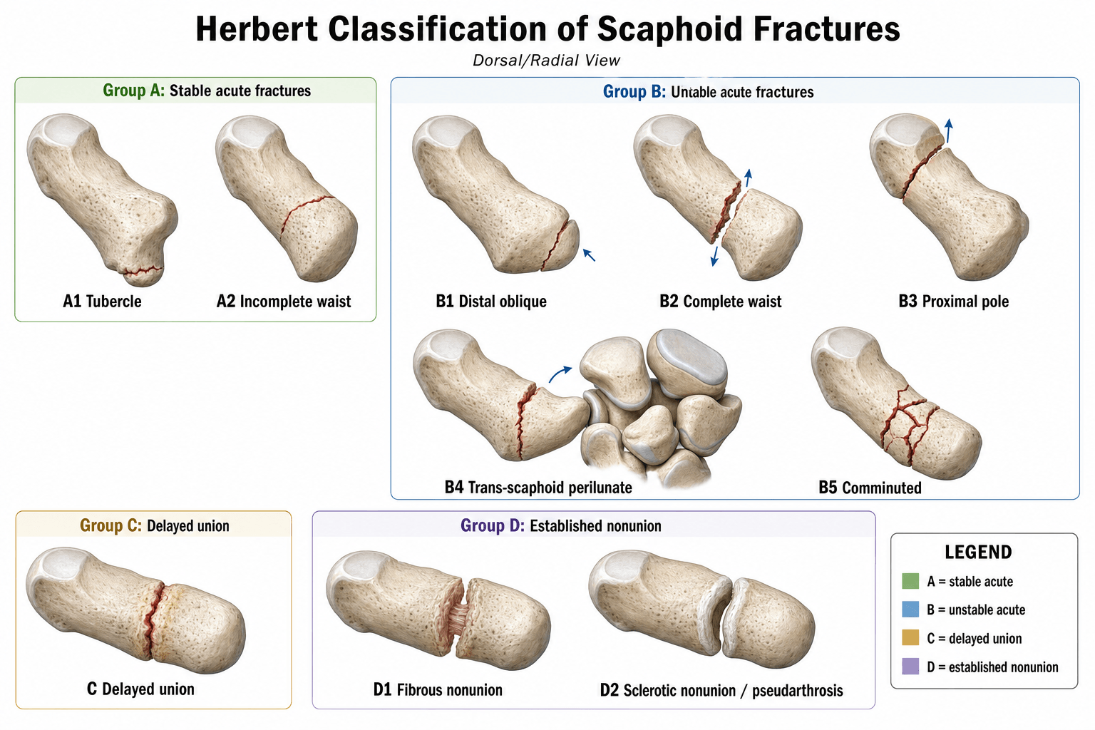
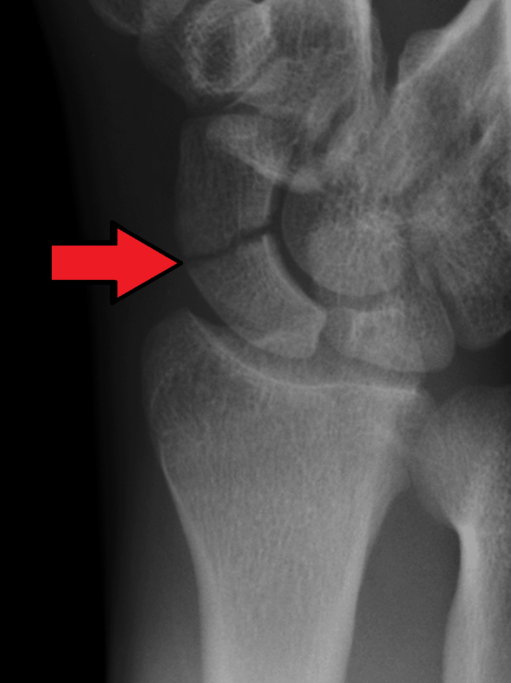
The Herbert system was introduced alongside the Herbert dual-pitch compression screw in 1984. It classifies scaphoid fractures into four types based on stability, chronicity, and displacement.
- Category
- Stable acute fracture
- Stability
- Stable
- Typical Treatment
- Below-elbow scaphoid cast for 6 to 8 weeks
- Category
- Unstable acute fracture
- Stability
- Unstable
- Typical Treatment
- Open or percutaneous screw fixation
- Category
- Delayed union
- Stability
- Uncertain
- Typical Treatment
- Screw fixation (plus bone graft if gap present)
- Category
- Established nonunion
- Stability
- Unstable
- Typical Treatment
- Open reduction, bone graft (Russe or vascularised), and screw fixation
A Alright · B Bad · C Cautious · D DisasterABCD — the four types
Hook:The whole system is a stability ladder: cast A, fix B, salvage C and D before the wrist collapses into SNAC arthritis.
Herbert type A2 (incomplete hairline fracture of the waist) is the only acute waist fracture considered stable. The key word is "incomplete" — if there is any displacement or complete cortical disruption, it upgrades to B2 and requires fixation. CT is the definitive test to confirm completeness and alignment.
Subtypes in detail
Each main type has subtypes that refine the classification further.
- Description
- Tubercle fracture
- Key Feature
- Distal pole cortex, corticocancellous
- Management
- Cast 4 to 6 weeks, excellent prognosis
- Description
- Incomplete waist fracture
- Key Feature
- Hairline, non-displaced, incomplete cortex
- Management
- Cast 6 to 8 weeks, confirm healing with CT
- Description
- Distal oblique fracture
- Key Feature
- Oblique pattern, prone to shear
- Management
- Percutaneous or open screw fixation
- Description
- Complete displaced waist fracture
- Key Feature
- Displaced or complete fracture through waist
- Management
- Open reduction and internal fixation
- Description
- Proximal pole fracture
- Key Feature
- Highest nonunion risk due to vascularity
- Management
- Operative fixation (percutaneous if non-displaced)
- Description
- Trans-scaphoid perilunate dislocation
- Key Feature
- High-energy, carpal instability
- Management
- Urgent open reduction, scaphoid fixation, ligament repair
- Description
- Comminuted fracture
- Key Feature
- Multiple fragments, high-energy
- Management
- Open reduction with bone graft and fixation
- Description
- Fibrous nonunion
- Key Feature
- No sclerosis, narrow gap
- Management
- Open fixation with bone graft
- Description
- Pseudarthrosis (sclerotic nonunion)
- Key Feature
- Sclerosis, cyst formation, resorption
- Management
- Vascularised bone graft (1,2-ICSRA) or Russe graft plus fixation
B1 Oblique · B2 Displaced waist · B3 Proximal pole · B4 Perilunate · B5 ComminutedType B subtypes
Hook:Roughly distal-to-proximal and increasingly severe — and every Type B needs fixation.
Retrograde: vessels enter DISTALLY and run proximallyBlood supply — why proximal-pole fractures fail
Hook:This is why even non-displaced proximal-pole fractures are often fixed operatively — to minimise time to union before the fragment dies.
Management by Herbert type
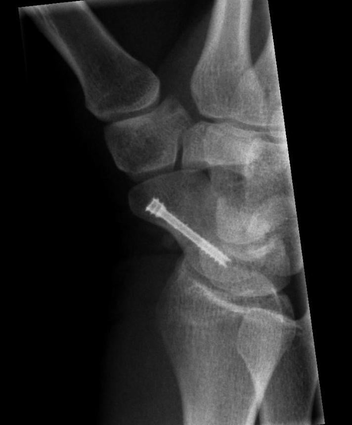
- 1Image — and don't miss itScaphoid-series radiographs. Up to ~20% are occult initially: if snuffbox + tubercle tenderness with normal films, immobilise and re-image with CT/MRI at 10–14 days.
- 2Classify on CTDedicated scaphoid CT (planes along the scaphoid axis) distinguishes incomplete non-displaced A2 from complete/displaced B2 — plain films can't reliably do this.
- 3Type A: cast (or offer fixation)A1/A2 → below-elbow scaphoid cast 6–8 weeks; the high-demand patient may choose percutaneous fixation for faster return.
- 4Type B/C/D: operativeB → headless compression screw (fix B3 even if non-displaced; B4 = urgent ORIF + ligament repair). C → fixation ± graft. D → bone graft (vascularised 1,2-ICSRA if proximal pole avascular) + correct humpback/DISI.
- Treatment
- Below-elbow scaphoid cast
- Immobilisation
- 4 to 6 weeks
- Union Rate
- Over 95 percent
- Return to Sport or Work
- 8 to 10 weeks
- Treatment
- Below-elbow scaphoid cast
- Immobilisation
- 6 to 8 weeks
- Union Rate
- 85 to 95 percent
- Return to Sport or Work
- 10 to 12 weeks
- Treatment
- Percutaneous or open Herbert screw
- Immobilisation
- 2 to 4 weeks post-op (thumb spica)
- Union Rate
- 85 to 95 percent
- Return to Sport or Work
- 8 to 12 weeks
- Treatment
- Open reduction, screw, ligament repair
- Immobilisation
- 6 to 8 weeks post-op
- Union Rate
- 70 to 85 percent
- Return to Sport or Work
- 4 to 6 months
- Treatment
- Screw fixation, consider bone graft
- Immobilisation
- 4 to 6 weeks post-op
- Union Rate
- 75 to 90 percent
- Return to Sport or Work
- 3 to 5 months
- Treatment
- Open fixation with cancellous bone graft
- Immobilisation
- 6 weeks post-op
- Union Rate
- 75 to 85 percent
- Return to Sport or Work
- 4 to 6 months
- Treatment
- Vascularised bone graft (1,2-ICSRA) or Russe graft plus fixation
- Immobilisation
- 6 to 8 weeks post-op
- Union Rate
- 60 to 80 percent
- Return to Sport or Work
- 6 to 9 months
Do not miss a scaphoid fracture on initial radiographs. Up to 20 percent of scaphoid fractures are occult on first X-rays. If a patient has anatomical snuffbox tenderness and scaphoid tubercle tenderness but normal radiographs, immobilise in a scaphoid cast and re-image with CT or MRI at 10 to 14 days. A missed scaphoid fracture that progresses to nonunion is a classic source of medicolegal claims and a preventable cause of wrist arthritis.
Clinical application and pitfalls
- The A2 versus B2 distinction hinges on displacement and completeness. An A2 hairline fracture must be genuinely non-displaced and incomplete on CT. Any displacement (even one millimetre of gapping or translation) upgrades the fracture to B2, which warrants fixation. Over-treating A2 with surgery is acceptable; under-treating B2 with a cast is not.
- Proximal pole fractures (B3) are inherently unstable. Even if non-displaced, the tenuous retrograde blood supply means prolonged casting carries an unacceptably high nonunion rate. Most hand surgeons now recommend operative fixation for all proximal pole fractures regardless of displacement.
- CT is the imaging modality of choice for classification. Plain radiographs underestimate displacement and cannot reliably distinguish complete from incomplete waist fractures. A dedicated scaphoid CT (sagittal and coronal reconstructions along the scaphoid axis) is essential before committing to non-operative treatment.
- Time to diagnosis matters. Scaphoid fractures diagnosed after a delay of more than 4 weeks have a higher nonunion rate. A patient presenting late with snuffbox tenderness and a radiographic waist fracture may already be a type C (delayed union) rather than an acute type B.
- The Herbert screw has largely been superseded by cannulated headless compression screws (e.g. Acutrak, Synthes HCS), but the classification remains the decision-making framework. The principle of rigid internal fixation with a headless compression screw spanning the fracture is unchanged.
- Type D2 nonunions with a humpback deformity and dorsal intercalated segment instability (DISI) require correction of the lunate angle in addition to bone grafting and fixation. A vascularised pedicle graft from the 1,2 intercompartmental supraretinacular artery (1,2-ICSRA) is a standard option for sclerotic proximal pole nonunions.
- Patient factors influence the decision. An elite athlete or manual worker with a type A2 fracture may reasonably choose percutaneous fixation to return to activity sooner, even though casting would likely succeed. Conversely, a low-demand patient with significant comorbidities may be managed more conservatively even for some type B fractures.
The classification depends entirely on imaging, so the examiner wants the strategy, not just "get a CT":
- The scaphoid radiographic series is more than a PA and lateral — it adds a dedicated scaphoid view (PA in ULNAR DEVIATION, which extends the scaphoid and elongates/profiles the waist) plus semi-pronated and semi-supinated obliques (typically four views). Even so, up to ~20% of fractures are occult on the initial films.
- MRI is the most sensitive (and highly specific) test for a radiographically-occult fracture and for bone-marrow oedema, and it shows proximal-pole vascularity / AVN (low T1 signal) — it is the first-line advanced image in many pathways for the clinically-suspected but film-normal scaphoid, and it avoids weeks of unnecessary casting in the patient who has no fracture.
- CT is the workhorse for classification and union — it best shows displacement and completeness (the A2-versus-B2 call), the humpback deformity / intrascaphoid angle, and bridging trabeculae to confirm union; reconstruct along the scaphoid's long axis. It is slightly less sensitive than MRI for a truly occult undisplaced fracture.
- (Bone scan was historically used for occult fractures — sensitive but non-specific — and has largely been superseded by MRI.)
Guidelines, registries and global practice
- No single society guideline replaces the classification. BOA/BSSH (UK), AAOS (US) and AO Foundation teaching all use the Herbert stability framework to triage cast versus fixation; the AAOS appropriate-use criteria and BOAST scaphoid pathways emphasise CT for equivocal injuries and early specialist referral for proximal-pole and displaced fractures.
- A widening role for early fixation. Internationally there is a trend toward percutaneous fixation of non-displaced waist fractures in high-demand patients (athletes, military, manual workers) to shorten time to union and return to work, balanced against the cost and small surgical risk — union rates are equivalent to casting, so the decision is functional, not about achieving union.
- Resource-setting variation. Where CT/MRI access is limited, presumptive casting with clinical follow-up and repeat radiographs remains standard; in well-resourced centres, immediate advanced imaging reduces both over-immobilisation of non-fractures and missed unstable fractures.
- Salvage for failed nonunion (proximal row carpectomy, four-corner fusion) is consistent across regions for SNAC-wrist arthritis when union cannot be achieved.
The topic keeps citing "SNAC wrist" as the consequence of untreated nonunion — here is the staging the examiner expects (it mirrors SLAC but starts from the scaphoid):
- Stage I — arthritis between the radial styloid and the distal scaphoid (styloscaphoid).
- Stage II — adds scaphocapitate arthritis.
- Stage III — adds capitolunate (midcarpal) arthritis.
- The radiolunate joint is characteristically SPARED even into late disease (as in SLAC) — which is the anatomical reason lunate-preserving salvage works.
Stage-dependent salvage: Stage I → radial styloidectomy (and address the nonunion); Stage II → proximal row carpectomy (PRC) or scaphoid excision + four-corner (midcarpal) fusion; Stage III → because the capitolunate joint is now involved, PRC is contraindicated (it depends on an intact capitate head articulating with the lunate fossa), so scaphoidectomy + four-corner fusion is preferred; an end-stage pancarpal wrist → total wrist arthrodesis (or arthroplasty in the low-demand). This is why staging the arthritis — not just diagnosing nonunion — drives the salvage choice.
Viva practice
- A stable acute (cast); B unstable acute (fix); C delayed union; D established nonunion.
- A2 (incomplete, non-displaced) is the only stable waist fracture — completeness/displacement (on CT) decides A2 vs B2.
- B3 proximal pole = highest nonunion from the retrograde (distal-entry) blood supply — fix even if non-displaced.
- B4 = trans-scaphoid perilunate — urgent ORIF + ligament repair.
- D2 with humpback/DISI — correct alignment, bone graft (vascularised 1,2-ICSRA if avascular).
- ~20% occult on first films — immobilise and re-image; untreated nonunion → SNAC wrist.
Exam viva
Practise clinical reasoning and management decisions out loud
“A 24-year-old man fell on his outstretched hand playing rugby. He has anatomical snuffbox tenderness. Initial scaphoid series radiographs show a non-displaced fracture through the scaphoid waist with no displacement. How do you classify and manage this?”
“A 38-year-old labourer presents with wrist pain 6 months after a fall. He was told at the time that X-rays were normal. Current radiographs show a sclerotic scaphoid waist fracture with cyst formation and a humpback deformity. The lunate is extended on the lateral view. Classify and outline your management.”
Exam cheat sheet
The four types
- Type A — stable acute (A1 tubercle, A2 incomplete hairline waist): cast treatment
- Type B — unstable acute (B1 distal oblique, B2 complete/displaced waist, B3 proximal pole, B4 perilunate dislocation, B5 comminuted): screw fixation
- Type C — delayed union (no union beyond 12 weeks despite adequate immobilisation): fix, add graft if gap
- Type D — established nonunion (D1 fibrous, D2 pseudarthrosis with sclerosis): open bone graft and fixation
Key decision points
- A2 versus B2: CT is mandatory — plain films cannot reliably distinguish incomplete from complete waist fractures
- Proximal pole (B3): fix operatively even if non-displaced due to retrograde blood supply and high nonunion risk
- Type D2 with DISI: correct lunate alignment, bone graft, fixation; use vascularised graft if proximal pole avascular
- Perilunate dislocation (B4): emergency reduction, open scaphoid fixation, scapholunate ligament repair
Vascular anatomy (why it matters)
- Main blood supply enters via the dorsal ridge (distal pole) and runs retrograde to the proximal pole
- Waist and proximal pole fractures disrupt this retrograde supply — risk of AVN of the proximal fragment
- Proximal pole AVN is assessed with MRI (low signal on T1) and intra-operative punctate bleeding (poke test)
- Vascularised bone graft (1,2-ICSRA from distal radius) is a standard option for avascular nonunions
Pitfalls and high-yield facts
- Up to 20 percent of scaphoid fractures are occult on initial radiographs — immobilise and re-image (CT or MRI at 10 to 14 days)
- Nonunion risk factors: proximal pole, displacement, delay to diagnosis, smoking, male sex
- SNAC wrist: radial styloscaphoid then scaphocapitate then capitolunate arthritis in untreated nonunion
- Even Herbert type A2 can be fixed percutaneously for faster return to work or sport (patient choice)
Evidence
Management of the fractured scaphoid using a new bone screw
- Introduced the Herbert classification (types A to D) and the original double-threaded (dual-pitch) compression screw.
- Prospective trial of 158 operations (1977–1981): union 100% for acute fractures and 83% overall (including nonunions).
- Rigid screw fixation often avoided post-operative plaster and allowed early return to work.
Herbert screw fixation of scaphoid fractures
- Review of 431 patients operated by a single surgeon over 13 years; average return to work 4.7 weeks after surgery.
- Healing rates for acute fractures were better than those reported for plaster immobilisation and were independent of fracture location.
- For established nonunions, healing depended on stage and location, but arthritis progression was halted and carpal collapse improved.
According to PubMed, the classification and dual-pitch screw are from Herbert & Fisher 1984 (DOI 10.1302/0301-620X.66B1.6693468; union 100% acute / 83% overall in 158 operations), with the larger single-surgeon series by Filan & Herbert 1996 (PMID 8682813; 431 patients, return to work ~4.7 weeks). The randomised screw-versus-cast data (union 7 vs 12 weeks; work 8 vs 15 weeks) are from Bond et al. 2001 (DOI 10.2106/00004623-200104000-00001), the natural history of nonunion (100% arthritis) from Lindström & Nyström 1992 (DOI 10.1016/0266-7681(92)90204-f), and the 1,2-ICSRA vascularised graft from Zaidemberg et al. 1991 (DOI 10.1016/0363-5023(91)90017-6).