Schwannoma | Meningioma | Neurofibroma | Myxopapillary Ependymoma
- Schwannoma vs Meningioma: Schwannoma is T2 hyperintense, meningioma is T2 iso/hypointense
- Dural tail sign: Suggestive of meningioma (contrast-enhancing dura adjacent to tumor)
- Dumbbell tumors: Neural foraminal extension, most commonly schwannoma (69%)
- Gross total resection: Curative for most benign IDEM tumors
- NF2 association: Multiple schwannomas/meningiomas, consider NF2 screening
- “T2 bright = schwannoma, T2 iso/dark = meningioma
- “Thoracic location + female = think meningioma
- “Dumbbell shape + cervical = think schwannoma
- “NF1 = neurofibromas, NF2 = schwannomas and meningiomas
Overview and Epidemiology
The compartment. Intradural extramedullary (IDEM) tumours lie within the dural sac but outside the spinal cord parenchyma. They account for 55-75% of all intradural spinal tumours and are predominantly benign.
- Frequency
- 40%
- Peak age
- 30-60
- Sex
- M = F
- Frequency
- 25%
- Peak age
- 40-70
- Sex
- F more than M (4:1)
- Frequency
- 15%
- Peak age
- 30-50
- Sex
- M = F
- Frequency
- 10%
- Peak age
- 20-40
- Sex
- M more than F
- Frequency
- 10%
- Peak age
- Variable
- Sex
- Variable
The patterns. Meningioma shows a strong female predominance (80%) and a thoracic predilection. Schwannoma affects males and females equally and occurs at any spinal level, often cervical or lumbar. Myxopapillary ependymoma of the filum terminale is the most common primary tumour of the conus and cauda equina region.
Pathophysiology
Schwannoma. Arises from the Schwann cells of a spinal nerve root, usually a single one, and grows eccentrically from it as an encapsulated mass. The nerve fibres are displaced around the capsule rather than infiltrated, which is why the nerve can often be preserved at surgery. Malignant transformation is extremely rare in sporadic schwannomas but may occur in NF2.
- Antoni A areas: cellular, organised palisading (Verocay bodies)
- Antoni B areas: loose, myxoid, less cellular
- S-100 protein positive
Meningioma. Arises from arachnoid cap cells in the spinal meninges, most commonly at the posterolateral aspect of the dura, and grows as a typically well-circumscribed mass with a broad dural attachment. It may calcify (psammoma bodies), and the dural tail seen on MRI reflects reactive dural thickening. There is an association with female sex and hormonal factors; progesterone receptors are often positive.
- Meningothelial: the most common spinal subtype
- Psammomatous: with calcifications
- Transitional
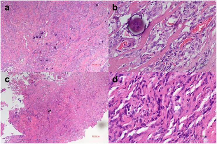
Neurofibroma. Contains Schwann cells, fibroblasts and collagen, and the nerve fibres pass through the tumour rather than around it. That infiltrative architecture is the practical difference from schwannoma: complete resection usually requires sacrifice of the nerve root. S-100 marks the Schwann cell component and CD34 the fibroblast component.
- Localised: a single nerve, similar to schwannoma
- Plexiform: "bag of worms" appearance, pathognomonic of NF1
- Diffuse: poorly circumscribed
Myxopapillary ependymoma. A distinct subtype arising from the ependymal cells of the filum terminale: well-circumscribed and encapsulated, with papillary architecture over myxoid cores, GFAP positive. Its mucin content makes it T2 hyperintense, and it may contain haemorrhagic areas. The histology is benign, but violating the capsule at surgery increases the risk of CSF seeding and recurrence.
Classification
- Location
- Outside dura
- Common tumours
- Metastases, primary bone tumours
- Location
- Inside dura, outside cord
- Common tumours
- Schwannoma, meningioma, neurofibroma
- Location
- Within spinal cord
- Common tumours
- Ependymoma, astrocytoma, haemangioblastoma
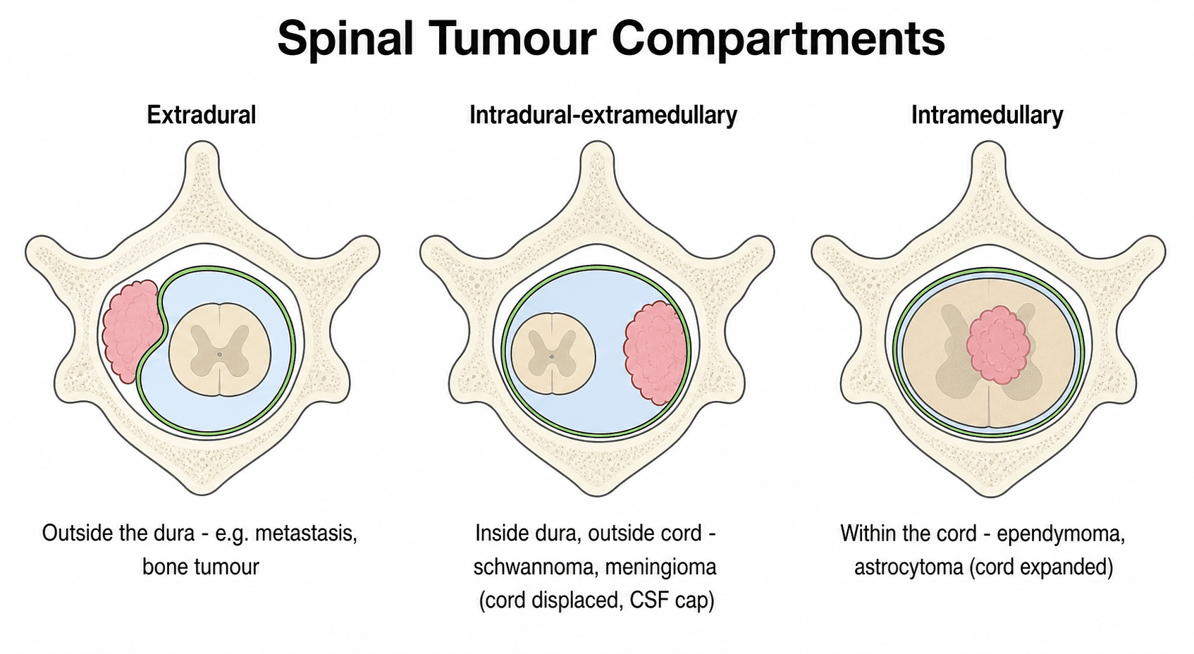
Localising on axial MRI. An extradural tumour sits between dura and bone and pushes the thecal sac and cord inward. An IDEM tumour displaces the cord to the opposite side while widening (capping) the CSF space on the tumour side. An intramedullary tumour expands the cord itself and thins the surrounding CSF all round.
Why it matters. IDEM tumours displace the spinal cord rather than infiltrate it, which generally allows cord function to be preserved at resection.
Clinical Presentation
Pain. The most common initial symptom: localised back pain in 70%, or radicular pain following the distribution of a nerve root, and it may precede any neurological deficit by months to years. Night pain, and pain that is worse on lying down, is the discriminator. Mechanical back pain is typically relieved by recumbency, so the patient whose pain wakes them or drives them out of bed to sit up has earned an MRI. The proposed mechanism is the rise in epidural venous pressure and the loss of the distraction of daytime activity when supine.
The trap. Because pain can precede any deficit by months to years, these patients are frequently treated as mechanical back pain through several cycles of physiotherapy before anyone images them.
Deficit. Sensory change (numbness, paraesthesiae), progressive motor weakness and gait disturbance; bladder and bowel dysfunction is late.
Each tumour presents in its own way.
- Schwannoma: radicular pain in a dermatomal distribution, sensory loss in the affected root, and motor weakness if the motor root is involved
- Meningioma: often progressive myelopathy with gait disturbance and spasticity, Brown-Sequard syndrome if the compression is lateral, and may have minimal radicular symptoms
- Myxopapillary ependymoma: low back pain and cauda equina symptoms (bowel and bladder, saddle anaesthesia), often with a long history before diagnosis
Examination. Cord compression gives upper motor neuron signs below the lesion: spasticity, hyperreflexia, clonus and a positive Babinski. Root compression gives lower motor neuron signs at the affected level: weakness in a myotomal distribution, hyporeflexia and muscle atrophy.
Tumours of the lumbar spine may cause cauda equina syndrome with saddle anaesthesia, urinary retention, and bilateral leg weakness. This is a surgical emergency requiring urgent decompression.
Investigations
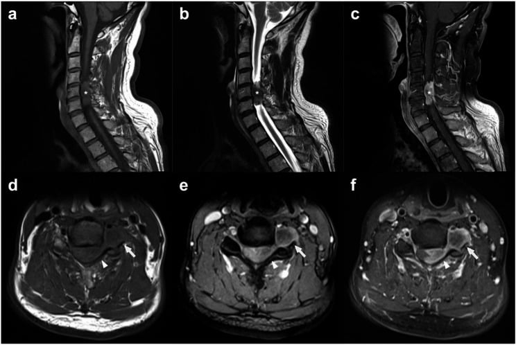
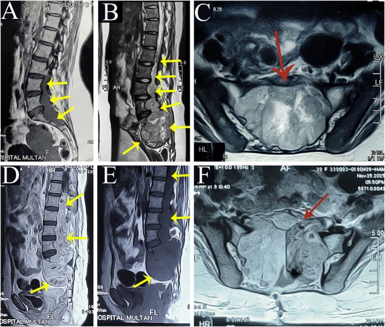
MRI is the gold standard, and each sequence answers a different question. T1 defines the anatomical relationships and gives the pre-contrast baseline; tumours are usually isointense to cord. T2 or STIR is the key differentiating sequence, schwannoma hyperintense and meningioma iso- or hypointense, and shows cord oedema. All IDEM tumours enhance after gadolinium: schwannoma heterogeneously, meningioma homogeneously and with a dural tail. Image the whole spine to exclude skip lesions, additional tumours and NF2.
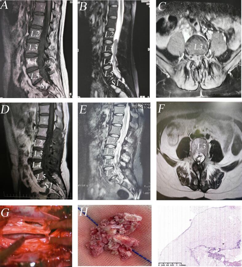
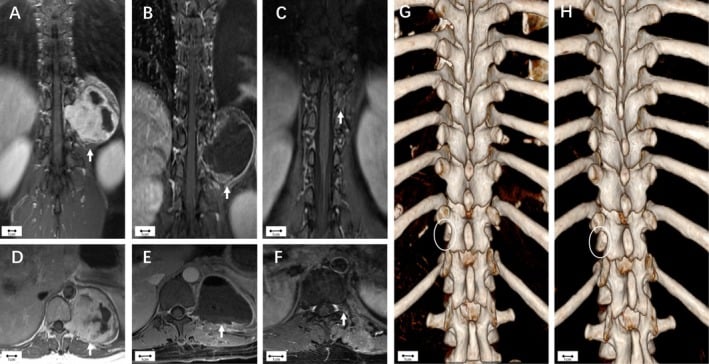
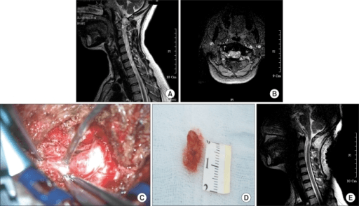
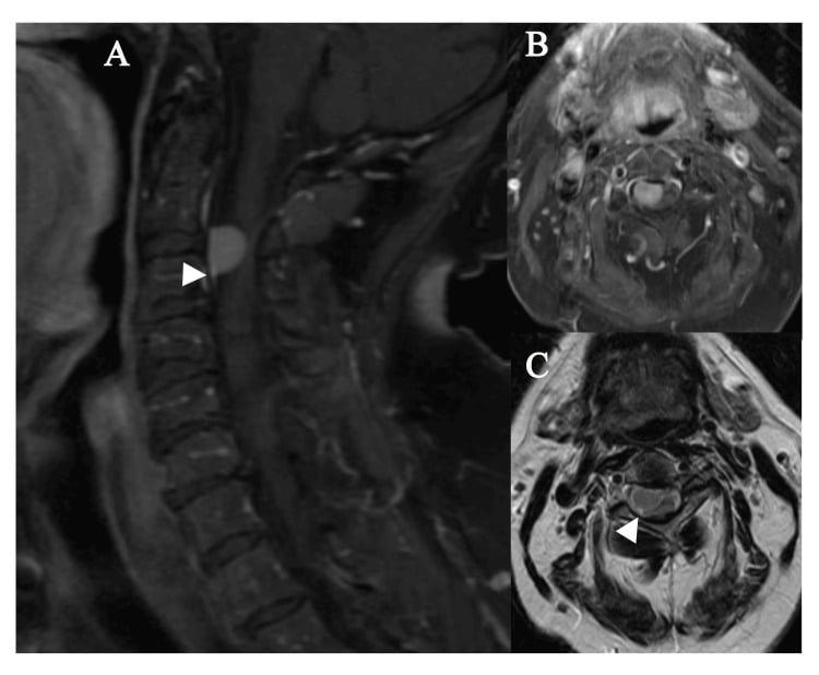
Schwannoma. Iso- or hypointense to cord on T1 and hyperintense on T2, round and eccentric to the nerve root, with heterogeneous enhancement that may have non-enhancing cystic areas. Foraminal extension and bone erosion mark the dumbbell tumour. The tumour-to-fat signal intensity ratio on T2 is significantly higher in schwannoma than in meningioma and differentiates the two reliably.
Meningioma. Isointense on T1 and iso- or hypointense on T2, with avid homogeneous enhancement from a broad dural base, a lateral or posterolateral position, and a ginkgo leaf shape. Calcification may be present and is dark on all sequences. The dural tail is suggestive but not pathognomonic.
- Schwannoma
- HYPERINTENSE (bright)
- Meningioma
- Iso/HYPOINTENSE (dark)
- Schwannoma
- Heterogeneous, may be cystic
- Meningioma
- Homogeneous
- Schwannoma
- Absent/rare
- Meningioma
- Present (64%)
- Schwannoma
- Any level, cervical common
- Meningioma
- 80% thoracic
- Schwannoma
- M = F
- Meningioma
- 80% female
- Schwannoma
- Common (29%)
- Meningioma
- Rare (3%)
- Schwannoma
- Common (96%)
- Meningioma
- Rare (24%)
- Schwannoma
- Yes (displaced/encased)
- Meningioma
- No (dural origin)
Neurofibroma. T2 hyperintense like a schwannoma, but with a target sign of central low T2 signal; plexiform lesions have the bag-of-worms appearance, and the tumours may be multiple in NF1.
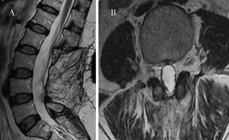
Myxopapillary ependymoma. T2 hyperintense and well-circumscribed at the filum terminale, with homogeneous enhancement; haemorrhage shows as T1 bright areas.
Drop metastases. Multiple nodular lesions coating the nerve roots. Consider a primary CNS tumour (medulloblastoma, ependymoma).
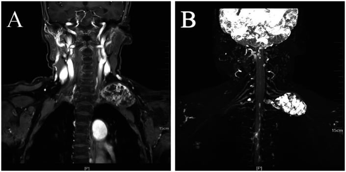
CT shows the bone erosion of a dumbbell tumour and the calcification of a meningioma, and is used for pre-operative planning. Blood tests are the routine pre-operative workup; consider genetic testing if NF is suspected.
- Compartment
- IDEM
- Discriminating Features
- T2 bright, cystic, foraminal extension, eccentric to root
- Key Pitfall
- Mistaking for neurofibroma when solitary
- Compartment
- IDEM
- Discriminating Features
- T2 iso/dark, broad dural base, dural tail, thoracic, female
- Key Pitfall
- Calcified plaque mimicking bone lesion
- Compartment
- IDEM
- Discriminating Features
- Target sign, nerve fibres pass through tumor, NF1, plexiform
- Key Pitfall
- Plexiform lesion harbouring MPNST
- Compartment
- IDEM (filum)
- Discriminating Features
- Filum terminale, sausage-shaped, mucin, may bleed
- Key Pitfall
- Piecemeal removal causing CSF seeding
- Compartment
- Intramedullary
- Discriminating Features
- Cord expansion, central location, syrinx
- Key Pitfall
- Calling an intramedullary tumor IDEM
- Compartment
- IDEM (multiple)
- Discriminating Features
- Multiple nodules, nerve-root coating, sugar-coating
- Key Pitfall
- Single deposit mimicking primary tumor
- Compartment
- IDEM
- Discriminating Features
- CSF signal, no enhancement, cord displacement
- Key Pitfall
- Cyst masquerading as cystic schwannoma
- Compartment
- Extradural
- Discriminating Features
- Bone destruction, epidural soft tissue, restricted diffusion (abscess)
- Key Pitfall
- Missing infection in a diabetic patient
Management
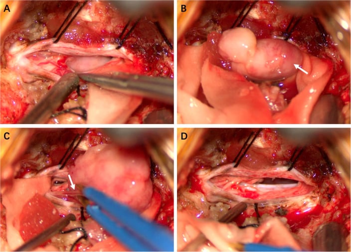
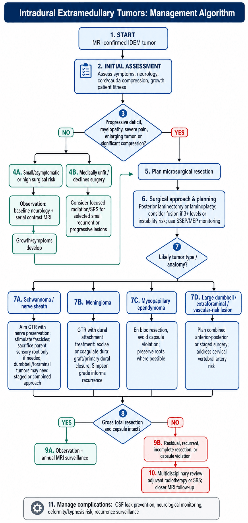
The standard operation is posterior. Most IDEM tumours are approached this way, with intraoperative neurophysiological monitoring (SSEP, MEP) recommended throughout. Laminectomy is the standard approach and laminoplasty the alternative that may reduce post-laminectomy kyphosis; whether the lamina is removed or replaced, and whether instrumentation is added, is decided on the stability grounds set out below.
- Posterior midline incision
- Laminectomy or laminoplasty
- Durotomy (midline or paramedian)
- Tumour identification
- Microsurgical resection
- Watertight dural closure
- Bone replacement if laminoplasty
Schwannoma. The goal is gross total resection with the nerve preserved where possible, and it is achievable in 90%+. Identify the functioning fascicles with stimulation, peel the tumour from the nerve capsule, and sacrifice the parent root only if necessary; a sensory root is often tolerated. Long-term control after gross total resection is excellent.
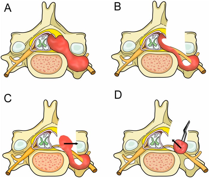
Dumbbell tumours. The approach follows the Eden type. Stage the surgery when the extraforaminal component is large, and in the cervical spine plan for the vertebral artery.
Meningioma. The goal is gross total resection with the dural attachment dealt with, achievable in 85-95% of spinal meningiomas. Identify the dural attachment, dissect circumferentially from the cord, and excise the involved dura with primary closure or a graft; where excision is not possible, coagulate the dural margin. Simpson grading predicts recurrence:
- Grade I: GTR + dural excision + bone removal
- Grade II: GTR + coagulation of the dural attachment
- Grade III: GTR without dural treatment
- Grade IV: subtotal resection
- Grade V: decompression only
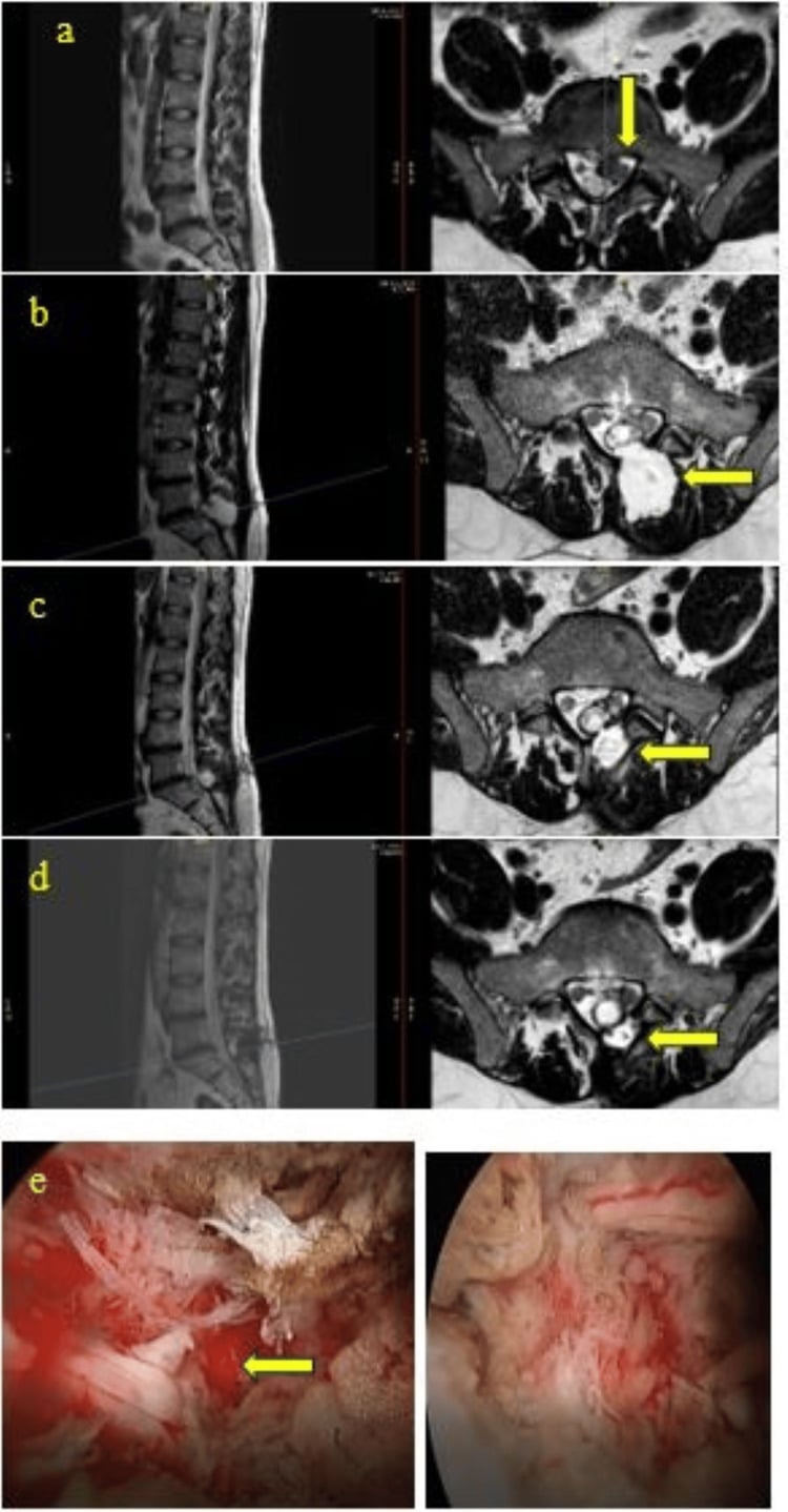
Myxopapillary ependymoma. The goal is en bloc resection without violating the capsule, preserving the nerve roots where possible. The prognosis after gross total resection is excellent, capsule violation increases the risk of CSF seeding, and en bloc removal is preferred over piecemeal. After a gross total resection the patient is observed; after a subtotal resection or a violated capsule, radiotherapy. Follow-up MRI is annual.
What the largest series shows. Weber's 183 patients: 10-year overall survival 92.4% and 10-year progression-free survival 61.2%. Quote both, because this tumour rarely kills and frequently returns. Treatment failed in about a third (31.7%), mostly locally (26.8%), but distant spinal failure in 9.3% and intracranial in 6.0% is why the whole neuraxis is imaged, not just the lumbar spine. Extent of resection and adjuvant radiotherapy were independent prognostic factors, as was age: patients under 36 were more likely to progress.
En bloc is the aim, not the outcome that matters. The outcome that separates good from catastrophic is whether the resection was total or partial, and whether an incomplete resection received adequate radiotherapy, not whether the specimen came out in one piece.
Observation suits small asymptomatic tumours, slow-growing tumours followed with serial MRI, and elderly or medically unfit patients. Radiation is rarely used for benign IDEM tumours: consider it for recurrent or incompletely resected meningioma, stereotactic radiosurgery for small recurrences, and as primary treatment if the patient is not a surgical candidate.
Calcified and Ventral Meningioma: The Difficult Subset
The typical spinal meningioma. Most are soft, posterolateral, WHO Grade I lesions that are gross totally resected in 85-95 percent of cases. A minority behave very differently, and recognising them preoperatively changes the consent and the surgical plan: a ventral calcified thoracic meningioma carries a higher recurrence and complication rate than the typical posterolateral lesion.
- Surgical challenge
- The spinal cord lies between the surgeon and the tumour, so aggressive dural resection risks cord injury
- Strategy
- Often accept Simpson Grade II (coagulate the dural attachment) over a hazardous Grade I; consider a more lateral approach and gentle cord handling
- Surgical challenge
- Hard, adherent and cannot be debulked or rolled off the cord; higher rate of subtotal resection and neurological worsening
- Strategy
- Ultrasonic aspirator, patient piecemeal removal, leave an adherent calcified plaque rather than injure the cord
- Surgical challenge
- Can mimic a bony lesion or ossification and be under-recognised as a meningioma
- Strategy
- CT to characterise calcification; correlate with the dural-based enhancing mass on MRI
For a ventral or ventrolateral calcified thoracic meningioma, do not chase a Simpson Grade I dural excision across the front of the cord. Coagulating the dural attachment (Grade II) accepts a marginally higher recurrence rate in exchange for avoiding catastrophic cord injury - the right trade-off when the dura is ventral.
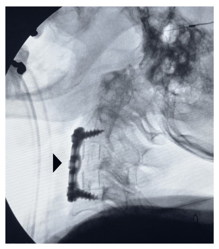
Spinal Stability and Post-Laminectomy Deformity
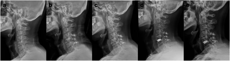
The approach itself can destabilise the spine. Resecting an IDEM tumour requires a laminectomy or laminoplasty, and post-laminectomy kyphosis (a swan-neck deformity in the cervical spine) is the chief late mechanical problem, so deciding when to add instrumented fusion is part of operative planning. Laminoplasty, replacing the lamina rather than removing it, preserves the posterior tension band and may reduce kyphosis.
- Why it destabilises
- Loss of the posterior tension band over a long segment
- Action
- Strongly consider instrumented fusion or laminoplasty
- Why it destabilises
- Facet joints are key stabilisers; unilateral complete facetectomy is destabilising
- Action
- Add fusion, especially with a large extraforaminal component
- Why it destabilises
- High mechanical demand and transition in alignment
- Action
- Lower threshold to fuse
- Why it destabilises
- Highest rate of post-laminectomy kyphosis
- Action
- Favour laminoplasty; close deformity surveillance
- Why it destabilises
- Surgery unmasks or worsens it
- Action
- Correct and stabilise as indicated
The highest risk of post-laminectomy kyphosis is in children and after cervical laminectomy. Preserve the facet joints where possible, favour laminoplasty over laminectomy in these groups, and add instrumented fusion when the laminectomy is long, crosses a junction, or requires more than half a facet.
Management Algorithm
Complications
Early. CSF leak is the most common, at 5-10%; the others are wound infection, a new neurological deficit in 2-5%, and haematoma.
Late. Post-laminectomy kyphosis, tumour recurrence, chronic pain and arachnoiditis.
Preventing the CSF leak. Meticulous dural closure, a dural graft if primary closure is not possible, fibrin sealant augmentation, and a watertight closure in layers. If a leak occurs:
- Wound care
- Lumbar drain for 3-5 days
- Re-exploration if conservative measures fail
Recurrence. The conventional figures, by extent of resection:
- GTR recurrence
- Less than 5%
- STR recurrence
- 30-40%
- GTR recurrence
- 5-10%
- STR recurrence
- 20-30%
- GTR recurrence
- Variable
- STR recurrence
- Higher with NF1
- GTR recurrence
- See the Weber figures under Management: resection extent was prognostic
- STR recurrence
- See Management
Read this table as orders of magnitude, not as sourced figures. The schwannoma, meningioma and neurofibroma rows are the conventional teaching estimates and no single series establishes them. For myxopapillary ependymoma the best figure available is Weber's 10-year progression-free survival quoted under Management, so roughly two in five progress by ten years; that series does not publish a clean GTR-versus-STR recurrence split, so quoting one would be inventing it.
Guidelines, Registries & Global Practice
Global Epidemiology
IDEM tumors comprise roughly 55-75% of all intradural spinal tumors. Incidence of primary spinal tumours overall is about 0.5-2.5 per 100,000 person-years. Schwannoma and meningioma together account for the majority; spinal meningioma shows a 3:1 to 4:1 female predominance with a thoracic predilection, while nerve sheath tumours are evenly distributed by sex and across spinal levels. Myxopapillary ependymoma is the commonest tumour of the conus/filum and presents in young adults.
Society Guidance & Frameworks (side by side)
- Scope
- Histopathology
- Practical recommendation
- Grades schwannoma/meningioma/neurofibroma (Gr I), atypical meningioma (Gr II); myxopapillary ependymoma reclassified to WHO Grade 2
- Scope
- Meningioma
- Practical recommendation
- Surgery first-line for symptomatic lesions; radiotherapy for atypical/incompletely resected disease
- Scope
- Ependymoma
- Practical recommendation
- Maximal safe resection; adjuvant RT after subtotal resection or anaplastic histology
- Scope
- Suspected spinal tumour / cord compression
- Practical recommendation
- Urgent whole-spine MRI and specialist referral for new myelopathy or cauda equina features
- Scope
- NF2-related schwannomatosis
- Practical recommendation
- Coordinated genetics, audiology, ophthalmology; consider bevacizumab for progressive vestibular schwannoma
There is no single arthroplasty-style registry for IDEM tumours; outcome evidence derives from institutional series, the Rare Cancer Network (myxopapillary ependymoma) and national cancer/CNS-tumour registries (e.g. CBTRUS in the US) which inform incidence and survival rather than implant survival.
High- vs Limited-Resource Practice Variation
In well-resourced centres, microsurgery with the operating microscope, intra-operative neurophysiological monitoring (SSEP/MEP/D-wave), ultrasonic aspiration and ready access to MRI surveillance underpin gross total resection rates comparable to international centres of excellence. In limited-resource settings, delayed presentation with established myelopathy is common, monitoring may be unavailable, and emphasis shifts to timely decompression and watertight closure to limit CSF leak. Genetic testing and dedicated NF2/schwannomatosis multidisciplinary clinics are concentrated in tertiary referral networks; telemedicine and regional referral pathways help extend specialist input where local capacity is limited.
Controversies & Areas of Uncertainty
1. Laminectomy vs laminoplasty vs instrumented fusion. Whether laminoplasty or routine instrumentation reduces post-operative deformity remains unsettled. Most series reserve fusion for multilevel laminectomy (3 or more levels), the cervicothoracic/thoracolumbar junctions, pre-existing deformity, and paediatric patients in whom post-laminectomy kyphosis is far more common.
2. Nerve root sacrifice in nerve sheath tumors. Traditional teaching preserves the parent root; contemporary data (Vandenbulcke et al., 2023) suggest the involved root in foraminal/dumbbell tumors is frequently non-functional and can be sacrificed to achieve gross total resection with a low risk of clinically significant deficit. Intra-operative stimulation to confirm a non-eloquent root is the pragmatic compromise.
3. Extent of dural handling for meningioma. Simpson Grade I (dural excision and duraplasty) lowers recurrence versus Grade II (coagulation only) but raises the risk of CSF leak and cord injury when the dura is ventral. Many surgeons accept Grade II for ventral/ventrolateral thoracic meningiomas where aggressive dural resection is hazardous.
4. Role of radiosurgery. Stereotactic radiosurgery is increasingly used for residual/recurrent meningioma and schwannoma and for NF2 tumors, but long-term spinal cord tolerance data are limited; surgery remains first-line for symptomatic compressive lesions.
5. Surveillance versus treatment of small asymptomatic tumors. Incidental small IDEM tumors (especially in NF2) are often observed with serial MRI; growth rate and symptom development, rather than size alone, drive the decision to operate.
Clinical Decision Scenarios
Practise clinical reasoning and management decisions out loud
“A 55-year-old woman presents with 6 months of progressive gait difficulty and band-like thoracic pain. MRI shows a T6-T7 intradural extramedullary mass that is isointense on T2, enhances homogeneously, and has a dural tail sign.”
“A 45-year-old man presents with right-sided neck pain and C6 radiculopathy. MRI shows a C5-C6 intradural extramedullary tumor that is T2 hyperintense with heterogeneous enhancement. There is foraminal extension with an extraspinal component.”
“A 28-year-old man presents with 18 months of low back pain and recent onset of urinary hesitancy. MRI shows a well-circumscribed L3-L4 intradural tumor arising from the filum terminale. It is T2 hyperintense with homogeneous enhancement.”
“A 32-year-old woman presents with progressive gait ataxia. MRI shows three intradural extramedullary tumors at C3, T6, and L2. She also reports hearing loss for 2 years.”
Schwannoma vs Meningioma
- Schwannoma: T2 BRIGHT, heterogeneous, cystic, foraminal extension
- Meningioma: T2 ISO/DARK, homogeneous, dural tail, thoracic
- Signal intensity ratio on T2 reliably differentiates
- Schwannoma = any level, Meningioma = 80% thoracic
Tumor Frequencies
- Schwannoma: 40% (most common)
- Meningioma: 25% (80% female)
- Neurofibroma: 15% (NF1 associated)
- Myxopapillary ependymoma: 10% (filum terminale)
Surgical Goals
- GTR is curative for most IDEM tumors
- Schwannoma: Nerve preservation often possible
- Meningioma: GTR with dural excision (Simpson Grade I) or coagulation of the attachment (Grade II)
- Ependymoma: EN BLOC essential (capsule violation = seeding)
Dumbbell Tumors
- 69% are schwannomas
- Eden Classification: I-III based on extension
- Combined approach may be needed
- Assess stability if facetectomy required
NF Association
- NF1: Neurofibromas, plexiform, cafe-au-lait, chr 17
- NF2: Schwannomas + meningiomas, bilateral VS, chr 22
- Multiple spinal tumors = screen for NF2
- NF2 requires multidisciplinary approach
Complications
- CSF leak most common (5-10%)
- Post-laminectomy kyphosis (consider fusion if 3+ levels)
- Recurrence depends on the tumour: schwannoma and meningioma are low after complete excision; myxopapillary ependymoma reaches 39% progression by 10 years (Weber, 10-year PFS 61.2%)
- Neurological deficit rare with microsurgical technique
Evidence Base
T2 Signal Intensity Ratio Differentiates Schwannoma from Meningioma
- 20 schwannomas vs 20 meningiomas, pathology-proven IDEM tumors
- Tumor-to-fat T2 signal-intensity ratio significantly higher in schwannoma (p=0.002)
- Optimal SI-ratio cutoff 0.420 (sensitivity 80%, specificity 70-75%)
- Area under ROC curve 0.78 for both independent readers
Long-Term Outcome After Spinal Schwannoma Removal
- 187 surgically treated spinal schwannomas, median follow-up 12.9 years
- THE HEADLINE THE PAPER OPENS WITH: only ONE-FIFTH of patients considered themselves free of symptoms at follow-up. The authors' first line asks whether spinal schwannomas are as benign as we think
- Neurological residua were common: paraparesis in 31%, radicular deficit in 28%, sensory deficit from a cord lesion in 27%, and difficulty voiding in 19%
- Life expectancy equivalent to the general population
- Late complications in 21%: arachnoiditis (6%), spinal deformity (6%), cystic myelopathy (2%)
- Residual local pain (46%) and radiating pain (43%) were the commonest late complaints
Spinal Meningiomas: Surgical Management and Outcome
- Single-institution series plus pooled review of 556 patients from six large series
- Spinal meningiomas can be completely resected in most cases
- Surgery associated with postoperative functional improvement
- Low recurrence rate after complete resection
Long-Term Outcome of Spinal Myxopapillary Ependymoma (Rare Cancer Network)
- 183 patients across MD Anderson and the Rare Cancer Network
- Estimated 10-year overall survival 92.4%; 10-year progression-free survival 61.2%
- Treatment failure in ~32%, predominantly local (27%) but distant spinal/brain relapse in 9-6%
- THREE independent prognostic factors, not two: extent of surgery, use of adjuvant radiotherapy - and AGE, where the finding runs against intuition. Patients UNDER 36 were significantly MORE likely to recur or progress, not less
- The 92.4% ten-year survival and the 61.2% ten-year progression-free survival are the two numbers to quote together: this tumour rarely kills and frequently comes back
Nerve Root Sacrifice for Foraminal/Dumbbell Nerve Sheath Tumors
- 26 foraminal/dumbbell benign nerve sheath tumors; involved root routinely sacrificed
- Gross total resection achieved in 84.6%
- Functional motor roots (C5-T1, L3-S1) involved in 14 cases
- Persistent new or worsened motor deficit in only 1/9 and 1/5 patients respectively at follow-up
What Actually Drives Myxopapillary Recurrence: Total versus Partial Resection
- 25 cauda equina myxopapillary ependymomas, mean post-operative follow-up 10.4 years
- THE CATASTROPHIC GROUP IS PARTIAL RESECTION: all SIX patients treated with partial resection and local radiotherapy alone DIED of CSF dissemination
- Total resection did well irrespective of whether it was en bloc or piecemeal - 14 of 15 totally resected patients were recurrence-free, and 9 of those 15 total resections were performed PIECEMEAL and then irradiated
- The single recurrence among total resections was the one case given NO radiotherapy after the capsule was violated intraoperatively - it recurred locally at 2 years
- Radiotherapy adequacy mattered as much as its use: after subtotal resection, 24 Gy craniospinal alone recurred in 2 of 2, while craniospinal plus a 46 Gy local boost recurred in 0 of 2
Unencapsulated Myxopapillary Tumours: When Chasing Total Removal Causes the Deficit
- Retrospective multicentre series of 20 surgically treated spinal myxopapillary ependymomas, median follow-up 72.9 months
- ALL FIVE POST-OPERATIVE NEUROLOGICAL DETERIORATIONS OCCURRED IN UNENCAPSULATED TUMOURS - two after piecemeal gross total removal and three after subtotal removal. None followed a clean encapsulated resection
- There was NO tumour recurrence and no progression of residual tumour anywhere in the series, including the subtotally resected patients who received radiotherapy
- Heterogeneous enhancement on MRI was significantly associated with an unencapsulated tumour - a preoperative warning that the plane will not be clean
- The authors' explicit conclusion: surgeons 'should not stick to total removal in infiltrated and adhering tumors', because subtotally resected tumours with postoperative radiotherapy have not always recurred
References
- Takashima H, Takebayashi T, Yoshimoto M, et al. Differentiating spinal intradural-extramedullary schwannoma from meningioma using MRI T2 weighted images. Br J Radiol. 2018;91(1092):20180262. doi:10.1259/bjr.20180262. PMID 30052467.
- Seppala MT, Haltia MJ, Sankila RJ, et al. Long-term outcome after removal of spinal schwannoma: a clinicopathological study of 187 cases. J Neurosurg. 1995;83(4):621-6. doi:10.3171/jns.1995.83.4.0621. PMID 7674010.
- Gottfried ON, Gluf W, Quinones-Hinojosa A, et al. Spinal meningiomas: surgical management and outcome. Neurosurg Focus. 2003;14(6):e2. doi:10.3171/foc.2003.14.6.2. PMID 15669787.
- Weber DC, Wang Y, Miller R, et al. Long-term outcome of patients with spinal myxopapillary ependymoma: treatment results from the MD Anderson Cancer Center and institutions from the Rare Cancer Network. Neuro Oncol. 2015;17(4):588-95. doi:10.1093/neuonc/nou293. PMID 25301811.
- Vandenbulcke A, D'Onofrio GF, Capo G, et al. Sacrifice of involved nerve root during surgical resection of foraminal and/or dumbbell spinal neurinomas. Brain Sci. 2023;13(1):109. doi:10.3390/brainsci13010109. PMID 36672090.