Kellgren-Lawrence Grading of Osteoarthritis
A Kellgren-Lawrence grade of 2 or greater defines radiographic osteoarthritis in most research definitions. Grade 1 is doubtful and not counted as OA. When reporting, always state the joint, the view, and whether an atlas was used for grading. Remember that radiographic severity alone does not dictate treatment — symptoms, function and patient factors matter more.
The Kellgren-Lawrence Grading System
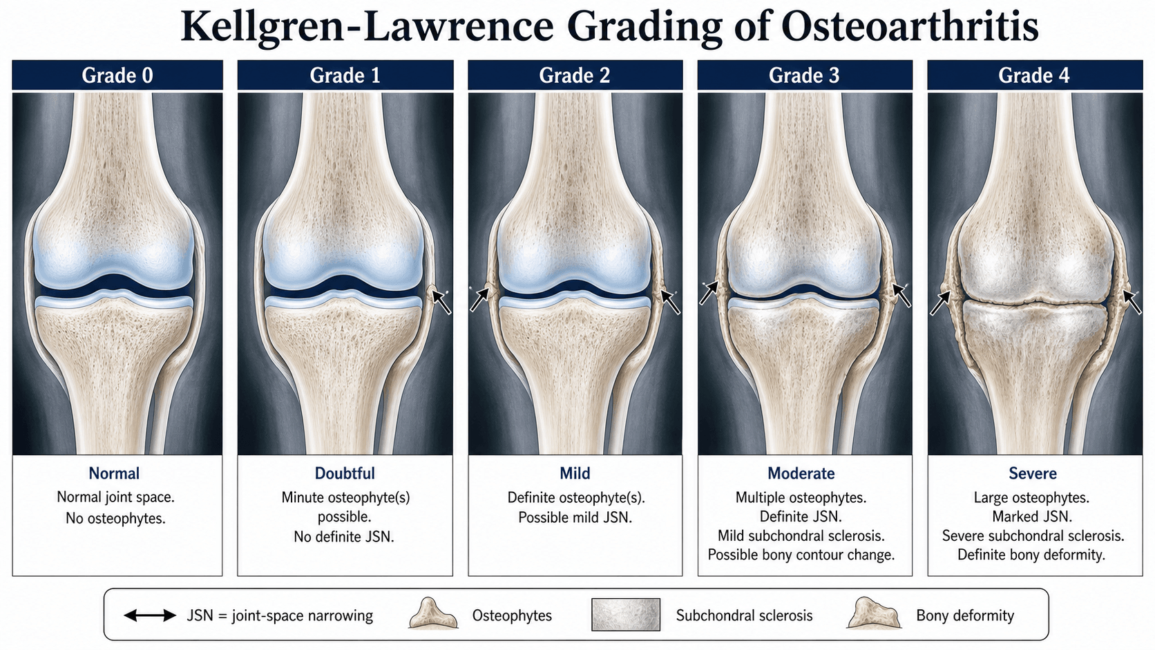
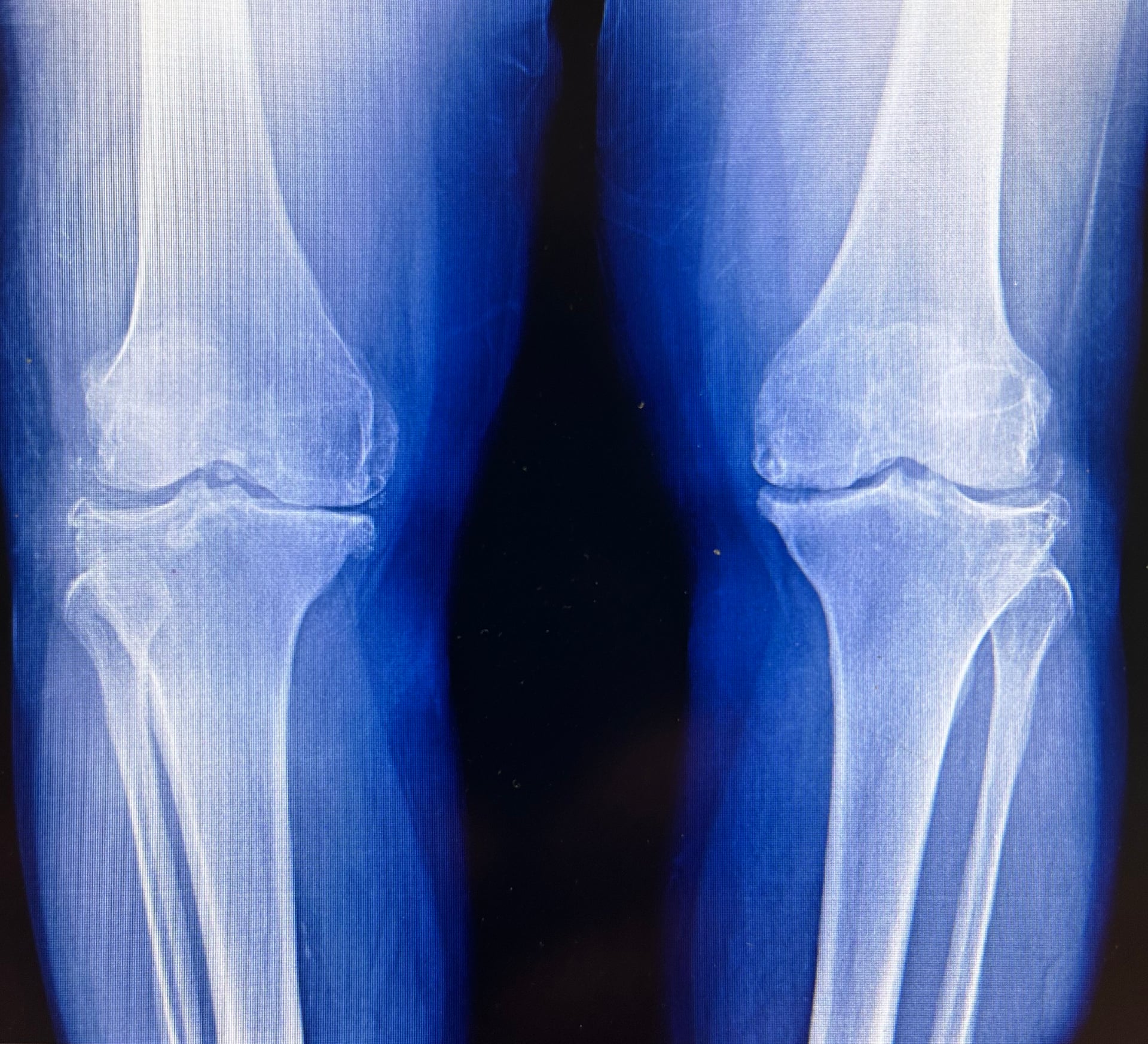
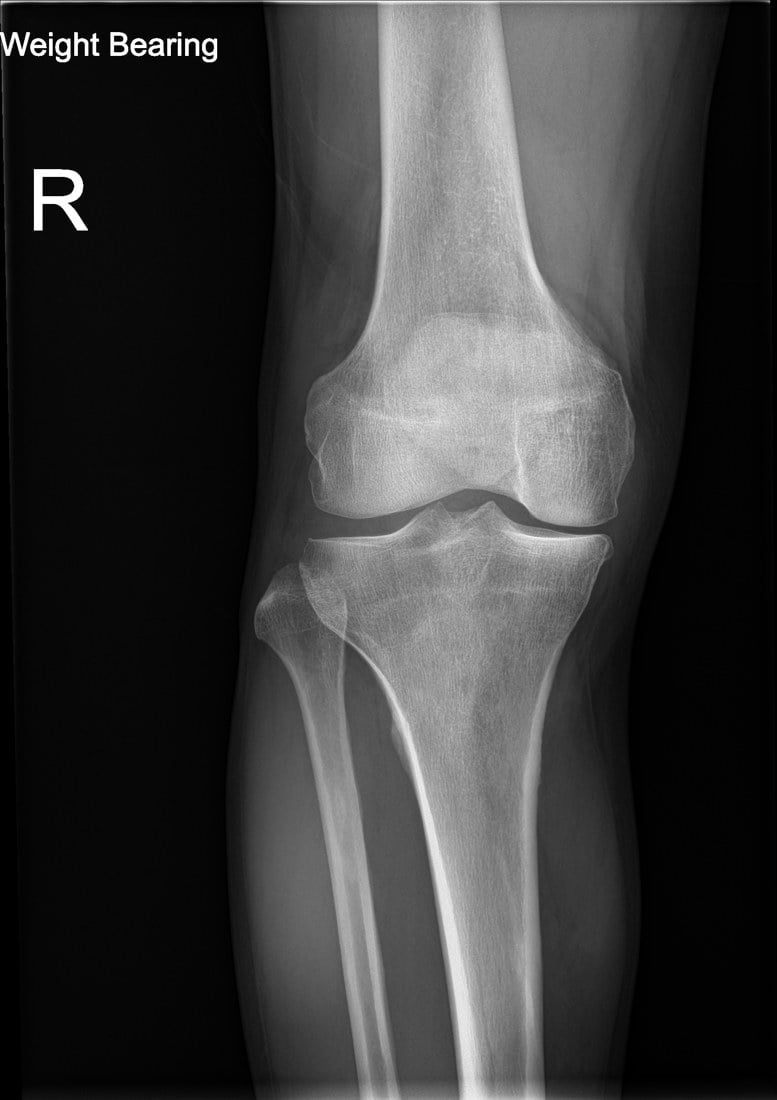
The five grades are defined by progressive radiographic features. The original description used an atlas for reference.
- Osteophytes
- None
- Joint-space narrowing
- None
- Sclerosis
- None
- Deformity
- None
- Osteophytes
- Possible
- Joint-space narrowing
- None
- Sclerosis
- None
- Deformity
- None
- Osteophytes
- Definite
- Joint-space narrowing
- Possible
- Sclerosis
- None
- Deformity
- None
- Osteophytes
- Multiple moderate
- Joint-space narrowing
- Definite
- Sclerosis
- Present
- Deformity
- Possible
- Osteophytes
- Large
- Joint-space narrowing
- Marked
- Sclerosis
- Marked
- Deformity
- Definite
Osteophytes • Narrowing • Sclerosis • DeformityThe four radiographic features
Hook:Look for osteophytes first (earliest), then narrowing, then sclerosis, then deformity — each higher grade adds or worsens a feature.
Grade 2 is the key cut-off. Definite osteophytes with or without possible narrowing equals mild but definite OA. This threshold is used in most epidemiological studies and trial inclusion criteria.
Joint-space narrowing — the feature that separates grades 2-4 — is view-dependent, so the radiographic protocol matters as much as the atlas:
- A supine film masks narrowing; OA must be assessed on a weight-bearing view (already noted in the comparator above).
- A standard extended (full-extension) weight-bearing AP can still miss early narrowing because the earliest cartilage loss is on the posterior femoral condyles, which only contact the tibia in flexion. The flexed weight-bearing PA views — the Rosenberg view (PA, knees flexed ~45°, beam 10° caudad) and the Lyon schuss / semiflexed views — load that posterior cartilage and are more sensitive for tibiofemoral joint-space narrowing; a knee that looks grade 1 in extension can be clearly grade 3 on a Rosenberg view.
- The patellofemoral compartment is invisible on the AP — add a skyline/Merchant (axial) and a true lateral view to grade it.
- A long-leg (hip-to-ankle) alignment view quantifies the mechanical axis (varus/valgus), which the KL grade does not capture but which drives realignment-versus-arthroplasty decisions.
So when reporting a KL grade, state the view — and if the clinical picture suggests OA but the extended AP looks normal, get a flexed weight-bearing (Rosenberg) view before calling it grade 0-1.
Score Interpretation and Clinical Use
- Meaning
- Normal or doubtful
- Clinical context
- No radiographic OA; investigate other causes of pain
- Research use
- Exclude from OA cohorts
- Meaning
- Mild definite OA
- Clinical context
- Early disease; often still good function; consider conservative care
- Research use
- Minimal inclusion threshold
- Meaning
- Moderate OA
- Clinical context
- Established disease; may need injections, realignment or arthroplasty discussion
- Research use
- Moderate disease cohort
- Meaning
- Severe OA
- Clinical context
- End-stage; bone-on-bone; arthroplasty usually indicated if symptomatic
- Research use
- Severe disease or failure endpoint
Grade 2 start • Grade 3 consider • Grade 4 replaceClinical decision thresholds
Hook:Grade 2 confirm, grade 3 consider advanced options, grade 4 replace — but only when symptoms (not the X-ray) justify it.
Radiographic grade must never be used in isolation to recommend surgery. A grade 4 knee with minimal symptoms may be managed non-operatively for years, while a grade 2 joint with severe functional limitation may warrant earlier intervention after failed conservative treatment.
Limitations and Modern Context
Symptoms rule • MRI sees more • Atlas mattersWhy KL alone is not enough
Hook:KL is a screening/research tool: symptoms drive treatment, MRI detects earlier disease, and an atlas keeps grading reproducible.
- Poor correlation with symptoms. Multiple studies show only weak association between KL grade and pain or function scores; up to 40 percent of people with grade 3-4 changes report little or no pain.
- Subjective elements. Osteophyte size and joint-space assessment have moderate inter-observer reliability even with atlases; MRI or CT can show earlier cartilage loss not captured on plain films.
- Atlas dependence. Without the original atlas or training, graders tend to over- or under-call borderline grades.
- Does not capture all joints equally. The original system was developed for the knee; hip and hand versions exist but weight the features differently. For the hip, many clinicians prefer the Tönnis grade (0–3, based on sclerosis, joint-space narrowing and cysts) or the Croft system, and the Tönnis grade is widely used for surgical decision-making in hip preservation and arthroplasty.
- Cartilage grading is separate. KL is a radiographic (bone/joint-space) grade; chondral surface damage is graded arthroscopically/by MRI with the Outerbridge or ICRS systems, which capture cartilage loss that plain films miss.
- Modern alternatives. The OARSI atlas (feature-specific, compartmental), WORMS and MOAKS on MRI, and quantitative joint-space width measurement on radiographs offer greater sensitivity for progression, yet KL remains the regulatory reference standard for defining OA in trials.
KL grades attritional (hypertrophic) osteoarthritis — osteophytes, narrowing, sclerosis. It does not represent several distinct entities examiners expect you to recognise as exceptions:
- Erosive (inflammatory) OA: a subset of hand OA (DIP/PIP) with an inflammatory component and central subchondral erosions giving the classic "gull-wing" / saw-tooth appearance, which can progress to ankylosis — a destructive, erosive pattern not described by the proliferative KL features (and overlapping with the inflammatory-arthritis differential).
- Rapidly destructive (progressive) OA: typically of the hip in older women, with rapid chondrolysis and femoral-head destruction over months (e.g. over 2 mm joint-space loss per year or over 50% in under a year) — a diagnosis of exclusion against septic arthritis, osteonecrosis and a neuropathic (Charcot) joint, none of which the KL grade distinguishes.
- Chondrocalcinosis / CPPD: calcium pyrophosphate deposition calcifies cartilage and coexists with or mimics OA, confounding low-grade KL scoring and producing a destructive "pyrophosphate arthropathy" with prominent subchondral cysts; spot the cartilage calcification rather than just assigning a KL grade.
So when the radiograph looks "too destructive," erosive, or rapidly changing for ordinary OA, step outside the KL framework and name the specific pattern.
Exam Viva
Practise clinical reasoning and management decisions out loud
“A 58-year-old woman presents with medial knee pain. Standing AP radiograph shows definite osteophytes, possible joint-space narrowing and no sclerosis. What is the Kellgren-Lawrence grade and how does it influence your management?”
“You are designing a randomised trial of a potential disease-modifying osteoarthritis drug. Which Kellgren-Lawrence grades would you include as entry criteria and why? What are the limitations of using KL as the primary outcome?”
The five grades
- Grade 0: normal radiograph, no features of OA
- Grade 1: possible osteophytes, no definite narrowing
- Grade 2: definite osteophytes, possible narrowing (threshold for definite OA)
- Grade 3: multiple moderate osteophytes, definite narrowing, some sclerosis, possible deformity
- Grade 4: large osteophytes, marked narrowing, severe sclerosis, definite deformity
Key clinical points
- Grade 2 or higher defines radiographic OA in research
- Correlation with pain and function is only modest — treat the patient, not the X-ray
- Use standardised atlas and state the radiographic view when reporting
- Limitations include subjectivity and insensitivity to early cartilage loss
Exam pitfalls
- Confusing possible versus definite osteophytes (grade 1 vs 2)
- Assuming higher grade always means worse symptoms or surgical indication
- Forgetting that KL was developed for knee but is applied to other joints with varying reliability
Evidence Base
All four citations were verified against PubMed. Kellgren & Lawrence (1957) is the original description — the reference standard that still defines radiographic OA in trials. The newer evidence reframes its value: Lo (2026, OAI cohort of 5,653 knees) shows baseline KL grade is a prognostic biomarker for the future onset of frequent knee pain (incidence rising 12% at KL0 to 37% at KL4), and Crema (MOST study) links KL-graded joint-space narrowing to MRI cartilage/meniscal damage — both answering the "KL correlates poorly with symptoms" critique by showing it still carries real prognostic and structural information. Ukachukwu illustrates a modern alternative (the OARSI atlas) for compartment-specific scoring. The balanced exam line: KL is insensitive and only modestly symptom-linked, yet remains the durable, regulator-accepted standard with genuine prognostic value.
Radiological assessment of osteo-arthrosis
- Original description of the five-grade system based on knee, hip and hand radiographs
- Emphasised osteophytes as the earliest reliable sign of OA
- Provided the first standardised atlas for epidemiological studies
Do Not Throw Out the Radiographs! Radiographic Disease Severity as a Prognostic Biomarker for the Onset of Frequent Knee Pain
- Osteoarthritis Initiative cohort of 5,653 knees initially without frequent symptoms, scored by baseline Kellgren-Lawrence grade
- Incidence of frequent knee symptoms at 12 months rose steadily with grade: 12.4% (KL0), 15.6% (KL1), 18.6% (KL2), 24.7% (KL3), 37.2% (KL4)
- Relative to KL0, the odds of frequent symptoms at 12 months were elevated at every grade (KL4 OR 4.18, 95% CI 2.69-6.48); 48-month results were similar
Progression of cartilage damage and meniscal pathology over 30 months is associated with an increase in radiographic tibiofemoral joint space narrowing in persons with knee OA--the MOST study.
- MOST study cohort of 276 KL grade-2 knees with baseline and 30-month MRI (scored by WORMS)
- Worsening of tibiofemoral cartilage damage, meniscal damage and meniscal extrusion were each independently associated with progression of radiographic joint-space narrowing
- The risk of joint-space-narrowing progression rose with the number of worsening MRI features (cumulative effect)
Clinical Significance of Medial Versus Lateral Compartment Patellofemoral Osteoarthritis: Cross-Sectional Analyses in an Adult Population With Knee Pain
- Cross-sectional study of 745 symptomatic adults that scored patellofemoral joint-space narrowing and osteophytes using the OARSI atlas (a KL alternative), not the KL grade itself
- Isolated lateral patellofemoral OA was more common than isolated medial, and more consistently linked to age, BMI and valgus malalignment
- Differences in pain and function between lateral and medial PF OA were modest and not statistically significant