The universal system for classifying acetabular fractures by pattern and guiding surgical approach selection
- The Letournel-Judet system is the universally accepted classification for acetabular fractures, dividing them into five elementary types (involving one column or the transverse plane) and five associated types (combinations of elementary patterns). Correct classification is the single most important step because it dictates the surgical approach, reduction strategy and prognosis.
- The acetabulum is conceptually an inverted Y of two columns: the anterior column runs from the iliac wing to the symphysis pubis (iliopubic), and the posterior column from the sacroiliac joint to the ischium (ilioischial). The walls are articular extensions that constrain the femoral head.
- Three radiographic views are mandatory: an AP pelvis, an obturator oblique view (profile of the obturator foramen, assesses the anterior column and posterior wall), and an iliac oblique view (profile of the iliac wing, assesses the posterior column and anterior wall). CT with 3D reconstruction is essential to characterise fragment size, impaction, and intra-articular debris.
- The classification drives the approach: posterior patterns are approached posteriorly (Kocher-Langenbeck), anterior patterns anteriorly (ilioinguinal or Stoppa), and transverse or both-column fractures may require an extensile or staged approach. An incorrect classification leads to the wrong approach and an irreducible fracture.
- “The spur sign on obturator oblique is pathognomonic for both-column fractures
- “Each oblique view profiles the OPPOSITE side: obturator oblique shows the posterior wall, iliac oblique shows the anterior wall
- “Transverse fractures cross both columns but leave the obturator foramen intact (unlike T-shaped)
- “Posterior wall fragments greater than 40 to 50 percent on CT plus an unstable hip require fixation
Examiners will show you an acetabular fracture and expect you to name the Letournel-Judet pattern before discussing anything else. The most commonly tested patterns are transverse with posterior wall, both-column, and posterior wall (by frequency in exams and in practice). If you cannot classify the fracture, you cannot choose the approach, and the viva ends quickly. Learn to trace each radiographic line on the AP pelvis (ilioischial line, iliopectineal or iliopubic line, anterior and posterior rim lines, teardrop, roof) and state what each disruption represents.
The Column-and-Wall Concept
The acetabulum is not a single bone but a complex articular surface formed where the ilium, ischium and pubis fuse at the triradiate cartilage. The Letournel system simplifies this into a two-column model.
the broad, curved strut extending from the sacroiliac joint through the iliac wing, across the pelvic brim, along the superior pubic ramus to the symphysis. It includes the anterior two-thirds of the acetabular roof.
the thick, vertical strut from the sacroiliac joint through the posterior ilium, the quadrilateral surface, the ischium, and the inferior pubic ramus. It includes the posterior one-third of the roof and the weight-bearing dome.
Walls are the articular extensions of the columns that form the cup around the femoral head:
- The posterior wall is the articular rim attached to the posterior column; it restrains posterior femoral head subluxation and is disrupted in posterior wall fractures and posterior fracture-dislocations.
- The anterior wall is the smaller articular extension of the anterior column; isolated anterior wall fractures are uncommon.
On the AP pelvis, the ilioischial line (Kocher line) represents the posterior column and the iliopectineal line represents the anterior column. If the ilioischial line is disrupted, the posterior column is fractured. If the iliopectineal line is disrupted, the anterior column is fractured. If BOTH lines are disrupted, you are dealing with an associated pattern (both-column, T-type, or transverse plus posterior wall).
The Five Elementary Fracture Patterns
Elementary fractures involve a single structural element: one wall, one column, or the transverse plane.
- Structures Involved
- Posterior wall articular fragment
- Key Radiograph
- Iliac oblique: disrupted posterior rim
- Mechanism
- Dashboard injury with posterior wall shear and posterior dislocation of the hip
- Structures Involved
- Entire posterior column from SI joint to ischium
- Key Radiograph
- AP and iliac oblique: disrupted ilioischial line, displaced ischium
- Mechanism
- Posterior force to flexed knee with hip internally rotated
- Structures Involved
- Anterior wall and part of the anterior column
- Key Radiograph
- Obturator oblique: disrupted anterior rim with femoral head subluxation anteriorly
- Mechanism
- External rotation force on the abducted hip
- Structures Involved
- Entire anterior column from iliac wing to symphysis
- Key Radiograph
- AP and obturator oblique: disrupted iliopectineal line, disrupted obturator ring
- Mechanism
- Lateral compression or external rotation of the hemipelvis
- Structures Involved
- Single fracture line across both columns at the acetabular level, splitting the innominate bone into superior and inferior halves
- Key Radiograph
- AP: both ilioischial and iliopectineal lines disrupted; obturator foramen intact
- Mechanism
- Lateral force to the greater trochanter at varying hip flexion angles
P-P-A-A-TThe Five Elementary Patterns
Hook:Two Posterior, Two Anterior, One Transverse: single-element breaks only
A transverse fracture is the only elementary pattern that crosses BOTH columns. The fracture line runs horizontally (or at a variable angle, classed as transtectal, juxtatectal, or infratectal depending on its relation to the acetabular roof). The obturator foramen remains intact because the fracture does not exit through the inferior pubic ramus. This distinguishes it from the T-type (associated), where a vertical split through the obturator foramen creates a T shape.
The Five Associated Fracture Patterns
Associated fractures combine two or more elementary patterns and are more complex to reduce and fix.
- Components Combined
- Posterior column fracture with separate posterior wall fragment
- Key Radiographic Clue
- Iliac oblique: disrupted ilioischial line AND posterior wall fragment
- Components Combined
- Transverse fracture with separate posterior wall fragment
- Key Radiographic Clue
- AP: both column lines disrupted PLUS posterior wall fragment on iliac oblique; often with posterior dislocation
- Components Combined
- Transverse fracture with vertical split through the obturator foramen dividing the inferior fragment
- Key Radiographic Clue
- AP: both column lines disrupted; obturator oblique: vertical fracture through the obturator foramen
- Components Combined
- Anterior column fracture with a transverse component crossing only the posterior half
- Key Radiographic Clue
- Obturator oblique: disrupted iliopectineal line; iliac oblique: disrupted ilioischial line posteriorly; anterior displacement dominant
- Components Combined
- Complete separation of all acetabular articular surface from the intact ilium; both columns fractured, articular fragments entirely detached from axial skeleton
- Key Radiographic Clue
- Obturator oblique: spur sign; iliac oblique: disrupted ilioischial line; CT: articular segments completely separate from the remaining ilium
PC-PW, T-PW, T-shape, AC-PHT, Both-ColumnThe Five Associated Patterns
Hook:Each combines at least two elementary patterns. Both column lines disrupted plus an extra feature pins the type.
- T-PW
- Disrupted at one level
- AC-PHT
- Disrupted along its length
- Both-Col
- Disrupted completely
- T-PW
- Disrupted at same level
- AC-PHT
- Disrupted posteriorly
- Both-Col
- Disrupted completely
- T-PW
- Intact
- AC-PHT
- May be disrupted
- Both-Col
- Disrupted vertically
- T-PW
- Absent
- AC-PHT
- Absent
- Both-Col
- Present and pathognomonic
- T-PW
- Present
- AC-PHT
- Present partially
- Both-Col
- Absent entirely
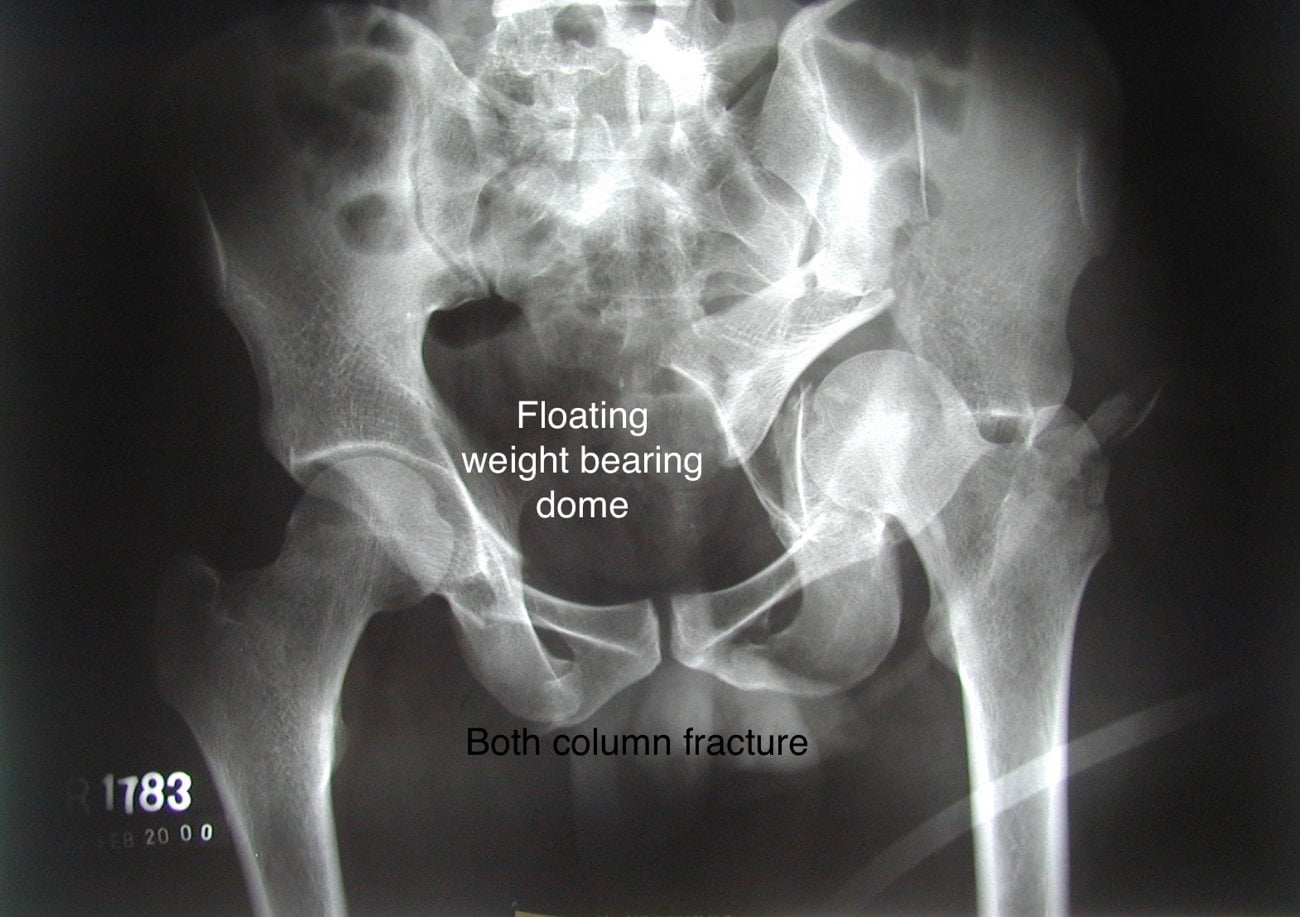
The spur sign on the obturator oblique view is pathognomonic for an associated both-column fracture. It represents the detached posterior iliac wing fragment that remains articulating with the sacrum while the entire acetabular articular surface is separated. If you see a spur sign, the fracture is a both-column, not an anterior column plus posterior hemitransverse. Misclassification here leads to the wrong surgical approach and potentially a secondary column fracture from an anterior-only exposure.
The AO/OTA System and How It Maps to Letournel
Examiners may ask for the AO/OTA classification, the alphanumeric system that runs in parallel with Letournel (the acetabulum is bone segment 62). It groups fractures by how much of the articular surface remains attached to the intact ilium.
- Definition
- Partial articular - ONE column or wall involved
- Corresponding Letournel patterns
- Posterior wall, posterior column, anterior wall, anterior column
- Definition
- Partial articular - transverse-type, part of the dome stays attached to the ilium
- Corresponding Letournel patterns
- Transverse, T-shaped, transverse plus posterior wall, anterior column plus posterior hemitransverse
- Definition
- Complete articular - the whole articular surface (both columns) detached from the ilium (a 'floating' acetabulum)
- Corresponding Letournel patterns
- Both-column
The conceptual divide is the same one the spur sign signals: in 62-A and 62-B some articular surface stays in continuity with the axial skeleton, whereas in 62-C none does.
Q: A both-column fracture is which AO/OTA type, and why? A: 62-C. It is the only pattern in which the entire articular surface is dissociated from the intact ilium (a complete articular fracture) - exactly what the pathognomonic spur sign represents. Single-element (one column or one wall) fractures are 62-A; transverse-family fractures that keep part of the dome attached are 62-B.
Radiographic Evaluation: Judet Views and CT
Accurate classification requires three specific radiographic views plus CT.
- Patient Position
- Supine, beam centred on symphysis
- What It Profiles
- Overview of both hemipelves and acetabuli
- Key Lines to Assess
- Iliopectineal line, ilioischial line, anterior rim, posterior rim, teardrop, roof
- Patient Position
- Rolled 45 degrees toward affected side, injured side UP
- What It Profiles
- Anterior column and posterior wall
- Key Lines to Assess
- Anterior column in profile, posterior wall rim, look for spur sign
- Patient Position
- Rolled 45 degrees away from affected side, injured side DOWN
- What It Profiles
- Posterior column and anterior wall
- Key Lines to Assess
- Posterior column in profile, anterior wall rim, iliac wing fractures visible
On the obturator oblique view, the posterior wall is seen in profile and the anterior column is outlined. On the iliac oblique view, the anterior wall is seen in profile and the posterior column is outlined. Each oblique view profiles the structures on the OPPOSITE side of what its name suggests: the obturator oblique profiles the posterior wall, and the iliac oblique profiles the anterior wall. This is a favourite examiner trick.
CT assessment is mandatory for every acetabular fracture and adds critical information:
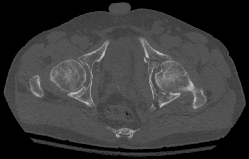
- Axial cuts: identify fracture planes, marginal impaction, intra-articular fragments (loose bodies), femoral head lesions, and the degree of posterior wall comminution.
- 2D reformats (coronal and sagittal): clarify the orientation of fracture lines and assess the weight-bearing dome.
- 3D surface rendering: invaluable for visualising the overall pattern and communicating with the team. Subtract the femoral head to see the articular surface from inside the acetabulum.
- Posterior wall fragment size: CT quantifies the percentage of the posterior wall involved. Fragments involving greater than 40 to 50 percent of the posterior wall in sagittal reconstruction are generally considered unstable and require fixation, though clinical assessment of hip stability under fluoroscopy remains the gold standard for borderline cases.
Classification-to-Approach: How the Pattern Dictates Surgery
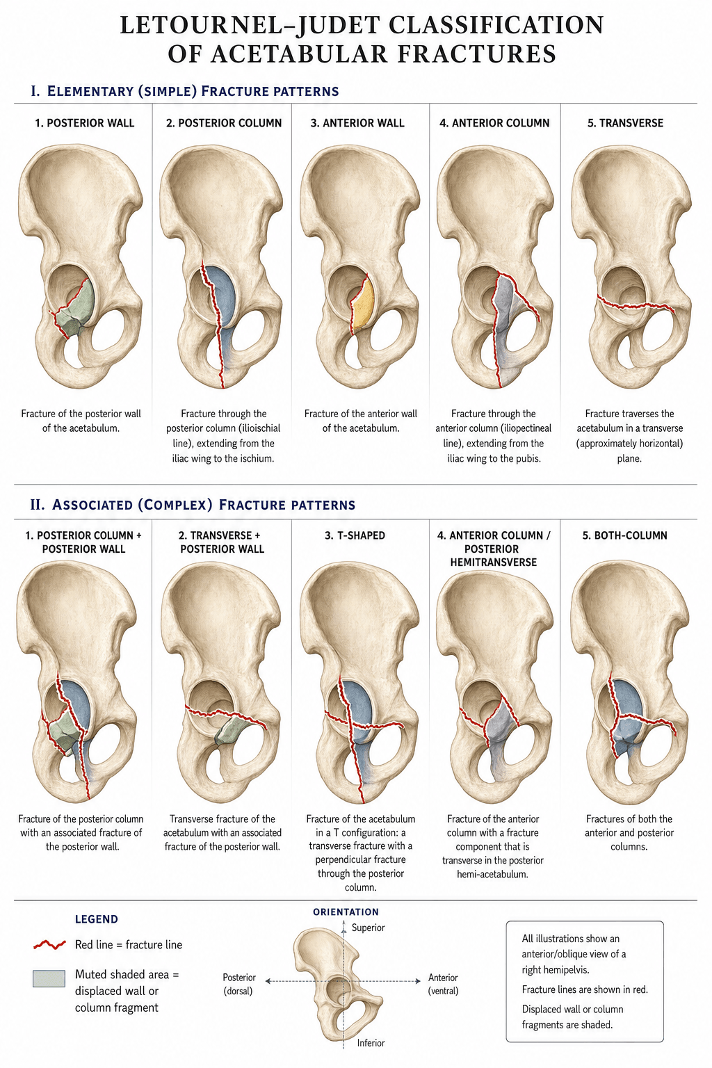
The Letournel-Judet classification directly determines the surgical approach because each pattern has a predictable displacement direction and a specific reduction strategy.
- Primary Approach
- Kocher-Langenbeck (prone or lateral)
- Key Access
- Posterior wall and column through short external rotators
- Rationale
- Direct visualisation and fixation of the wall fragment; capsular attachment preserved for vascularity
- Primary Approach
- Kocher-Langenbeck
- Key Access
- Posterior column via gluteal interval
- Rationale
- Access to posterior column for reduction and plate fixation
- Primary Approach
- Ilioinguinal (supine)
- Key Access
- Anterior column and wall through three windows
- Rationale
- Anterior access for buttress plating of the wall
- Primary Approach
- Ilioinguinal or iliofemoral
- Key Access
- Anterior column from iliac wing to symphysis
- Rationale
- Access to reduce and plate the entire anterior column
- Primary Approach
- K-L or ilioinguinal depending on displacement direction
- Key Access
- Approach the side with greater displacement
- Rationale
- Fracture crosses both columns at one level; approach the dominant displacement
- Primary Approach
- Kocher-Langenbeck
- Key Access
- Posterior wall fixation plus transverse reduction from behind
- Rationale
- Posterior wall component demands posterior approach; transverse line can be reduced from behind
- Primary Approach
- K-L or ilioinguinal or combined
- Key Access
- Depends on which column component is dominant
- Rationale
- Both columns separated; may require extensile or staged approach
- Primary Approach
- Ilioinguinal (supine)
- Key Access
- Anterior column reduction first; posterior hemitransverse reduced indirectly through middle window
- Rationale
- Dominant displacement is anterior; reducing anterior column often brings posterior hemitransverse into alignment
- Primary Approach
- Ilioinguinal preferred, or extensile if posterior column needs direct access
- Key Access
- Comprehensive access to both columns
- Rationale
- Most challenging pattern; anterior approach preferred because extensile approaches carry high complication rates
Posterior patterns go posterior. Anterior patterns go anterior. Transverse follows the displacement.Approach Selection Rule of Thumb
Hook:If the dominant fragment faces posterior, open from behind (Kocher-Langenbeck). If anterior, open from in front (ilioinguinal or Stoppa).
The ilioinguinal approach (Letournel) has three windows: lateral (between iliac wing and iliacus, giving access to the iliac wing and sacroiliac joint), middle (between iliopectineal fascia and external iliac vessels, giving access to the quadrilateral surface and posterior column), and medial (medial to the vessels, giving access to the symphysis and superior pubic ramus). The modified Stoppa (intrapelvic) approach accesses the quadrilateral surface and posterior column from within the pelvis and has become a popular alternative or supplement to the middle window of the ilioinguinal.
Secondary Congruence in Both-Column Fractures
A both-column fracture has a unique escape route from surgery: secondary congruence (Letournel's original term was "apparent congruence"). Because the detached articular fragments stay tethered to the femoral head by intact capsule and labrum, the displaced columns can rotate around the head and settle into a congruent - though medially displaced - relationship with it, even though every articular fragment is separated from the ilium.
- Why it matters: if the femoral head sits congruently within the secondarily-congruent acetabulum and the patient is elderly or low-demand, a both-column fracture may be managed non-operatively with protected weight-bearing, accepting some medialisation, rather than undergoing major reconstruction.
- The caveat: congruence must be present on all three Judet views - congruence on one view but not the others is not acceptable and mandates surgery. The roof fragments must not leave an incongruent, subluxating joint, and a young, high-demand patient is still better served by anatomic open reduction.
Q: Why can some both-column fractures be treated non-operatively when the pattern looks so severe? A: Secondary (apparent) congruence. The capsulolabral attachments hold the detached articular fragments around the femoral head, so the joint stays congruent (albeit medialised) despite complete dissociation from the ilium. In a low-demand or elderly patient with a head congruent on all three views, this permits non-operative management; a young patient still warrants anatomic reduction.
Limitations, Pitfalls and Modern Context
- Inter-observer reliability is moderate, not perfect. Studies show that experienced pelvic surgeons agree on the Letournel classification in about 70 to 80 percent of cases. Disagreement is greatest for the associated patterns, particularly distinguishing anterior column plus posterior hemitransverse from both-column. CT and 3D reconstruction improve agreement significantly over plain films alone.
- The classification does not account for marginal impaction, femoral head injury, or cartilage damage, all of which influence prognosis independently. A perfectly reduced both-column fracture with a femoral head impaction may still do poorly.
- Roof-arc angle measurements (medial, anterior, posterior roof-arc angles on the three views) supplement the classification by quantifying how much of the weight-bearing dome remains intact, guiding non-operative versus operative management for minimally displaced patterns. A roof-arc angle greater than 45 degrees on all three views suggests sufficient dome integrity for non-operative treatment if the femoral head is congruent.
- Modern trends favour anterior approaches (ilioinguinal, modified Stoppa) over extensile posterior approaches because of lower morbidity. The ilioinguinal approach is now the workhorse for most anterior and both-column patterns. Percutaneous screw fixation (column screws) is increasingly used for minimally displaced patterns or as adjunct fixation, but demands intra-operative CT or stereotactic navigation.
- Time to surgery matters. Open anatomic reduction is technically easier within the first 10 to 14 days. After three weeks, the fracture begins to consolidate and reduction becomes substantially harder, often requiring more extensile approaches and osteotomies.
Exam Viva
Practise clinical reasoning and management decisions out loud
“A 34-year-old man is involved in a high-speed motor vehicle collision. The AP pelvis shows disruption of both iliopectineal and ilioischial lines on the left. The obturator oblique view shows a spur sign. There is no disruption of the obturator foramen ring. Classify this fracture and outline your management plan.”
“A 45-year-old woman sustains a dashboard injury. AP pelvis shows disruption of both column lines on the right with a separate posterior wall fragment visible on the iliac oblique view. The obturator foramen ring is intact. There was a posterior dislocation of the hip reduced in the emergency department. Classify the fracture and discuss your surgical plan.”
The 10 fracture patterns
- 5 elementary: posterior wall, posterior column, anterior wall, anterior column, transverse
- 5 associated: posterior column plus posterior wall, transverse plus posterior wall, T-shaped, anterior column plus posterior hemitransverse, both-column
- Elementary means single structural element. Associated means combination of elementary patterns
Column and wall anatomy
- Anterior column equals iliopectineal (iliopubic) line on AP pelvis; runs from iliac wing to symphysis
- Posterior column equals ilioischial line on AP pelvis; runs from SI joint to ischium
- Posterior wall constrains the femoral head posteriorly (dashboard mechanism); anterior wall is smaller, less commonly injured in isolation
Three mandatory views
- AP pelvis: overview, assess both column lines, teardrop, roof, rims
- Obturator oblique (injured side UP): profiles anterior column and posterior wall; look for spur sign
- Iliac oblique (injured side DOWN): profiles posterior column and anterior wall
Classification-to-approach rules
- Posterior patterns (posterior wall, posterior column, transverse plus posterior wall): Kocher-Langenbeck
- Anterior patterns (anterior wall, anterior column, AC plus posterior hemitransverse): ilioinguinal or Stoppa
- Transverse: approach from the side of dominant displacement
- Both-column: ilioinguinal preferred, or extensile if posterior column cannot be reduced anteriorly
Key differentiators and red flags
- Spur sign on obturator oblique equals both-column (pathognomonic)
- T-shaped versus transverse: T-shaped splits through the obturator foramen; transverse does not
- AC plus PHT versus both-column: spur sign absent in AC-PHT; articular surface remains partially attached to ilium
- Aim for surgery within 10 to 14 days; anatomic reduction (less than 2 mm) is the strongest predictor of good outcome
- Posterior wall greater than 40 to 50 percent on CT plus unstable hip equals operative fixation
Evidence Base
Acetabulum fractures: classification and management
- Distils Letournel's large operative experience into the principle that a PERFECT open reduction is the method of choice for displaced acetabular fractures, with outcome dependent on the congruence achieved
- The fracture pattern (the elementary/associated column-and-wall framework) dictates the surgical approach
- Defines total, partial and apparent incongruence and how each guides the operative-versus-conservative decision
The treatment of acetabular fractures through the ilioinguinal approach
- 195 acetabular fractures via the ilioinguinal approach (alone in 90 percent), giving total access to the anterior column from the sacroiliac joint to the pubic symphysis
- Perfect reduction in 85 percent of anterior-column/posterior-hemitransverse and 73 percent of both-column fractures (87 percent across anterior wall/column patterns)
- Very low complication rate with no external-iliac-fossa heterotopic ossification
Operative treatment of acetabular fractures through the ilioinguinal approach. A 10-year perspective
- The ilioinguinal approach was used for 119 of 373 acetabular fractures (about 33 percent) operated over 10 years - for anterior wall/column, anterior column + posterior hemitransverse, and selected both-column and transverse patterns
- Clinical results were excellent in 37 percent, good in 47 percent, fair in 14 percent and poor in 2 percent (excellent/good ~84 percent)
- Low complication rate: ~3 percent surgical-site infection, ~2 percent iatrogenic nerve palsy, ~1 percent significant ectopic bone
Fractures of the acetabulum: accuracy of reduction and clinical results in patients managed operatively within three weeks after the injury
- 262 displaced fractures operated within 21 days (both-column the commonest pattern, 35 percent); anatomical reduction achieved in 71 percent
- Anatomical reduction was the key determinant of outcome, and the rate of anatomical reduction fell with increasing fracture complexity, older patient age and longer injury-to-surgery interval
- Overall clinical result excellent in 40 percent, good in 36 percent, fair in 8 percent, poor in 16 percent, correlating closely with the radiographic reduction
Results of operative treatment of fractures of the posterior wall of the acetabulum
- 100 operatively fixed posterior-wall fractures; anatomic reduction achieved in 97, yet clinical outcome was excellent in only 55, very good 25, good 9, fair 1 and POOR in 10 - 'simple' posterior-wall fractures belie a real risk of poor results
- Clinical outcome correlated strongly with the final radiographic grade
- Risk factors for an unsatisfactory result: delay greater than 12 hours before reducing the associated hip dislocation, age 55 or older, intra-articular comminution and osteonecrosis
Letournel classification for acetabular fractures: assessment of interobserver and intraobserver reliability
- Interobserver reliability was substantial for surgeons trained under Letournel (kappa 0.70-0.74) and for acetabular specialists (0.69-0.71), but only moderate for general trauma surgeons (0.51)
- Adding axial CT to plain radiographs (AP + Judet views) did NOT meaningfully improve classification agreement (e.g. 0.70 vs 0.74 in the trained group)
- Intraobserver reliability was higher than interobserver (0.80-0.83 trained); overall radiographic agreement with the intraoperative pattern was 74 percent