Measurement, maturity prediction and treatment timing
- Separate structural limb length discrepancy from functional discrepancy caused by pelvic obliquity, scoliosis, contracture, pain, equinus or foot height.
- Measure the discrepancy clinically with blocks, then radiographically with calibrated standing imaging that separates femoral and tibial contributions and assesses mechanical axis.
- Base treatment on predicted discrepancy at skeletal maturity, not the current measurement alone.
- Epiphysiodesis slows the longer limb. It is appropriate only when the responsible physis is open, enough growth remains and the predicted discrepancy is within its correction capacity.
- Lengthening is not just bone distraction. It is a prolonged reconstruction affecting regenerate bone, joints, nerves, muscles, school, sport, family capacity and psychological tolerance.
- “A shoe lift is a treatment, not a failure of surgical planning.
- “The block test answers a functional question: how much correction levels the pelvis and improves gait today?
- “Prediction methods are estimates, not guarantees. A mature plan acknowledges error and uses serial review.
- “Do not plan epiphysiodesis without knowing which segment is long and how much growth remains in that segment.
- “A congenital deficiency needs a life-plan, not a single centimetre target.
- “A modern lengthening nail improves patient experience, but it does not make lengthening a low-risk procedure.
Overview and Epidemiology
Limb length discrepancy is a difference in functional or anatomical length between the lower limbs. Mild asymmetry is common and often asymptomatic, so the clinical question is not simply whether one leg is shorter but whether the discrepancy affects gait, pelvis, spine, sport, pain, shoe wear, cosmesis, joints, family burden or long-term function.
Why it matters. A discrepancy can increase the energy cost of walking and alter gait, and both limbs compensate for it in ways described under Clinical Presentation. Pelvic obliquity can create a functional scoliosis that improves when the pelvis is levelled. A post-traumatic physeal arrest may produce shortening and angular deformity together, and a congenital deficiency may involve length, foot rays, ankle stability, knee stability, hip morphology and prosthetic options.
The evidence is thin and the decision is elective. Published reviews consistently make two points: high-quality evidence for exact treatment thresholds is limited, so many thresholds are consensus-based, and treatment is usually elective. That matters: the indication is not the X-ray measurement alone. The indication is the predicted discrepancy plus symptoms, gait, maturity, treatment risk and the family's priorities. A child with a 2 cm discrepancy, a compensating pelvis and no symptoms is not the same clinical problem as a child with a 2 cm discrepancy, pain, a progressive physeal bar or a complex congenital deficiency.
The four questions. Every assessment should answer four linked questions, and each has a characteristic error.
- What to prove
- Blocks and examination show whether the pelvis levels and whether contracture, scoliosis or foot height is driving the asymmetry.
- Common error
- Treating pelvic obliquity or contracture as bony shortening.
- What to prove
- Femur, tibia, foot height and mechanical axis are measured separately.
- Common error
- Planning distal femoral epiphysiodesis when the tibia or foot is the main contributor.
- What to prove
- Bone age, serial measurements and a recognised prediction method estimate the final discrepancy.
- Common error
- Treating today's measurement in a growing child.
- What to prove
- Lift, epiphysiodesis, shortening, lengthening and prosthetic pathways are matched to function and family goals.
- Common error
- Offering lengthening before proving that simpler options are inadequate.
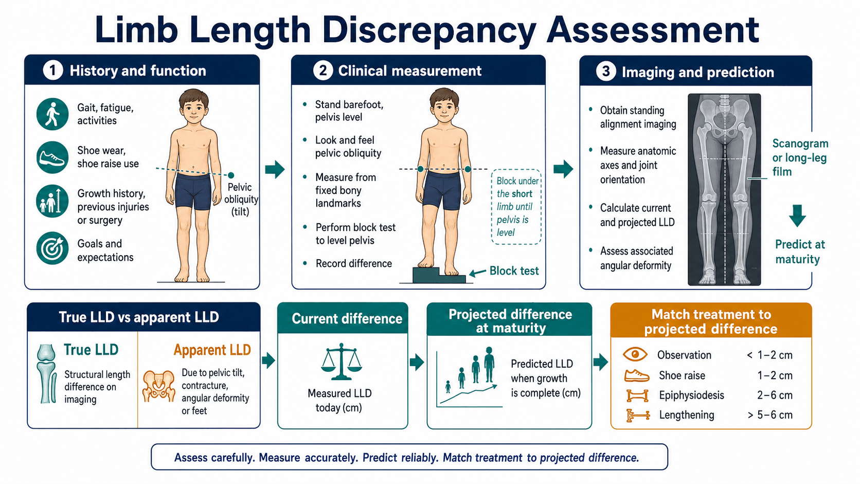
What success looks like. The goal is functional equalisation, not a mathematical promise of exactly equal limbs. A residual discrepancy of about 1 cm is often acceptable if gait, pelvis and symptoms are satisfactory. Conversely, a smaller discrepancy may still need treatment when it is progressive, symptomatic, associated with deformity or part of a congenital deficiency plan.
Pathophysiology
True discrepancy. The bones or the foot height are genuinely different: the femur may be short, the tibia may be short, the foot may contribute, or all three may. Segmental localisation is essential because the operation must match the segment.
- Congenital femoral deficiency produces a femoral-dominant discrepancy, often with hip and knee instability
- Fibular hemimelia may combine tibial shortening, foot ray deficiency, ankle deformity and valgus instability
- Post-infective proximal femoral damage may combine shortening, deformity and joint damage
- Post-fracture overgrowth may make the injured limb longer, especially after paediatric femoral shaft fracture
- Physeal arrest can produce both length inequality and angular deformity
Apparent discrepancy. Posture, contracture, deformity or pain can make equal bones look unequal, and this is the common trap. A hip adduction contracture makes the limb appear short and a hip abduction contracture makes it appear long, with the pelvis tilted in each case; a knee flexion contracture functionally shortens the limb; equinus on the short side is a compensation; and a difference in foot height from cavus, planovalgus, vertical talus or partial foot deficiency is real height that epiphysiodesis does not correct. Pelvic obliquity from scoliosis is another driver, and a painful limp can be mislabelled as discrepancy. If a contracture is missed, the calculation and the operation may both be wrong.
Growth behaviour. Congenital discrepancies often behave proportionally: the difference grows as the child grows, which is the basis for multiplier-style prediction. Acquired discrepancies may not behave predictably. A complete physeal arrest, partial bar, fracture overgrowth, vascular malformation, infection or tumour treatment can change the trend, which is why serial measurement is more valuable than a single radiograph.
Growth around the knee. The knee physes are the usual epiphysiodesis targets because they provide most of the clinically useful remaining growth: the distal femur contributes about 9 mm per year and the proximal tibia about 6 mm per year, roughly 15 mm per year together, although individual growth depends on skeletal maturity, sex, puberty and pathology. The proximal femur and distal tibia contribute to total limb length but are not routine simple arrest targets; they matter in prediction, congenital deficiency planning, ankle-level deformity and complex reconstruction.
Why epiphysiodesis works. Epiphysiodesis slows or stops growth in the longer limb; the shorter limb does not lengthen faster. Equalisation occurs because the long limb stops gaining length while the short limb continues to grow, so the operation works only if:
- The child is skeletally immature
- The physes that are stopped still have enough growth remaining
- The predicted discrepancy is within the correction capacity of growth arrest
- The segment treated matches the segment responsible
- The timing is correct
The operation is technically simple compared with lengthening; the planning is not.
Why lengthening is different. Lengthening is distraction osteogenesis: a corticotomy or osteotomy creates a biological regenerate that is gradually distracted. The bone can lengthen, but bone is rarely the only limit, and the questions that decide whether lengthening is safe are about everything around it.
- Why it matters
- Hip, knee and ankle must tolerate gradual distraction.
- Risk if ignored
- Subluxation, contracture or loss of function.
- Why it matters
- A longer limb still needs a useful end-bearing lever.
- Risk if ignored
- Longer limb with poor shoe wear or poor prosthetic compatibility.
- Why it matters
- Angular and rotational deformity may need simultaneous or staged correction.
- Risk if ignored
- Lengthening magnifies malalignment.
- Why it matters
- Nerves, vessels, muscles and skin often limit safe correction.
- Risk if ignored
- Pain, nerve stretch, stiffness and treatment abandonment.
- Why it matters
- Lengthening requires months of monitoring, therapy and device care.
- Risk if ignored
- Technically successful surgery with poor practical outcome.
Classification
By type. A true anatomical discrepancy is a real difference in bone length or foot height, and it needs clinical and radiographic measurement before any plan is made. An apparent functional discrepancy is an inequality produced by the pelvis, spine, joint contracture, ankle position, foot deformity or pain, and the treatment is the driver rather than growth arrest. A mixed discrepancy is common in congenital deficiency and neuromuscular disease, where true shortening coexists with pelvic obliquity, angular deformity, joint instability and foot-height difference.
By cause. The diagnosis determines the growth behaviour: a congenital proportional discrepancy behaves differently from a post-traumatic bar, a vascular overgrowth syndrome or a post-infective joint-damaging lesion.
- Congenital or developmental. Congenital femoral deficiency, fibular hemimelia, tibial deficiency, hemihyperplasia, skeletal dysplasia, neurofibromatosis, vascular malformation and idiopathic asymmetrical growth. These children need early longitudinal planning.
- Post-traumatic. Malunion with shortening, physeal arrest, growth stimulation after femoral shaft fracture, or partial bar formation.
- Infective or inflammatory. Neonatal osteomyelitis, septic arthritis, juvenile inflammatory disease or physeal injury.
- Tumour-related. Resection, radiation, chemotherapy effects, benign aggressive lesions or limb salvage reconstruction.
- Neuromuscular and functional. Cerebral palsy, spina bifida, pelvic obliquity, scoliosis, hip contracture, knee flexion, equinus, foot deformity or muscle imbalance. Function may matter more than bony length.
For any acquired discrepancy, whether from fracture, overgrowth, physeal injury, infection, tumour treatment, radiation or surgery, the serial trend is especially important.
By predicted size. The working bands are a predicted discrepancy at maturity of less than 2 cm, 2 to 5 cm and more than 5 cm, with partial physeal arrest and complex deformity treated as categories of their own. The treatment each band points to is set out under Management; the thresholds are guides, not absolute rules.
Clinical Presentation
How they present. Children arrive by several routes:
- A parent notices asymmetric gait, hip height or toe walking
- Screening identifies pelvic tilt or functional scoliosis
- The child reports fatigue, tripping, sport limitation or shoe-wear difficulty
- An adolescent reports back pain, hip pain, knee pain or cosmetic concern
- A known congenital deficiency is followed through growth
- A post-traumatic or post-infective child develops progressive inequality
Compensations. The short side compensates with toe walking or equinus, pelvic drop on that side and, in severe cases, vaulting or circumduction. The long side compensates with knee flexion, hip and knee flexion during stance, pelvic hiking, shoe modification or altered foot posture.
History. The history should identify the cause, its progression and whether treatment would improve the child's function:
- Onset. Present from birth, noticed with walking, or acquired after fracture, infection or surgery
- Progression. Stable, increasing proportionally, or changing unpredictably
- Function. Walking distance, running, sport, falls, fatigue, pain and school participation
- Symptoms. Back, hip, knee, ankle or foot pain, callosities and shoe intolerance
- Known diagnosis. Congenital femoral deficiency, fibular hemimelia, Perthes disease, SCFE, tumour treatment, infection or skeletal dysplasia
- Previous treatment. Lifts, orthoses, physiotherapy, guided growth, reconstruction, lengthening or prosthetic discussion
- Family goals. Avoid limp, avoid a large shoe lift, participate in sport, reduce pain, avoid repeated operations, or plan a congenital deficiency pathway
Red flags. Any of these changes the pathway from length planning to diagnosis:
- Painful limp that does not improve with blocks
- Night pain, systemic symptoms or tumour or infection features
- Neurological signs, progressive weakness or abnormal reflexes
- Rapidly changing discrepancy after physeal injury
- Pelvic obliquity that does not correct when the limbs are levelled
- Joint instability in a child being considered for lengthening
Clinical Examination
Standing inspection. Observe the child barefoot from front, side and back:
- Shoulder and pelvic levels
- Lumbar scoliosis or compensatory curve
- Iliac crest height
- Knee flexion on the long side
- Toe standing on the short side
- Hindfoot valgus or varus
- Foot size and foot height
- Calf and thigh girth
- Skin scars from trauma, infection or previous reconstruction
Gait. Look for:
- An antalgic component suggesting pain rather than discrepancy alone
- Short-leg gait with pelvic drop
- Toe walking on the short side
- Knee flexion on the long side
- Circumduction or vaulting
- Compensatory trunk lean
- A Trendelenburg pattern suggesting hip abductor weakness or dysplasia
- Neuromuscular features such as spasticity, foot drop or crouch
The block test. Place measured blocks under the short limb until the pelvis is level. This is the key clinical test: it tells you the functional correction needed to level the pelvis, whether gait improves with correction, whether spinal or pelvic obliquity is flexible, whether the patient tolerates correction and whether the symptoms are likely related to the discrepancy. Do not assume the block height equals the final surgical target; it is a clinical correction estimate, not a maturity prediction.
Localising the short segment. Before imaging, localise which segment is short, because the operation must match the responsible segment. The Galeazzi (Allis) sign compares knee heights with the child supine, both knees flexed to 90 degrees, feet flat and heels level on the couch: a knee that sits lower viewed from the foot end suggests a shorter tibia, and a knee set further back viewed from the side suggests a shorter femur. Tape measurement is useful but not definitive. True length runs from the anterior superior iliac spine to the medial malleolus and apparent length from the umbilicus to the medial malleolus, so a true-length difference is structural while an apparent-only difference points to pelvic obliquity or contracture. Compare femur and tibia clinically, compare plantar foot height, foot length and deformity, and include the foot in the standing block test: a foot-height discrepancy adds to functional length but is not corrected by epiphysiodesis, so it needs a shoe raise or foot surgery. Calibrated standing imaging then quantifies the femoral, tibial and foot contributions the clinical tests suggested.
Joints, spine and neurology. Assess:
- Spine: scoliosis, pelvic obliquity and flexibility
- Hip: abduction, adduction contracture, flexion contracture, rotation, Trendelenburg and stability
- Knee: flexion contracture, recurvatum, valgus or varus, instability and patellar height
- Ankle and foot: equinus, calcaneus, cavus, planovalgus, subtalar stiffness and braceability
- Neurology: tone, reflexes, power, sensation and selective motor control when indicated
Investigations
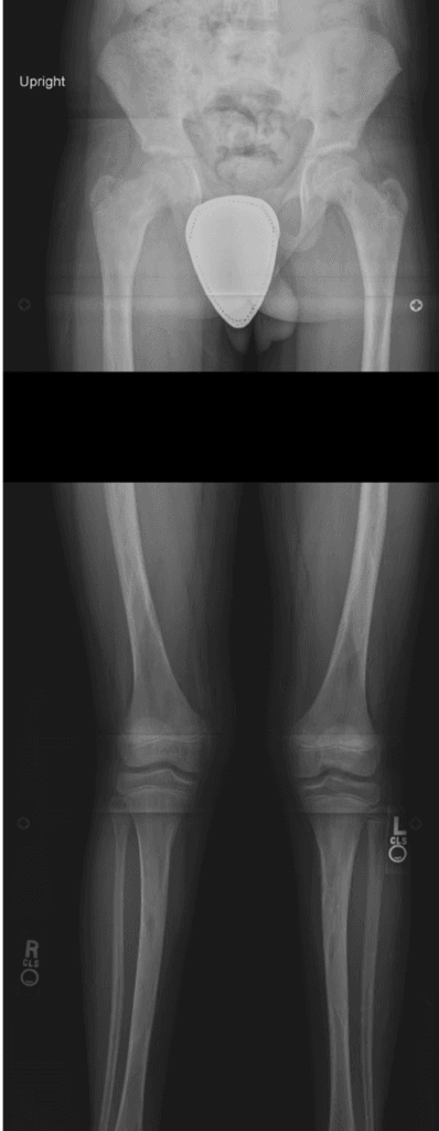
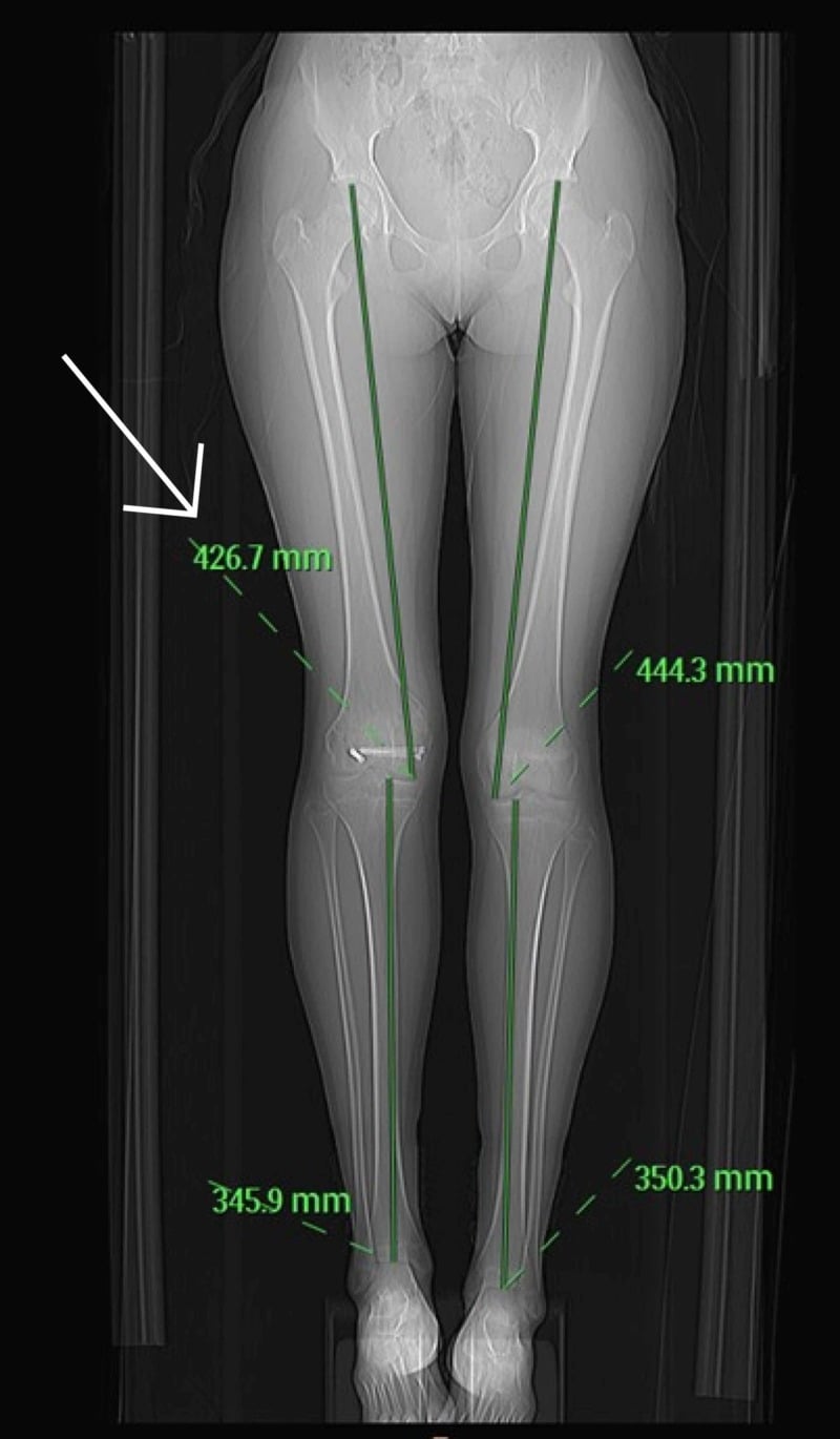
Imaging. Use calibrated imaging, and make the report state total length, femoral length, tibial length, mechanical axis, angular deformity, foot-height contribution when relevant, and whether the pelvis was levelled. A standing long-leg radiograph is often preferable as the first study because it assesses length and mechanical alignment together on one weight-bearing image, with the physes visible for maturity assessment. EOS, where available, suits repeated low-dose assessment; a scanogram gives the classic length measurement; a CT scanogram is for complex rotation or flexion deformity, or when standing imaging is difficult to interpret, and should be reserved for a question that weight-bearing radiographs or EOS cannot answer.
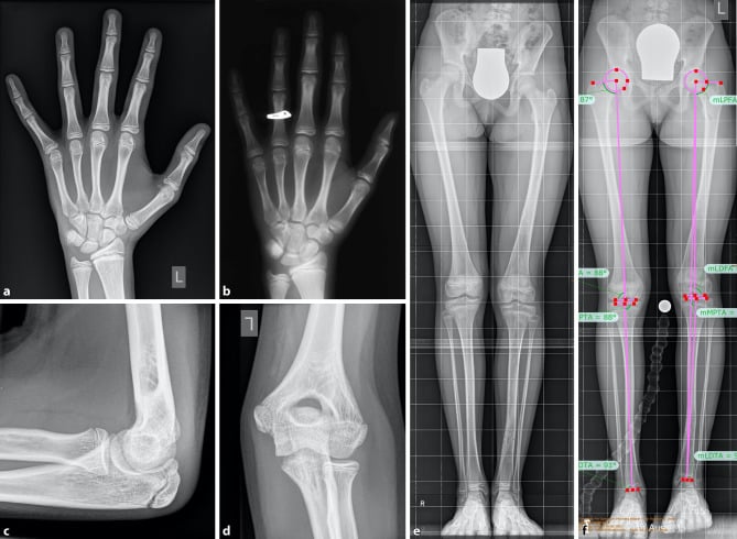
Skeletal maturity. Chronological age is not enough. Use hand and wrist bone age, compared with a reference atlas and cross-checked at the elbow where needed, together with pubertal history, height velocity, growth chart trend and radiographic physeal appearance. Knee skeletal maturity systems are increasingly useful because the knee is already imaged in a discrepancy work-up, but they still require clinical judgement and local familiarity. Bone age rather than chronological age is what determines how much correction a timed epiphysiodesis can still deliver, because the operation works only through remaining growth.
Predicting the discrepancy at maturity. Use at least one recognised method and check it against the serial clinical trend. No method is perfect, so counsel error and repeat the measurements.
- How it works
- Uses skeletal age and expected remaining growth of the distal femur, proximal tibia and other physes.
- Best use
- Segment-specific planning when deciding distal femur versus proximal tibia epiphysiodesis.
- Limitation
- Needs accurate bone age; chart-based estimates can be wrong in abnormal growth.
- How it works
- Plots serial limb lengths against skeletal maturity to forecast final discrepancy and timing.
- Best use
- Excellent when serial measurements are available and growth appears linear.
- Limitation
- Less useful from a single data point or in unpredictable acquired physeal injury.
- How it works
- Uses age, sex and multipliers to predict mature length and final discrepancy.
- Best use
- Quick, widely used method for both congenital and acquired discrepancy planning.
- Limitation
- Can be inaccurate if physiological age differs from chronological age.
- How it works
- Uses remaining years of growth and approximate annual growth around the knee.
- Best use
- Rapid clinical cross-check.
- Limitation
- Too approximate to be the sole basis for surgery.
The Menelaus mental check. Take skeletal maturity as roughly 14 years in girls and 16 years in boys, adjusted for bone age and puberty, and multiply the years remaining by the knee growth rates given under Pathophysiology. Distal femoral arrest alone therefore corrects less than combined distal femoral and proximal tibial arrest over the same time, and if the correction required exceeds the remaining growth capacity, epiphysiodesis alone cannot solve the problem. It is a sense check on the plan, not a substitute for formal prediction.
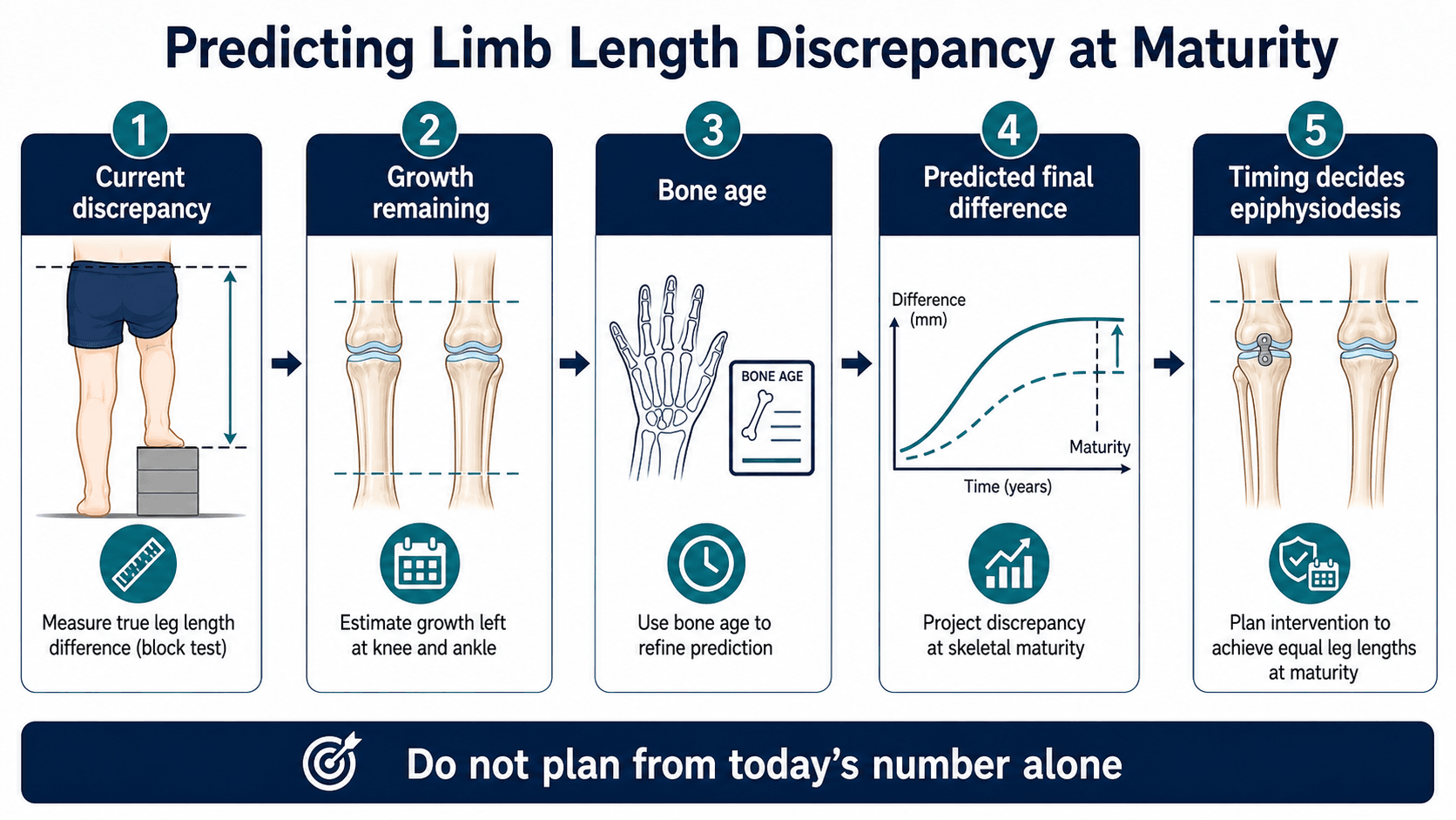
The workflow in clinic. The prediction is built in a fixed order, and the treatment band falls out at the end:
- Measure the current total discrepancy and segmental discrepancy.
- Decide whether the short limb is truly short, the long limb is overgrown, or both are contributing.
- Determine skeletal maturity using bone age or a validated knee maturity method where appropriate.
- Estimate the predicted discrepancy at maturity using a recognised method.
- Check the estimate against prior measurements. A single radiograph is weaker than a trend.
- Calculate whether the chosen physis has enough remaining growth to close the gap.
- Counsel the family that prediction error can leave residual discrepancy or overcorrection, then place the child in the less than 2 cm, 2 to 5 cm or more than 5 cm pathway.
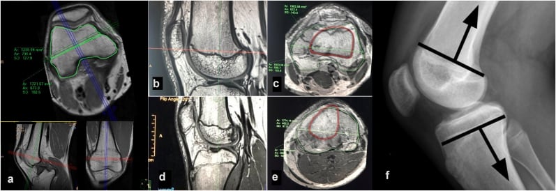
Physeal bar. If a partial growth arrest is suspected, map the bar with MRI or CT to define its size, its location and how much normal physis remains. The question is not only length: a bar may create progressive angular deformity, joint-line obliquity and mechanical-axis shift. Bar resection is considered when there is enough growth remaining, the bar is technically resectable and the deformity can be corrected or guided.
Using the Paley Multiplier
Most candidates can name the multiplier method; far fewer can do the arithmetic when asked, and the arithmetic is where the marks are. The multiplier (M) is the ratio of a bone's length at maturity to its length now (M = mature length divided by current length), read from published age- and sex-specific tables. How you apply it depends on whether the discrepancy is proportional, and its great strength, speed from a single visit, is bought by assuming that chronological age tracks skeletal maturity.
- How to use the multiplier
- Predicted discrepancy at maturity = current discrepancy multiplied by M (both limbs scale by the same multiplier, so their difference scales too)
- Caveat
- Assumes the discrepancy stays proportional - typical of congenital cases
- How to use the multiplier
- Predict each limb (or segment) separately as current length multiplied by M, then take the difference; do NOT simply multiply the current discrepancy
- Caveat
- Acquired/arrest discrepancies are not proportional, so the simple shortcut over- or under-estimates
- How to use the multiplier
- Growth remaining in a physis is about the predicted mature length minus the current length; use it to check whether arrest can close the predicted gap and to time surgery
- Caveat
- Based on chronological age - correct with bone age if skeletal and chronological age diverge
- How to use the multiplier
- Fast - needs only age, sex and a single current measurement
- Caveat
- Still cross-check against the serial clinical trend; abnormal growth breaks the proportionality assumption
Differential Diagnosis
The differential is about avoiding a false diagnosis of bony discrepancy. The key discriminators are the response to the standing block test, whether the inequality is in the bone or in soft tissue and posture, and whether one limb is genuinely short or the other is overgrown.
- Distinguishing feature
- Real difference in femur, tibia or foot height.
- Best discriminating test
- Calibrated standing radiograph or scanogram with segmental measurement.
- Why it changes management
- Eligible for lift, epiphysiodesis, shortening or lengthening.
- Distinguishing feature
- Pelvic obliquity and curve correct when blocks level the limbs.
- Best discriminating test
- Block test plus standing spine radiograph levelled and unlevelled.
- Why it changes management
- Treat the length, not the spine; curve resolves with equalisation.
- Distinguishing feature
- Curve and rotation persist despite block correction.
- Best discriminating test
- Standing radiograph with Adams forward bend and Cobb angle.
- Why it changes management
- Spinal deformity needs its own pathway; do not arrest growth for the curve.
- Distinguishing feature
- Limb appears short (adduction) or long (abduction) with pelvis tilted.
- Best discriminating test
- Examination of hip range with Thomas and abduction in extension.
- Why it changes management
- Releasing contracture can change the apparent measurement and the operation.
- Distinguishing feature
- Functional shortening with fixed flexion on the affected side.
- Best discriminating test
- Prone or supine knee extension assessment.
- Why it changes management
- Correcting contracture may remove most of the apparent inequality.
- Distinguishing feature
- Cavus, planovalgus, vertical talus or partial foot deficiency alters functional height.
- Best discriminating test
- Standing foot examination and weight-bearing foot/ankle radiographs.
- Why it changes management
- Foot height is not corrected by epiphysiodesis; needs shoe or foot surgery.
- Distinguishing feature
- Limp does not improve when the pelvis is levelled with blocks.
- Best discriminating test
- Targeted hip/knee imaging for Perthes, SCFE, infection or tumour.
- Why it changes management
- A painful pathology, not LLD, is driving gait; treat the cause.
- Distinguishing feature
- Tone, weakness and pelvic obliquity dominate gait more than length.
- Best discriminating test
- Neurological examination, tone and selective motor control assessment.
- Why it changes management
- Function and contracture, not the centimetre value, often lead management.
- Distinguishing feature
- One limb is long rather than the other being short; may have skin or girth asymmetry.
- Best discriminating test
- Whole-limb examination, segment measurement and overgrowth screen.
- Why it changes management
- Overgrowth syndromes need surveillance and may need tumour screening pathways.
Management
Everything here assumes the clinic workflow under Investigations has been completed: the discrepancy is true, measured, localised and predicted. The remaining decisions are which rung of the ladder fits the predicted discrepancy, whether contracture, angular deformity, joint instability or foot deficiency changes the plan, and how to explain prediction error, residual inequality and treatment burden so that the family chooses with open eyes. A level pelvis is not success if the limb is painful or unstable.
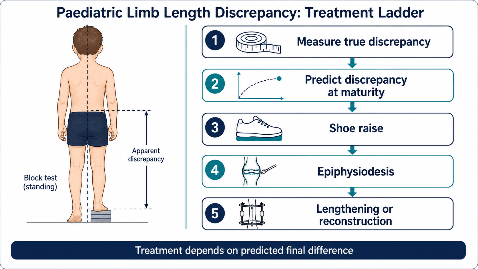
- Usual strategy
- Observe, insole, internal heel raise, external shoe raise if symptomatic.
- Key decision factors
- Symptoms, gait response to blocks, sport, back pain, cosmesis and family preference.
- Usual strategy
- Timed epiphysiodesis if growth remains; shortening or lift if near maturity.
- Key decision factors
- Bone age, growth remaining, femur versus tibia contribution, acceptable residual LLD.
- Usual strategy
- Lengthening, staged reconstruction, combined procedures or prosthetic pathway.
- Key decision factors
- Joint stability, soft tissues, diagnosis, family capacity, number of stages required.
- Usual strategy
- Bar resection, guided growth, osteotomy, epiphysiodesis or lengthening depending bar size and growth remaining.
- Key decision factors
- Bar location, deformity, remaining growth, joint line, mechanical axis and family expectations.
- Usual strategy
- Treat deformity and function, not the centimetre number alone.
- Key decision factors
- Contracture, malalignment, foot height, hip/knee/ankle stability and neuromuscular control.
When. Observation and lifts are usually first-line for a predicted discrepancy of less than 2 cm, uncertain early measurements, minimal symptoms, or families who prefer non-operative care. A lift may be definitive, temporary while awaiting maturity, or a bridge to surgery, and in a growing child the prediction is repeated because today's mild discrepancy may not remain mild.
What to offer. The correction is matched to the shoe and the symptoms:
- An insole or heel raise for a small discrepancy
- An internal shoe lift when shoe volume allows; a small correction can usually be placed inside the shoe
- An external shoe raise for larger correction
- Physiotherapy only if compensatory tightness, weakness or gait adaptation exists
- Surveillance imaging while growing, because the predicted discrepancy may change
How to prescribe it. Do not prescribe a lift blindly: test with blocks, observe gait, and ask whether pain or fatigue improves. Full correction is not always tolerated immediately, especially after long-standing compensation, so a long-standing discrepancy may need gradual lift introduction. A lift corrects function; it does not treat progression.
Operative Technique
Timing over technique. Epiphysiodesis is not one operation but a family of techniques that slow or stop growth in the longer limb. The operative method matters; the timing matters more, and equalisation occurs gradually during the remaining growth, not immediately, which the family must hear before the operation rather than after it.
Preoperative checklist. Before the child reaches theatre:
- Confirm current discrepancy, predicted maturity discrepancy and acceptable residual inequality
- Confirm bone age and growth remaining
- Confirm the long limb, the long segment and the exact physis
- Review femoral and tibial lengths, mechanical axis and joint status
- Decide whether distal femur, proximal tibia, both, fibula, or an associated angular correction is needed
- Mark the operative side carefully: wrong-side surgery is a catastrophic error
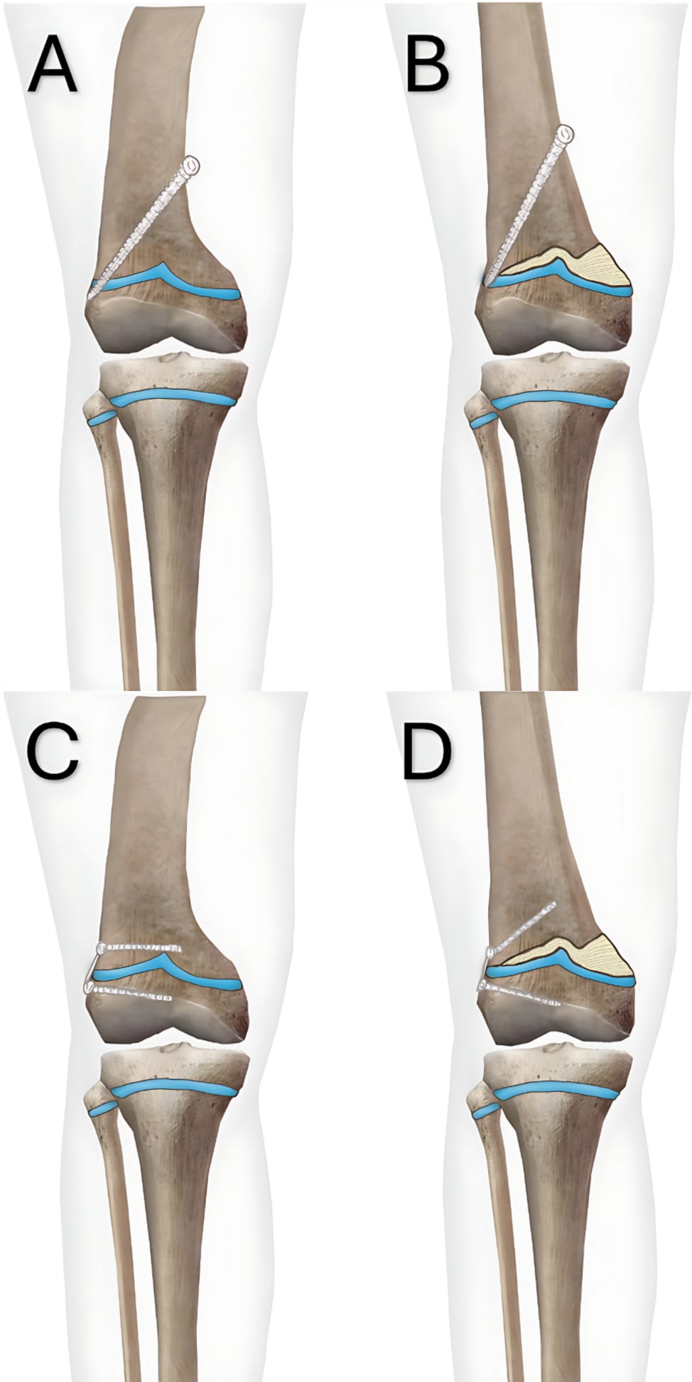
Permanent percutaneous epiphysiodesis. The aim is controlled physeal ablation: enough destruction, symmetrically placed, to produce a physeal bridge.
- General anaesthesia or appropriate paediatric anaesthesia; supine with fluoroscopy
- AP and lateral image confirmation of the physis
- Small medial and lateral incisions or percutaneous access, depending on technique
- Drill, burr or curette the physis sufficiently to create a physeal bridge, ablating medial and lateral sides symmetrically when the aim is pure length arrest
- Confirm physeal ablation radiographically and document fluoroscopic confirmation of level, side and ablation position
- Close and allow protected or early mobilisation according to local protocol
The pitfalls are the wrong side, the wrong physis, insufficient ablation with continued growth, and asymmetric ablation producing varus, valgus, procurvatum or recurvatum. Keep the joint line in view and avoid intra-articular penetration; in the proximal tibia respect the tibial tubercle apophysis and the lateral neurovascular anatomy. Radiographic surveillance continues afterwards, because growth arrest is a biological response, not a completed operation.
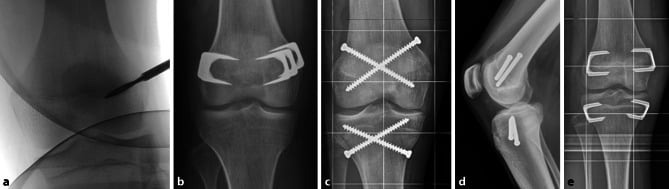
Percutaneous transphyseal screws (PETS). Screws crossing the physis slow growth. The approach is minimally invasive, with small incisions and a short operative time, commonly permits rapid mobilisation, and the hardware can sometimes be removed if clinically required; it is attractive around the distal femur and proximal tibia. It still needs accurate timing, careful screw placement and follow-up, and its problems are its own:
- Hardware irritation
- Incomplete arrest
- Rebound or continued growth after removal
- Angular deformity if screw placement or the physeal response is asymmetric
- A proximal tibial screw trajectory that must respect the nearby deep peroneal nerve
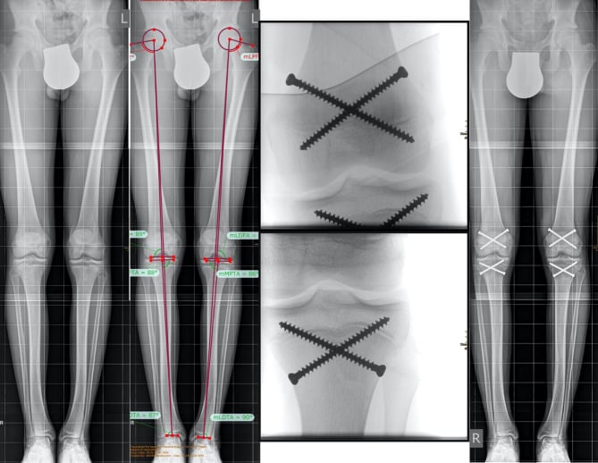
Temporary tension-band plates or staples. Temporary implants may slow growth and can later be removed. They are more commonly used for guided growth of angular deformity but can be considered in selected length problems, where their attractions are reversibility, usefulness when angular correction is also required, and avoidance of complete physeal destruction. Against that they are slower and less predictable for pure length correction, they require removal, rebound growth may occur, and hardware irritation or migration can occur. For pure length discrepancy, temporary tethering should not be presented as automatically equivalent to definitive epiphysiodesis: it can work, but some series report lower efficiency and a revision burden, especially where a predictable definitive arrest is needed.
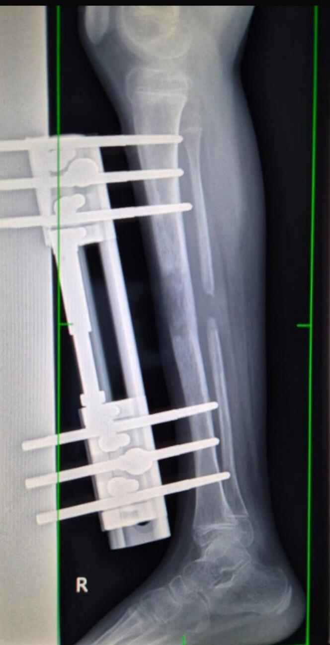
Lengthening. The surgeon manages bone, soft tissue and joints together, and each stage has a characteristic failure.
- Key action
- Decide femur, tibia or both; correct angular or rotational deformity simultaneously or in a planned stage.
- Failure to avoid
- Lengthening the wrong segment or magnifying malalignment.
- Key action
- Perform low-energy osteotomy or corticotomy and stabilise with frame, nail or combined construct.
- Failure to avoid
- Poor regenerate biology or unstable fixation.
- Key action
- Wait through latency, then distract gradually while monitoring regenerate and joints.
- Failure to avoid
- Fast distraction, nerve symptoms, joint subluxation or poor regenerate.
- Key action
- Protect until regenerate is strong enough for progression.
- Failure to avoid
- Fracture or deformity through immature regenerate.
- Key action
- Remove frame or implant when safe and continue rehabilitation until gait, strength and range recover.
- Failure to avoid
- Stopping follow-up when the bone unites but function remains poor.
The hardware. Externally, a monolateral fixator serves simple lengthening, a circular frame lengthening with stability needs, small bone segments or deformity, and a hexapod frame multiplanar deformity correction with lengthening. Internally, a motorised intramedullary nail suits appropriate older children and adolescents, requires adequate bone size, consideration of open growth plates, joint protection and implant availability, and is avoided when canal size, age, deformity, infection risk or joint instability makes it unsafe.
Technical priorities during distraction. The soft tissues and joints are protected throughout:
- Prevent hip, knee and ankle contracture, with physiotherapy from the start
- Monitor regenerate density and shape
- Slow or pause distraction if the regenerate is poor or nerves and joints become symptomatic
- Consider prophylactic joint spanning or soft-tissue release in high-risk congenital deficiency
Physeal bar resection. This is a reconstruction of growth, not a length-equalisation operation, and it is particularly unforgiving: incomplete bar removal may fail, and excessive resection can damage the remaining growth potential.
- Why it matters
- Complete arrest or inaccessible bars are poor candidates.
- Common failure
- Attempting restoration where meaningful growth cannot return.
- Why it matters
- The child must have enough growth left for resection to matter.
- Common failure
- Technically successful surgery with no clinical benefit.
- Why it matters
- MRI or CT defines bar size, location and approach.
- Common failure
- Incomplete resection or damage to normal physis.
- Why it matters
- Remove the bar while protecting remaining physis.
- Common failure
- Recurrent arrest or iatrogenic growth damage.
- Why it matters
- Guided growth, osteotomy or epiphysiodesis may still be needed.
- Common failure
- Treating growth restoration but leaving angulation or predicted LLD.
Complications and Follow-up
Prediction and timing. The failures of planning are residual discrepancy at maturity, undercorrection because surgery was too late, overcorrection because it was too early, the wrong segment treated, bone age or pubertal timing not accounted for, or a prediction never updated with serial data.
Epiphysiodesis. The operative complications are:
- Continued growth from incomplete arrest
- Angular deformity from asymmetric arrest
- Hardware irritation or screw prominence
- Knee pain or stiffness
- Infection or wound problem
- Reduced final height
- Need for a contralateral or revision procedure if the prediction proves wrong
Shortening. Shortening carries:
- Nonunion or delayed union
- Hardware irritation
- Malrotation
- Weakness from excessive shortening
- Loss of height
Lengthening. Lengthening carries the longest list:
- Pin-site infection or implant-related pain
- Poor regenerate or delayed consolidation
- Regenerate fracture
- Joint stiffness
- Hip, knee or ankle subluxation
- Nerve stretch symptoms
- Muscle contracture
- Psychological fatigue and family burnout
- Residual discrepancy or recurrent deformity
Follow-up. It continues beyond the operation and to skeletal maturity. At each review recheck the clinical block height, repeat calibrated imaging, monitor axis as well as length, confirm that the physis has responded, adjust any shoe lift during growth and watch for overcorrection.
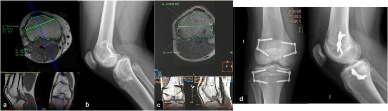
A successful wound review does not mean successful epiphysiodesis. The endpoint is acceptable limb length and alignment at skeletal maturity.
Clinical Pitfalls
Common pitfalls:
- Starting with "I would get a scanogram" before saying true versus apparent discrepancy
- Forgetting the block test
- Treating the current discrepancy instead of the predicted maturity discrepancy
- Using chronological age alone
- Forgetting the femur versus tibia contribution
- Ignoring foot height
- Ignoring angular deformity
- Offering lengthening without a joint stability assessment
- Calling epiphysiodesis "simple" without discussing timing error
- Assuming equal legs is always the goal
Guidelines, Registries & Global Practice
There is no single high-level international guideline that dictates limb length discrepancy (LLD) thresholds. Practice is built on consensus, narrative reviews and registry-style cohorts rather than randomised trials, and the major bodies (AAOS, the British Society for Children's Orthopaedic Surgery, EFORT/EPOS in Europe, and the deformity-correction community around the AO and Paley schools) converge on a similar treatment ladder while differing in emphasis and the strength of the evidence they cite.
Global epidemiology
Mild anatomical inequality is near universal; it is the magnitude and the symptoms, not the mere presence of inequality, that define a clinical problem.
Anatomical leg-length inequality is near universal but usually small
- Using accurate radiographic methods, anatomical leg-length inequality was present in about 90 percent of people.
- The mean magnitude was only 5.2 mm (SD 4.1 mm), so most inequality is small.
- Inequality did not appear clinically significant for most people until it reached approximately 20 mm.
Shoe lifts: low-quality evidence for symptom relief, guidelines inconsistent
- Across non-randomised studies, about 88 percent of treated adults reported partial or complete pain relief with a shoe lift.
- Overall study quality was low or poor, and only one randomised controlled trial was identified.
- Of nine guidelines reviewed, only two recommended shoe lift use, and those were based on consensus.
How guidance differs across regions
- Position on LLD management
- Treatment-ladder consensus: observe or lift for predicted discrepancy under 2 cm, epiphysiodesis for predicted 2 to 5 cm with growth remaining, lengthening or reconstruction for over 5 cm.
- Evidence strength
- Consensus and review level; no Grade A trial evidence for thresholds.
- Position on LLD management
- Same broad ladder with strong emphasis on multidisciplinary deformity units, calibrated EOS or long-leg imaging and family-centred counselling about treatment burden.
- Evidence strength
- Consensus; centralisation of complex lengthening to specialist centres is custom rather than published mandate.
- Position on LLD management
- Endorses prediction-based timing (Green-Anderson, Moseley, multiplier) and individualised, elective decision-making; the cited German consensus states the decision to treat is always elective.
- Evidence strength
- Narrative review and consensus (Vogt et al, Dtsch Arztebl Int 2020).
- Position on LLD management
- Multiplier method for prediction; structured selection for distraction osteogenesis with frames or motorised nails; stable joints and plantigrade foot are prerequisites.
- Evidence strength
- Validation and Level IV cohort evidence; technique superiority unproven.
- Position on LLD management
- External fixators remain the dominant lengthening tool where motorised nails are unavailable; shoe raises and timed epiphysiodesis are the most accessible interventions.
- Evidence strength
- Practice variation driven by access and cost, not by differing evidence.
Registry and practice-variation evidence
There is no dedicated international LLD registry equivalent to arthroplasty registries. The best registry-style evidence comes from large single-centre and institutional cohorts that prospectively track lengthening and epiphysiodesis to maturity, and these show that method selection - motorised intramedullary nails, epiphysiodesis around the knee, or external fixation - varies widely even within a single expert centre. Practice variation between centres and countries reflects implant availability, cost, family capacity for prolonged rehabilitation and surgeon training far more than disagreement about the underlying treatment ladder.
The treatment ladder (observe/lift under 2 cm, epiphysiodesis 2 to 5 cm, lengthening or reconstruction over 5 cm) is internationally consistent, but it rests on consensus, not randomised trials. A strong exam answer states the ladder and then acknowledges that thresholds are consensus-based and decisions are elective and individualised.
Controversies and Areas of Uncertainty
Almost every number in limb length discrepancy work is a consensus figure rather than a trial result, and a candidate who can say which ones are soft, and why they have stayed soft, is answering at a different level from one who recites thresholds.
Which prediction method to trust
Green-Anderson, Moseley and the Paley multiplier applied to the same child can give materially different predictions of the final discrepancy, and there is no agreed hierarchy between them.
Why it stays unresolved. Validating a prediction method requires following every child to skeletal maturity, so a definitive comparison takes fifteen years to produce and is obsolete by the time it appears. The methods were also derived on different populations and make different assumptions - the multiplier assumes chronological age tracks skeletal maturity, Moseley assumes growth has been linear and needs serial points, Green-Anderson depends entirely on the accuracy of a bone age that is itself observer-dependent. Every one of them breaks in exactly the situation where the prediction matters most: abnormal, non-proportional growth after a physeal injury. The defensible position is to use one formal method, cross-check it against the serial trend, and treat a single-visit prediction as a hypothesis.
Temporary versus permanent epiphysiodesis for pure length
Reversible constructs (tension-band plates, transphyseal screws) are attractive because a wrong timing decision can be undone. But for pure limb length discrepancy, as opposed to angular correction, temporary guided growth has proved less efficient than expected, with plate-related pain and unplanned revision surgery.
Why it stays unresolved. The theoretical advantage - reversibility - is real and matters most in exactly the children whose growth is least predictable, which is the group in whom any technique performs worst. So the comparison is confounded by indication: surgeons choose the reversible option for the harder cases, then the reversible option looks worse. Untangling that would need randomisation within a single diagnosis, and the numbers do not exist.
Lengthening nails versus external fixation
Magnetically driven nails remove the pin sites, the frame and much of the social burden, and modern series report accurate, reliable distraction. They do not remove the biology: temporary joint stiffness is common and a substantial minority need unplanned additional surgery.
Why it stays unresolved. A randomised comparison is close to impossible - patients and families have strong preferences, the devices differ in cost by an order of magnitude, and the indications only partly overlap (a nail needs a canal, adequate bone stock and no need for simultaneous multiplanar correction). What is genuinely settled is narrower than the marketing: the nail improves the experience of lengthening, and the evidence does not show that it makes lengthening a low-risk operation.
Whether the thresholds themselves are real
The 2 cm and 5 cm figures organise every discussion of limb length discrepancy, and neither has ever been tested against an outcome.
Why it stays unresolved. No trial has randomised children by discrepancy magnitude, and the long-term consequences of a small discrepancy in adult life - back pain, hip and knee arthritis, gait cost - remain genuinely disputed, with cohort data pointing in both directions. The thresholds persist because they are useful for organising decisions, not because they mark a biological transition. Treat them as bands for discussion rather than as indications.
The height that epiphysiodesis costs
Equalising by arresting the long limb necessarily makes the child shorter at maturity than they would otherwise have been, by approximately the amount of correction achieved. Lengthening the short limb does the opposite.
Why it stays unresolved. There is no evidence base at all on how much that height matters to the person it happens to, and the trade-off is rarely put to families in those terms. It is a values question wearing the clothes of a technical one - which is precisely why it belongs in the consent conversation rather than being settled by the surgeon in advance.
Viva Scenarios
Practise clinical reasoning and management decisions out loud
“A 10-year-old child is referred with a limp and suspected limb length discrepancy. How do you assess them?”
“A growing child has a predicted 3 cm discrepancy at maturity. When would you consider epiphysiodesis?”
“A child with fibular hemimelia has a large predicted discrepancy. How does your approach differ?”
“A teenager has residual discrepancy after epiphysiodesis. What went wrong and what do you do?”
Opening Line
- True versus apparent first
- Block test levels pelvis
- Measure femur and tibia
- Predict maturity discrepancy
- Apply less than 2, 2 to 5, more than 5 cm thresholds
Investigations
- Standing blocks
- EOS or scanogram
- Long-leg alignment
- Bone age
- Green-Anderson, Moseley, multiplier, serial measurements
Treatment Ladder
- Less than 2 cm: observe/lift
- 2 to 5 cm: epiphysiodesis if growing
- Near maturity: lift or shortening
- More than 5 cm: lengthening/reconstruction
- Complex deficiency: whole-limb life-plan
Do Not Miss
- Pelvic obliquity
- Hip contracture
- Foot height
- Angular deformity
- Joint instability
Complications
- Residual LLD
- Overcorrection
- Undercorrection
- Angular deformity
- Lengthening stiffness and regenerate problems
Evidence Base
Treatment indications are elective and individualised
- The evidence base for exact LLD treatment indications is limited.
- Prediction algorithms estimate final discrepancy but have clinically relevant error.
- Treatment should be individualised; the discrepancy size is not the sole determinant.
Multiplier method validation
- Sixty patients. The multiplier method's mean error for predicting BONE LENGTH DISCREPANCY at maturity was 0.6 cm (SD 0.6)
- For predicting the length of the EPIPHYSIODESED limb, the mean error was 1.6 cm (SD 1.2) - IDENTICAL for the multiplier and Moseley methods, so neither is better at the harder task
- For RESIDUAL discrepancy after epiphysiodesis the errors were 0.9 cm (multiplier, chronologic age), 1.0 cm (multiplier, skeletal age) and 1.3 cm (Moseley); the difference between methods was significant at P=0.0008
- Note the counterintuitive detail: using CHRONOLOGIC age was marginally more accurate than using skeletal age, so bone-age assessment did not improve the prediction here
Prediction variability
- Sixteen patients (mean age 14) treated by percutaneous epiphysiodesis using transphyseal screws, with three prediction methods compared against the actual discrepancy at maturity
- MEAN differences were tiny - Green-Anderson 0.2 cm, Paley -0.1 cm, Moseley 1.4 cm - BUT THE PAPER'S TITLE IS 'HIGH VARIABILITY AMONG GROWTH PREDICTOR MODELS', and that is the point: a mean error near zero with wide scatter means a method is UNBIASED BUT IMPRECISE, so it cannot be trusted for the individual child in front of you
- The correction actually achieved was modest: mean pre-operative discrepancy 3.1 cm became 1.7 cm at maturity (p less than 0.001) - a gain of only 1.4 cm
- Six of 16 had pain over the screw heads, but there were no infections and no angular deformity
- The authors' own conclusion is that results would have been better had PETS been done at an earlier skeletal age