PLIF vs TLIF vs ALIF vs Lateral | Approach-Specific Advantages | Circumferential Support
- TLIF = unilateral posterior approach, less nerve retraction than PLIF
- ALIF = anterior approach, best for L5-S1, risk of vascular/retrograde ejaculation
- LLIF/XLIF = lateral through psoas, lumbar plexus injury risk, avoid L5-S1
- Circumferential fusion = highest fusion rate (anterior + posterior)
- Pedicle screw fixation essential for most interbody techniques
- “TLIF: Unilateral approach, less nerve retraction than PLIF
- “ALIF at L5-S1: Great vessels bifurcate, better access
- “Lateral approach: Avoid L5-S1 (iliac crest), watch lumbar plexus
- “Vascular surgery standby for ALIF if needed
Overview
Lumbar fusion creates a bony bridge between vertebral segments to eliminate motion, and is used to treat instability and degenerative conditions. Interbody techniques place bone graft or a cage in the disc space for anterior column support, typically combined with posterior pedicle screw instrumentation, which provides immediate stability while the fusion mass consolidates.
Where the approaches came from. The first lumbar fusions were posterolateral, with bone graft laid between the transverse processes. Interbody techniques followed to improve fusion rates through anterior column support: PLIF was introduced in the 1950s-60s, ALIF in the 1980s, TLIF in the 1990s and the lateral approaches (XLIF/LLIF) in the 2000s.
Where practice is now. TLIF has become the most commonly performed interbody fusion because of its balance of visualisation, fusion rates and complication profile. Minimally invasive techniques are increasingly used, while multi-level constructs and adult deformity correction remain specialised applications.
The five routes at a glance. Each approach trades access for a particular risk, and the level being fused decides which trade is worth making.
- Approach
- Posterior bilateral
- Best Levels
- L4-S1
- Key Advantage
- Direct visualisation
- Main Risk
- Nerve retraction, dural tear
- Approach
- Posterior unilateral
- Best Levels
- L3-S1
- Key Advantage
- Less nerve retraction than PLIF
- Main Risk
- Learning curve
- Approach
- Anterior (retroperitoneal)
- Best Levels
- L4-S1 (best L5-S1)
- Key Advantage
- Large cage, lordosis restoration
- Main Risk
- Vascular injury, retrograde ejaculation
- Approach
- Lateral (transpsoas)
- Best Levels
- L1-L4 (avoid L5-S1)
- Key Advantage
- Indirect decompression, large cage
- Main Risk
- Lumbar plexus injury, psoas weakness
- Approach
- Oblique anterior to psoas
- Best Levels
- L1-L5
- Key Advantage
- Avoids psoas/plexus
- Main Risk
- Vascular injury
Anatomy and Biomechanics
Why the anterior column matters. About 80% of the axial load passes through the anterior column and 20% through the posterior elements, the facets and pedicles. An interbody cage restores disc height and anterior column support, so the construct shares that load, and the disc space itself becomes the site of fusion.
What makes a fusion take. Compressive forces promote fusion, decorticated endplates expose vascular bone, bone graft or a substitute fills the disc space, and instrumentation eliminates motion.
The posterior route (PLIF/TLIF) passes through the paraspinal muscles, lamina and facet joints to reach the dura, the cauda equina and the exiting and traversing nerve roots. Epidural veins lie in the way and can bleed significantly.
The anterior route (ALIF) crosses the retroperitoneal space. The structures in the way are the great vessels (aorta and IVC, and the iliac vessels), the sympathetic plexus (the hypogastric plexus at L5-S1), the ureter laterally and the psoas laterally.
The lateral route (LLIF/XLIF) traverses the psoas muscle directly. The lumbar plexus lies within or behind the psoas, the genitofemoral nerve runs on its anterior surface, and the segmental vessels are the other structures met on the way to the disc. The plexus does not sit in the same part of the psoas at every level, which is why the nerve at risk changes with the level being fused:
- Plexus Position
- More posterior in psoas
- Clinical Significance
- Femoral nerve higher risk with anterior approach
- Plexus Position
- Middle third of psoas
- Clinical Significance
- Moderate risk zone
- Plexus Position
- Anterior third of psoas
- Clinical Significance
- Genitofemoral more at risk
- Plexus Position
- Variable position
- Clinical Significance
- Most challenging level for lateral
- Plexus Position
- Not accessible laterally
- Clinical Significance
- Iliac crest blocks approach
The aorta and IVC lie directly anterior to the lumbar spine. At L5-S1, vessels have bifurcated, providing more working room. Above L4, the great vessels may require significant retraction. Vascular surgery involvement or standby should be considered.
Classification Systems
By approach. The five routes in the Overview table are the working classification. PLIF is posterior and bilateral: both sides are retracted and two cages are placed. TLIF is posterior and unilateral: a single cage passes through the foramen. ALIF is a direct anterior, retroperitoneal exposure that takes a large cage. LLIF/XLIF is a direct lateral route through the psoas, and OLIF an oblique route anterior to the psoas that avoids both the psoas and the lumbar plexus. Each has specific indications based on the target level and the patient's anatomy.
By cage. Cage selection depends on the approach, the level and the lordosis correction wanted.
- PEEK (polyetheretherketone): radiolucent, elastic modulus similar to bone
- Titanium: strong, radiopaque, which makes fusion assessment difficult
- 3D-printed titanium: porous surface for bone ingrowth
- Carbon fibre: radiolucent, historical
- Shape follows approach: banana or curved for TLIF, straight rectangular for PLIF, large lordotic for ALIF and lateral, expandable for various approaches
By extent. A posterolateral fusion has no interbody cage; an interbody fusion alone has a cage without posterior instrumentation; a circumferential (360°) fusion combines an interbody cage with posterior instrumentation; and a staged anterior/posterior fusion does the two as separate procedures. Circumferential fusion offers the highest fusion rates for complex cases.
Clinical Assessment
Who needs a fusion. The indications share a theme: instability or deformity.
- Fracture with instability: unstable burst or Chance fractures
- Unstable spondylolisthesis: Grade II or higher, or dynamic instability
- Stenosis with instability, where the decompression itself destabilises the segment
- Infection requiring stabilisation, after debridement
- Deformity: scoliosis or kyphosis correction
- Neoplasm requiring stabilisation, after resection
- Typical Approach
- TLIF, PLIF
- Key Consideration
- Reduce slip if needed
- Typical Approach
- ALIF + posterior, TLIF
- Key Consideration
- May need anterior support
- Typical Approach
- TLIF
- Key Consideration
- With decompression
- Typical Approach
- ALIF, TLIF
- Key Consideration
- Controversial indication
- Typical Approach
- TLIF, PLIF
- Key Consideration
- Salvage fusion
- Typical Approach
- Multi-approach
- Key Consideration
- May need lateral + posterior
- Typical Approach
- Approach depends on injury
- Key Consideration
- Anterior column support often needed
- Typical Approach
- Varies
- Key Consideration
- After debridement/resection
The most common error is choosing fusion when the indication is unclear. Degenerative disc disease alone, without instability, is a controversial fusion indication. Clear structural pathology (spondylolisthesis, instability, deformity) provides the best outcomes.
The differential. Before committing a patient to fusion, exclude the conditions that present with overlapping back or leg pain but are managed very differently. Misattributing pain to a degenerative segment is the commonest reason for a poor fusion outcome.
- Discriminating Features
- Neurogenic claudication, slip on standing film, dynamic instability
- Key Test
- Standing flexion-extension radiographs
- Why It Changes Management
- Instability supports fusion (SLIP trial)
- Discriminating Features
- Claudication, no slip, stable on dynamic films
- Key Test
- MRI plus standing radiographs
- Why It Changes Management
- Decompression alone often sufficient (Swedish Spinal Stenosis Study)
- Discriminating Features
- Acute dermatomal radiculopathy, positive SLR
- Key Test
- MRI
- Why It Changes Management
- Microdiscectomy, not fusion
- Discriminating Features
- Axial pain, no neural compression
- Key Test
- MRI Modic changes, diagnostic block
- Why It Changes Management
- Fusion for axial pain alone is controversial
- Discriminating Features
- Calf pain relieved by standing still, absent pulses
- Key Test
- ABPI, arterial duplex
- Why It Changes Management
- Vascular referral, not spine surgery
- Discriminating Features
- Groin pain, restricted internal rotation, C-sign
- Key Test
- AP pelvis radiograph, intra-articular block
- Why It Changes Management
- Hip arthroplasty, not lumbar fusion
- Discriminating Features
- Rest and night pain, fever, raised CRP/ESR
- Key Test
- MRI with contrast, inflammatory markers
- Why It Changes Management
- Antibiotics +/- debridement before any fusion
- Discriminating Features
- Night pain, weight loss, pathological collapse
- Key Test
- MRI whole spine, staging
- Why It Changes Management
- Oncological work-up; stabilise only after diagnosis
Choosing an approach. Once fusion is indicated, the pathology and the level point to the route:
- TLIF for a single- or two-level fusion, radiculopathy that needs decompression, moderate loss of disc height, and revision discectomy with instability
- ALIF when L5-S1 is the primary level, when significant disc collapse needs height restoration, when the posterior structures are to be preserved, and for a failed posterior fusion (pseudarthrosis)
- Lateral (LLIF) for L1-L4 (avoiding L5-S1), multi-level degenerative disease, when indirect decompression is wanted, and when coronal or sagittal correction is needed
Contraindications. Active infection, uncontrolled medical comorbidity and the absence of a clear structural indication are absolute. The relative ones are approach-specific: previous anterior surgery or retroperitoneal scarring for ALIF, the L5-S1 level or previous psoas surgery for the lateral approach, and, for every approach, severe osteoporosis and unrealistic expectations.
Investigations
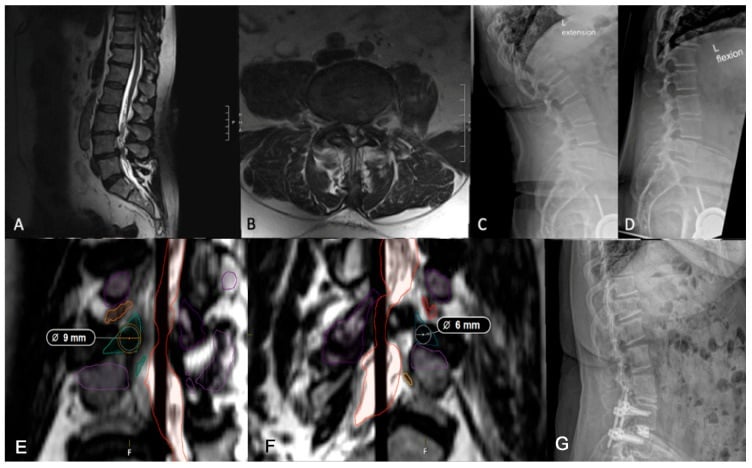
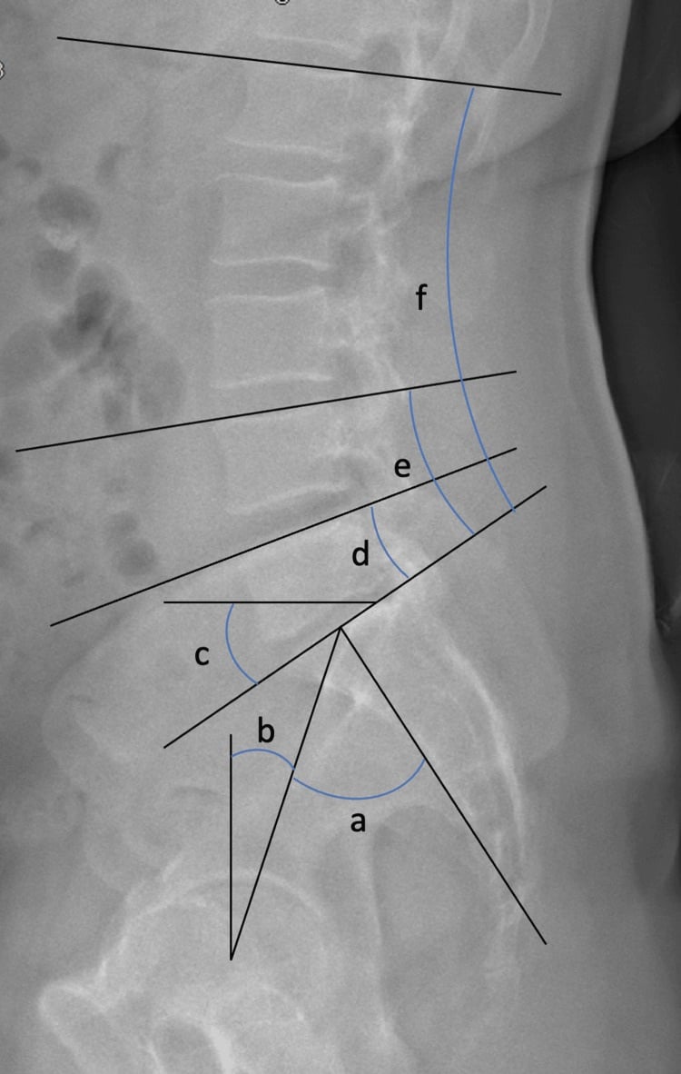
Standing radiographs. Standing AP and lateral films with flexion-extension views give what a supine MRI cannot: sagittal balance, pelvic incidence and lumbar lordosis, dynamic instability and the grade of any spondylolisthesis. They are essential because alignment and instability may not be visible on the supine study.
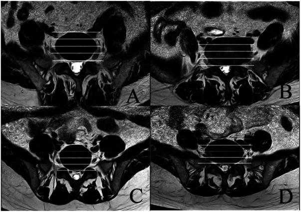
MRI is the primary modality for soft-tissue pathology and neural compression, and is essential for planning. It grades disc degeneration (Pfirrmann), shows the neural compression and any Modic endplate changes, assesses the adjacent levels, and maps the psoas and vascular anatomy for a lateral approach. In planning terms it identifies the pathology that needs decompression, gives the disc height for cage sizing, and shows the potential approach corridors.
CT gives the bone detail: pedicle anatomy for screw placement, the state of the facet joints and any bony stenosis. It is also the study for assessing fusion postoperatively. CT angiography maps the position of the great vessels and any aberrant vessels for ALIF planning.
Bone density. DEXA is essential in elderly patients. A T-score below -2.5 is osteoporosis, and it changes the fixation strategy towards cement augmentation.
Vascular surgery consultation. Consider it for ALIF at L4 and above, after previous vascular surgery, and where the vessel anatomy is aberrant.
Management Overview
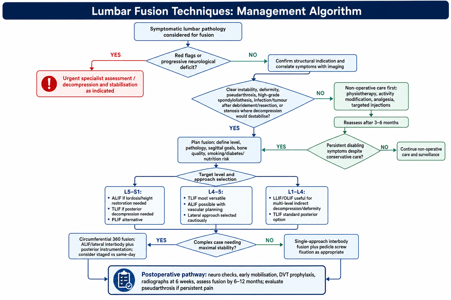
Conservative treatment first. Most degenerative conditions are treated non-operatively to begin with, typically for 3-6 months: physiotherapy and activity modification, with epidural or facet injections. Most patients should complete this before fusion is considered.
When to operate. Surgery is considered when there is a clear structural indication, conservative treatment has failed, there is a progressive neurological deficit, or the functional limitation is significant.
Matching the approach to the level. Approach selection depends on the target level, the pathology and the surgeon's experience:
- L5-S1: ALIF is excellent for height restoration and lordosis; TLIF is good when decompression is needed; PLIF is the alternative to TLIF
- L4-5: TLIF is the most versatile; ALIF is possible but the vascular concern is greater; the lateral approach is technically possible but L4-5 is the challenging level for it
- L1-L4: lateral (LLIF/OLIF) is good for multi-level disease; TLIF is the standard posterior approach; ALIF is limited by the vascular anatomy
Patient factors that change the plan. Previous surgery favours a different approach; ALIF planning needs the vascular anatomy on a CT angiogram; osteoporosis means planning augmentation; and a smoker must stop before elective fusion. Put together, the common decisions are these:
- Single level with radiculopathy: TLIF
- L5-S1 with significant collapse: ALIF plus posterior fixation
- Multi-level deformity: lateral plus posterior
- Revision for pseudarthrosis: consider circumferential fusion
Circumferential (360°) fusion combines ALIF with posterior instrumentation and gives the highest fusion rate. It is indicated for pseudarthrosis, high-grade spondylolisthesis and deformity, and is reserved for the complex cases that need maximum stability. Doing both stages on the same day means longer anaesthesia and more blood loss; staging means two recoveries but is possibly safer.
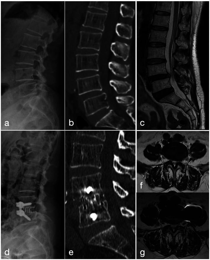
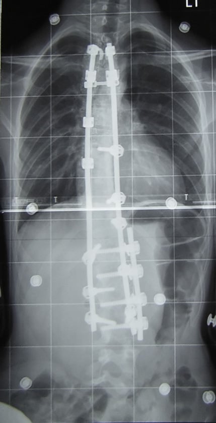
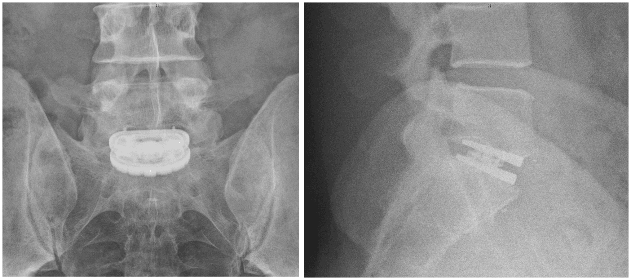
The lumbosacral junction. L5-S1, and any long fusion ending on the sacrum, is a special case. The junction is a high-stress cantilever with a notoriously high rate of S1-screw failure and L5-S1 pseudarthrosis, and failure to support it is a common cause of distal construct failure and pseudarthrosis. Tricortical or bicortical S1 screw purchase and good sacral fixation are the foundation, and two additions protect it:
- Anterior column support at L5-S1 (an interbody cage, by ALIF or TLIF) whenever fusing across it, and especially in a long construct: "in front and behind" loads the graft in compression and dramatically improves the fusion rate at this junction
- Sacropelvic fixation (S2-alar-iliac or iliac screws) for long constructs to the sacrum, because S1 pedicle screws alone are insufficient: it protects the S1 screws from cantilever pull-out and supports the lumbosacral fusion until it unites. The longer the construct above, the more the bottom must be anchored
Management Algorithm
Surgical Technique
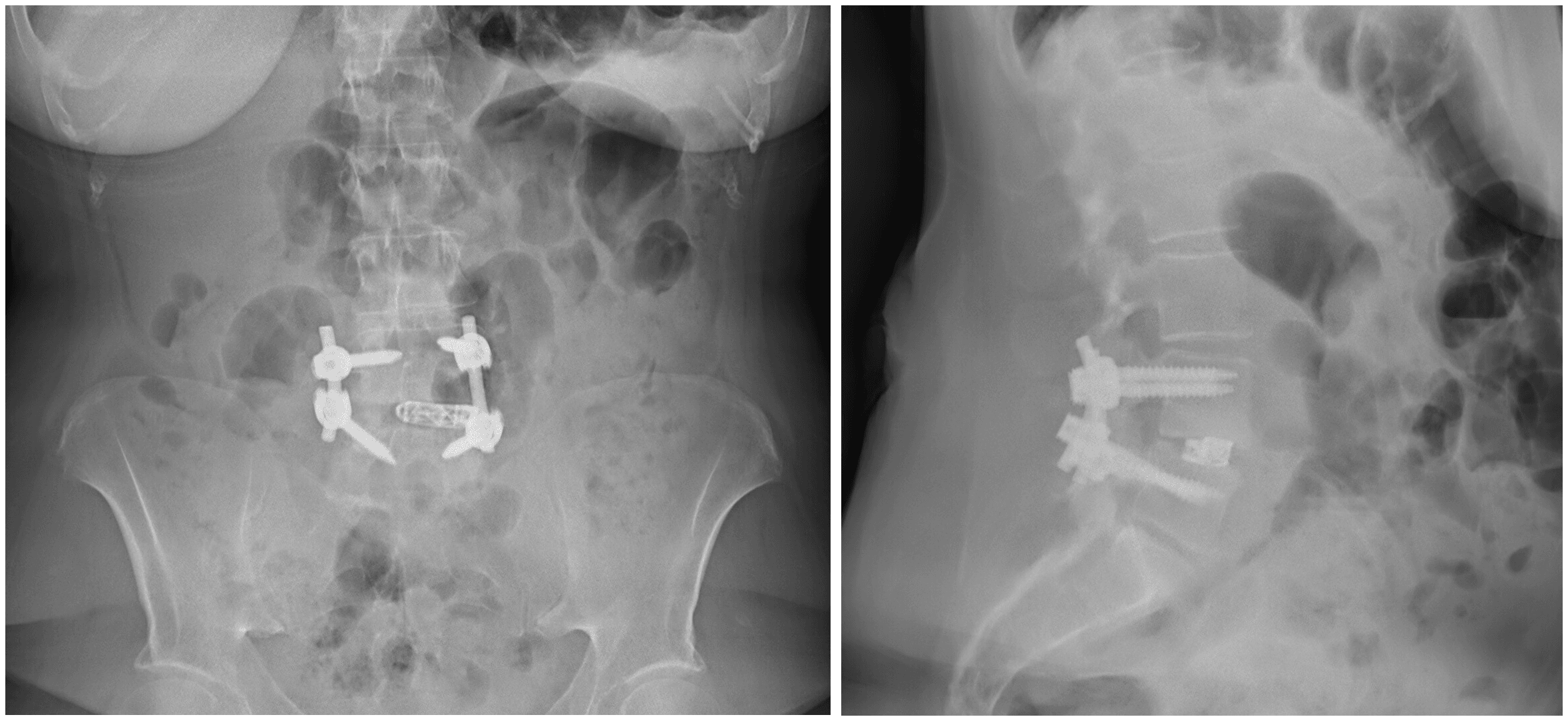
Transforaminal lumbar interbody fusion. The patient is prone on a Wilson frame or Jackson table and the exposure is midline, taken further on the symptomatic side. A complete facetectomy on the approach side is what opens the transforaminal window to the disc, and the exiting and traversing roots are identified and protected before the discectomy begins.
- Midline incision, exposing the symptomatic side more extensively
- Pedicle screws at the planned levels
- Complete facetectomy on the approach side
- Identify and protect the exiting and traversing roots
- Discectomy through the transforaminal window
- Prepare the endplates with curettes and shavers, and trial for size
- Pack bone graft into the disc space and the cage
- Insert the cage obliquely across the midline
- Compress the pedicle screws to engage the cage
Why it has become the workhorse. The unilateral approach reduces nerve retraction compared with PLIF, and with it the nerve complication rate; the cage angled across the midline gives central support; the contralateral facet is preserved where possible; and neuromonitoring is recommended. Interbody cage plus pedicle screws makes a circumferential construct through a single posterior exposure, which is the versatility that has put it ahead of PLIF.
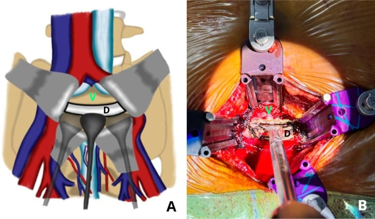
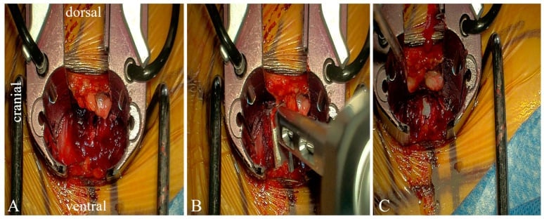
Minimally invasive fusion. The minimally invasive versions of these operations cause less muscle damage and less blood loss, and give a shorter hospital stay and a faster recovery. The costs are a steep learning curve, more radiation exposure, limited visualisation, and cases for which they are not suitable.
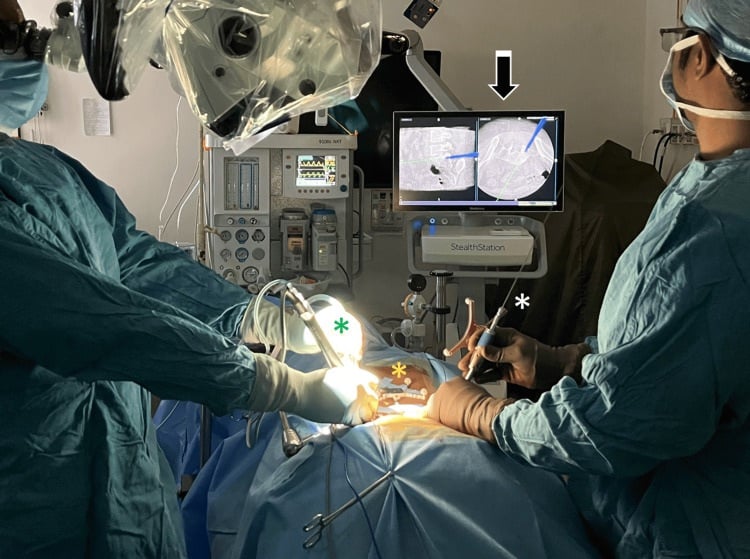
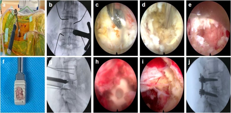
Graft. Fusion is a race between healing and hardware failure, so know the graft hierarchy. A successful graft needs osteogenic cells, an osteoinductive signal and an osteoconductive scaffold, with mechanical stability and a viable host (the "diamond").
- Autograft is the biological gold standard. Iliac crest has all three properties but carries donor-site morbidity and is rarely used now; local bone from the laminectomy and facetectomy is the workhorse for posterolateral fusion and TLIF
- Allograft is structural (femoral ring and similar) or particulate (chips, DBM). Allograft, DBM and ceramics are osteoconductive extenders with, at most, weak osteoinduction
- Synthetics: BMP-2, tricalcium phosphate and hydroxyapatite
- Cage filling is usually a combination of local autograft with allograft or DBM; BMP may be used, but it is controversial and off-label in some applications
rhBMP-2 (INFUSE) is FDA-approved only for single-level ALIF with a specific LT (tapered) cage. Its very common use in TLIF, PLIF and posterolateral fusion is off-label, and the harms follow the approach:
- Anterior (ALIF): associated with retrograde ejaculation (a sympathetic/hypogastric plexus effect) and anterior soft-tissue swelling
- Posterior (TLIF/PLIF): postoperative radiculitis, heterotopic or ectopic bone in the canal or foramen, and vertebral endplate osteolysis or resorption causing cage subsidence if placed near the dura or root
The earlier cancer-risk signal has been largely not substantiated, but dose and placement discipline and informed consent are essential. Use it deliberately, not routinely.
Complications
Each route has a signature complication, and the prevention column is the operative discipline that route demands.
- Major Complication
- Dural tear
- Incidence
- 3-5%
- Prevention
- Careful technique, protect dura
- Major Complication
- Nerve root injury
- Incidence
- 1-2%
- Prevention
- Identify roots, gentle retraction
- Major Complication
- Vascular injury
- Incidence
- 1-3%
- Prevention
- Vascular surgeon, careful retraction
- Major Complication
- Retrograde ejaculation
- Incidence
- 0-4%
- Prevention
- Avoid hypogastric plexus injury
- Major Complication
- Lumbar plexus injury
- Incidence
- 15-25% transient
- Prevention
- Neuromonitoring, safe zone approach
- Major Complication
- Psoas weakness
- Incidence
- 10-20% transient
- Prevention
- Minimise retraction time
In theatre. The intraoperative risks are wrong-level surgery, which is why fluoroscopic confirmation is essential; bleeding from the epidural veins and the segmental vessels; and cage malposition.
Afterwards. Infection in 1-3%, pseudarthrosis in 5-15% depending on risk factors, hardware failure (loosening or breakage) and cage subsidence.
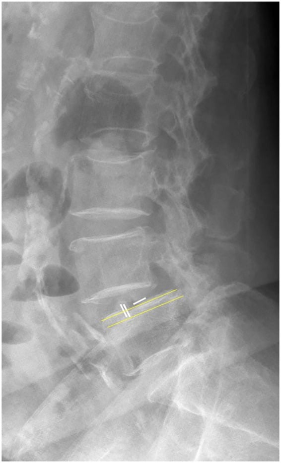
Adjacent segment disease (2-3% per year). The principal long-term cost of fusion, and the counterweight to the techniques above. A fused segment is rigid, so the load and motion it used to absorb are transferred to the neighbouring mobile segments, which then degenerate faster; the longer the fusion, the greater the effect. About 2-3% per year is the quoted rate, but the number that matters is cumulative: over a decade a meaningful proportion of fused patients develop symptomatic degeneration at the next level, and some come back for an extension of the fusion.
The motion-preservation answer. This stress transfer is the rationale for total disc replacement (lumbar arthroplasty) at single levels: by preserving motion rather than abolishing it, the implant aims to avoid the transfer that drives adjacent-segment degeneration. The trade-off is that disc replacement has its own failure modes and is unsuitable where there is facet arthropathy, instability or deformity. Fusion is the right operation for instability and deformity, but every fusion is also a bet against the next level, and choosing the shortest construct that achieves stability, and reserving fusion for patients who genuinely need it, is the main lever the surgeon controls.
Pseudarthrosis. Smoking is the most significant modifiable risk factor; diabetes, obesity, multi-level fusion, osteoporosis and poor nutrition add to it. The diagnosis is persistent pain with no bridging bone on CT at 1 year, hardware loosening or breakage, or motion on flexion-extension films. Management is to optimise the modifiable factors and revise with improved fixation and graft augmentation, considering a different approach.
Smoking cessation is the most important modifiable factor for fusion success - a meta-analysis found smoking nearly doubled the nonunion risk (relative risk 1.91). Nicotine impairs osteoblast function and vascular ingrowth. Many surgeons require smoking cessation before elective fusion.
Postoperative Care
The first days. On day 0-1 the priorities are a neurological assessment with particular attention to motor function, multimodal analgesia, DVT prophylaxis and early mobilisation. After ALIF, assess bowel function because ileus is possible; after a lateral approach, assess the hip flexors.
- Single-Level Fusion
- 1-3 days
- Multi-Level Fusion
- 3-5 days
- Single-Level Fusion
- Day 0-1
- Multi-Level Fusion
- Day 1-2
- Single-Level Fusion
- Variable (surgeon preference)
- Multi-Level Fusion
- Often for 6-12 weeks
- Single-Level Fusion
- 4-6 weeks
- Multi-Level Fusion
- 6-12 weeks
- Single-Level Fusion
- 4-6 weeks
- Multi-Level Fusion
- 6-12 weeks
- Single-Level Fusion
- 3-6 months
- Multi-Level Fusion
- 6-12 months
- Single-Level Fusion
- 6-12 months
- Multi-Level Fusion
- 12+ months
Watching for union. The imaging schedule is genuinely sequential, and each visit asks a different question:
Standing radiographs. You are looking for maintained alignment and correctly seated implants, not for fusion — bridging bone is not expected this early.
Radiographs to confirm nothing has shifted. Early screw loosening or cage migration declares itself here, and is the point at which activity restriction may need to be tightened rather than relaxed.
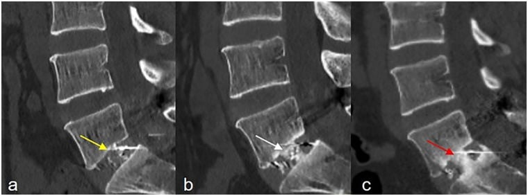
Do not order CT routinely. Reserve it for persistent or recurrent pain, or a radiographic suspicion of pseudarthrosis — plain films substantially overcall union, so CT is the arbiter when the question is actually being asked.
The point at which absent union is called a nonunion rather than a delay. Note that radiographic union and symptom relief are separate endpoints: a solidly fused segment can still hurt, and a radiographic nonunion can be asymptomatic and need no revision.
Signs of solid fusion. Bridging bone on CT, no motion on flexion-extension, no hardware loosening, and resolution of symptoms.
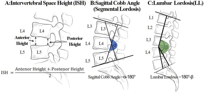
Rehabilitation. Core strengthening waits until the fusion is consolidating; flexibility, conditioning and functional training follow. Smoking cessation is mandatory, and weight optimisation and activity modification are part of the plan.
Outcomes and Prognosis
Fusion rates climb as anterior column support and posterior instrumentation are added, which is the biomechanical argument of the whole topic in a single table.
- Fusion Rate
- 70-80%
- Notes
- Without interbody support
- Fusion Rate
- 90-95%
- Notes
- With pedicle screws
- Fusion Rate
- 90-95%
- Notes
- With pedicle screws
- Fusion Rate
- 90-95%
- Notes
- With posterior instrumentation
- Fusion Rate
- 95-100%
- Notes
- Highest rate
Clinical success (symptom improvement) depends on the indication: 70-80% good or excellent for spondylolisthesis, 60-70% for DDD with instability, a controversial indication, and a variable 50-70% for revision and pseudarthrosis surgery.
Prognostic factors. The two lists are near mirror images.
- Favourable: clear structural indication, non-smoker, normal BMI, single level, good psychosocial status, no workers compensation claim
- Unfavourable: smoking (the most important), obesity, multi-level fusion, osteoporosis, depression, workers compensation
Guidelines, Registries & Global Practice
Global Epidemiology and Practice Variation
Elective lumbar fusion rates have risen steadily across high-income countries over the past two decades, with marked variation by region, hospital type and surgeon. In a US Nationwide Inpatient Sample analysis of degenerative spondylolisthesis (2001-2010), the choice of fusion technique varied significantly with geographic region, teaching status and hospital size, and combined anterior/posterior or interbody constructs carried higher complication and mortality risk than posterolateral fusion alone in the acute phase. In Norway, the rate of lumbar spinal stenosis surgery more than tripled between 1999 and 2013, with surgery in those over 65 years more than quadrupling, although the proportion involving fusion actually fell from 19.3% to 10.9%. The take-home for any examination: fusion utilisation reflects health-system and surgeon factors as much as pathology, and "more fusion" has not consistently meant "better outcome".
- Finding
- Technique choice varies by region/hospital; complex constructs raise complication and mortality risk
- Exam-relevant message
- Procedure selection is partly non-clinical; simpler constructs are safer acutely
- Finding
- Stenosis surgery tripled 1999-2013; fusion proportion fell to ~11%
- Exam-relevant message
- Decompression-led practice is increasing; routine fusion is being de-emphasised
- Finding
- National registries track patient-reported outcomes and reoperation after fusion
- Exam-relevant message
- Outcome and reoperation rates, not just fusion rates, define success
Guidelines Side by Side
Major bodies converge on the same core principle: fusion needs a structural indication (instability, deformity, neural compression requiring destabilising decompression), and is not supported for axial low back pain from degenerative disc disease alone.
- Position
- Do not offer fusion for low back pain unless within a randomised trial; consider for confirmed instability/deformity
- Evidence level / basis
- Systematic review of RCTs
- Position
- Fusion supported for spondylolisthesis with instability and selected deformity; not for uncomplicated DDD
- Evidence level / basis
- Level I-II evidence synthesis
- Position
- Fusion adds benefit over decompression alone in Grade I degenerative spondylolisthesis with stenosis
- Evidence level / basis
- Level I RCT
- Position
- Adding fusion to decompression gives no benefit for stenosis +/- spondylolisthesis
- Evidence level / basis
- Level I RCT (counters SLIP)
- Position
- Approach (PLIF/TLIF/ALIF/LLIF) chosen by level, sagittal goals and surgeon experience; no single superior interbody route
- Evidence level / basis
- Expert consensus + meta-analyses
The unresolved SLIP versus Swedish Spinal Stenosis Study controversy is a classic viva discussion point: SLIP supports fusion for unstable Grade I spondylolisthesis, while the Swedish trial found no benefit from adding fusion to decompression. The pragmatic synthesis is to fuse when there is demonstrable instability or when decompression itself will destabilise the segment, and to decompress alone when the segment is stable.
Approach Choice and Modifiable Risk - Global Standard of Care
- Technique: TLIF is the most widely used posterior interbody technique; meta-analysis favours it over PLIF on complication grounds. ALIF, LLIF/XLIF and OLIF are valid alternatives selected by level and sagittal goals, with comparable fusion rates.
- Anterior approaches: an access surgeon (vascular/general) for ALIF and continuous EMG neuromonitoring for transpsoas LLIF are widely regarded as standard safeguards.
- Modifiable risk: smoking cessation is the single most important modifiable factor (nonunion relative risk 1.91); diabetic control, weight optimisation and treatment of osteoporosis (including screw cement augmentation) are recommended before elective fusion regardless of health system.
Special Considerations
Revision fusion. Scarring obscures the anatomy, hardware may need removal, bone has been lost, and the pseudarthrosis rate is higher. The strategies answer each of those: a different approach (anterior if the previous surgery was posterior), augmented fixation (longer constructs, cement) and an optimised graft (BMP, autograft).
The osteoporotic spine. Poor screw purchase, cage subsidence and a higher failure rate are the problems. The solutions are cement augmentation of the screws, larger or expandable cages, extended fixation and optimising bone health.
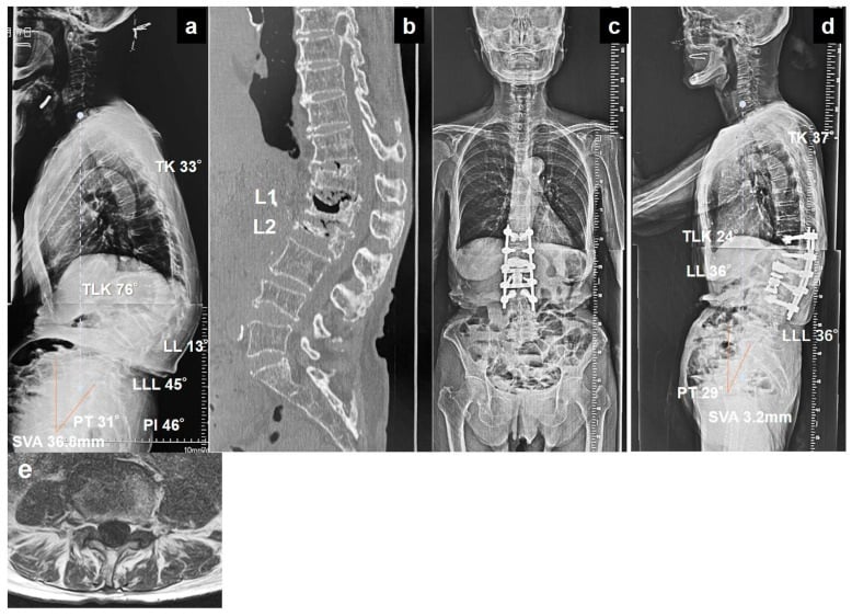
Multi-level fusion. The complication rate is higher and the adjacent segment disease risk greater; combined approaches are considered, and staged procedures may be safer.
MCQ Practice Points
Q: What is the main advantage of TLIF over PLIF?
A: TLIF uses a unilateral approach with less nerve retraction than PLIF's bilateral approach. This results in lower dural tear and nerve root injury rates. TLIF has largely replaced PLIF as the workhorse posterior interbody technique.
Q: What is the best level for ALIF and why?
A: L5-S1 is ideal for ALIF because the great vessels bifurcate at this level, providing more working space. Above L4, vascular structures require significant retraction and vascular surgery involvement is recommended.
Q: Why should you avoid lateral (LLIF/XLIF) approach at L5-S1?
A: The iliac crest blocks lateral access to L5-S1. Additionally, the lumbar plexus runs within/behind the psoas muscle and is at risk during transpsoas approaches (15-25% transient neurological symptoms).
Q: What fusion rate is achieved with circumferential (360°) fusion?
A: Circumferential fusion achieves 95-100% fusion rate. Compare to posterolateral alone (70-80%) and interbody with instrumentation (90-95%). Interbody support provides anterior column load-sharing.
Q: What is the most important modifiable risk factor for pseudarthrosis?
A: Smoking. A meta-analysis found smoking nearly doubled the nonunion risk (relative risk 1.91). Nicotine impairs osteoblast function and vascular ingrowth. Many surgeons require smoking cessation before elective fusion.
Summary
Key Takeaways
-
TLIF Has Replaced PLIF: The unilateral transforaminal approach provides adequate disc access with less nerve retraction compared to bilateral PLIF. TLIF is now the most commonly performed interbody fusion technique.
-
ALIF Best at L5-S1: The great vessels bifurcate at L5-S1, providing safe access. Above this level, vascular surgery involvement is recommended. Know the retrograde ejaculation risk from sympathetic plexus injury.
-
Lateral Approach Avoids L5-S1: The iliac crest blocks lateral access to L5-S1. The lumbar plexus runs within the psoas and is at risk - neuromonitoring is essential. Transient symptoms are common (15-25%).
-
Interbody Fusion Improves Fusion Rates: Adding an interbody cage to pedicle screw fixation increases fusion rates from 70-80% to 90-95%. Circumferential fusion provides the highest fusion rate.
-
Smoking is the Most Important Modifiable Factor: A meta-analysis found smoking nearly doubled the nonunion risk (relative risk 1.91). Cessation before elective fusion should be required.
-
Know the Indications: Clear structural indications (spondylolisthesis, instability, deformity) produce the best outcomes. Fusion for degenerative disc disease alone remains controversial.
-
Approach Selection Matters: Match the approach to the level, pathology, and patient factors. L5-S1 suits ALIF or TLIF; L1-L4 suits lateral or TLIF; multi-level deformity may need combined approaches.
Clinical Decision Scenarios
Practise clinical reasoning and management decisions out loud
“A 55-year-old woman has L5-S1 Grade I spondylolisthesis with bilateral L5 radiculopathy. She has failed 6 months of conservative treatment. What surgical options would you consider?”
“You are planning an L4-5 TLIF. Describe the key steps and how you would avoid complications.”
“What are the advantages and disadvantages of ALIF versus TLIF at L5-S1?”
“During a lateral interbody fusion at L3-4, the patient develops thigh weakness postoperatively. How do you assess and manage this?”
TLIF vs PLIF
- TLIF: Unilateral approach, single cage oblique
- PLIF: Bilateral approach, bilateral cages
- TLIF: Less nerve retraction, lower complications
- TLIF has largely replaced PLIF
ALIF
- Best level: L5-S1 (vessels bifurcate)
- Large cage, excellent lordosis restoration
- Vascular injury risk 1-3%
- Retrograde ejaculation 0-4% (sympathetic)
Lateral (LLIF/XLIF)
- Avoid L5-S1 (iliac crest blocks)
- Transpsoas approach - lumbar plexus at risk
- Transient neurological symptoms 15-25%
- Neuromonitoring essential
Fusion Rates
- Posterolateral alone: 70-80%
- Interbody + instrumentation: 90-95%
- Circumferential (360 degree): 95-100%
- Smoking is biggest modifiable risk factor
Evidence-Based Practice
SLIP Trial: Laminectomy plus Fusion vs Laminectomy Alone (RCT, NEJM 2016)
- 66 patients, Grade I degenerative spondylolisthesis with stenosis randomised
- Adding posterolateral instrumented fusion gave greater SF-36 physical gain at 2 years (15.2 vs 9.5; difference 5.7, 95% CI 0.1 to 11.3, P=0.046) - note the interval's lower bound of 0.1, which only just excludes zero
- ODI improvement did not differ significantly (-26.3 fusion vs -17.9 decompression, P=0.06)
- Reoperation lower with fusion (14% vs 34%, P=0.05)
- Fusion: more blood loss and longer hospital stay
Swedish Spinal Stenosis Study: Fusion vs Decompression Alone (RCT, NEJM 2016)
- 247 patients with lumbar stenosis (135 with degenerative spondylolisthesis) randomised
- No difference in ODI at 2 years (27 fusion vs 24 decompression-alone, P=0.24)
- No difference in 6-minute walk test or 5-year outcomes
- Results similar with or without spondylolisthesis
- Fusion: longer stay (7.4 vs 4.1 days), more bleeding, higher cost