MT2 Marching Fracture | MT5 High-Risk Zones | Female Athlete Triad | MRI Gold Standard | Activity Modification
- Second metatarsal is most common site (marching fracture) - usually low-risk, conservative treatment
- Fifth metatarsal Zone 2-3 are HIGH-RISK locations - 50%+ nonunion rate, often need surgery
- MRI is gold standard for early diagnosis - X-rays negative for 2-3 weeks after symptom onset
- Female athlete triad (energy deficiency, amenorrhoea, low BMD) increases stress fracture risk
- Activity modification is cornerstone - address training errors, biomechanics, nutrition
- “MT2 stress fracture = 'marching fracture' - most common, usually conservative, good prognosis
- “MT5 Zone 2-3 stress fractures = high nonunion risk = surgical fixation in athletes
- “X-ray delay: symptoms appear 2-3 weeks before radiographic changes (MRI earlier)
- “Bone scan vs MRI: MRI more specific, shows fracture line and oedema pattern
- “Return to sport: 6-8 weeks for low-risk, 8-10 weeks after fixation for high-risk (MT5 Zone 2-3), 10-12 weeks if bone grafted
Overview and Epidemiology
Metatarsal stress fractures are among the most common stress fractures in athletes and military recruits. Two sites organise the topic. The second metatarsal is the most frequent, the "marching fracture", and it is typically low-risk with an excellent prognosis after conservative treatment. Fifth metatarsal Zone 2-3 fractures are less common but sit in a watershed blood supply, carry a nonunion rate quoted at 50% or more, and in athletes are fixed. Site determines risk, not frequency.
Who. Runners, dancers and military recruits, peaking in young adults from the late teens to the 30s; track, basketball, ballet and marching are the usual settings. Overall incidence is similar in male and female athletes. The female-specific risk is mediated by the triad of low energy availability, menstrual dysfunction and low bone mineral density, not by sex itself.
What it costs. Training is interrupted for 6-12 weeks, the injury recurs if the underlying causes are not addressed, and for the professional athlete the career impact is why early surgery is needed.
Anatomy and Pathophysiology
How a stress fracture forms. With overuse, osteoclast activity exceeds osteoblast activity, microfractures accumulate faster than the bone can repair them, and the bone fails by fatigue. The cortical breach starts on the tension side, the inferior cortex; if the activity continues it becomes a complete fracture.
Why the second metatarsal. It is the longest metatarsal and the most rigidly fixed at the tarsometatarsal joint, so the repetitive loading of running, jumping and marching concentrates on the inferior cortex of its shaft.
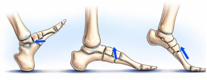
The base of the second metatarsal. The classic marching fracture is in the shaft. A distinct and easily missed entity is the stress fracture of the proximal second metatarsal base, characteristically seen in ballet dancers, and it behaves very differently. The base is recessed and rigidly keystoned into the cuneiforms, the Lisfranc "mortise", which makes MT2 the least mobile metatarsal, and en pointe or demi-pointe the load concentrates at this proximal base.
Unlike the benign shaft fracture it is higher-risk and slow to heal, lying in the relatively avascular proximal metaphyseal region near the Lisfranc complex; it is frequently missed because attention is on the shaft, and it can progress to delayed union or nonunion if the dancer keeps loading it. Suspect it when the pain and tenderness are at the proximal MT2 or midfoot rather than the shaft, in a dancer with a spike in training or pointe work; look specifically at the base on imaging and use MRI early, because radiographs lag and the base is hard to read. Management is more cautious than for the shaft: genuine offloading and relative rest with prolonged restriction from pointe work and a graded return, with surgery occasionally needed for nonunion.
The fifth metatarsal zones. The proximal fifth metatarsal has three zones, and the zone is the first thing to assign:
- Zone 1, the tuberosity: the peroneus brevis avulsion, a different injury from a stress fracture, with a good blood supply and reliable healing
- Zone 2, the metaphyseal-diaphyseal junction: the true Jones fracture
- Zone 3, the proximal diaphysis: where recurrent stress fractures occur, the chronic overload lesion
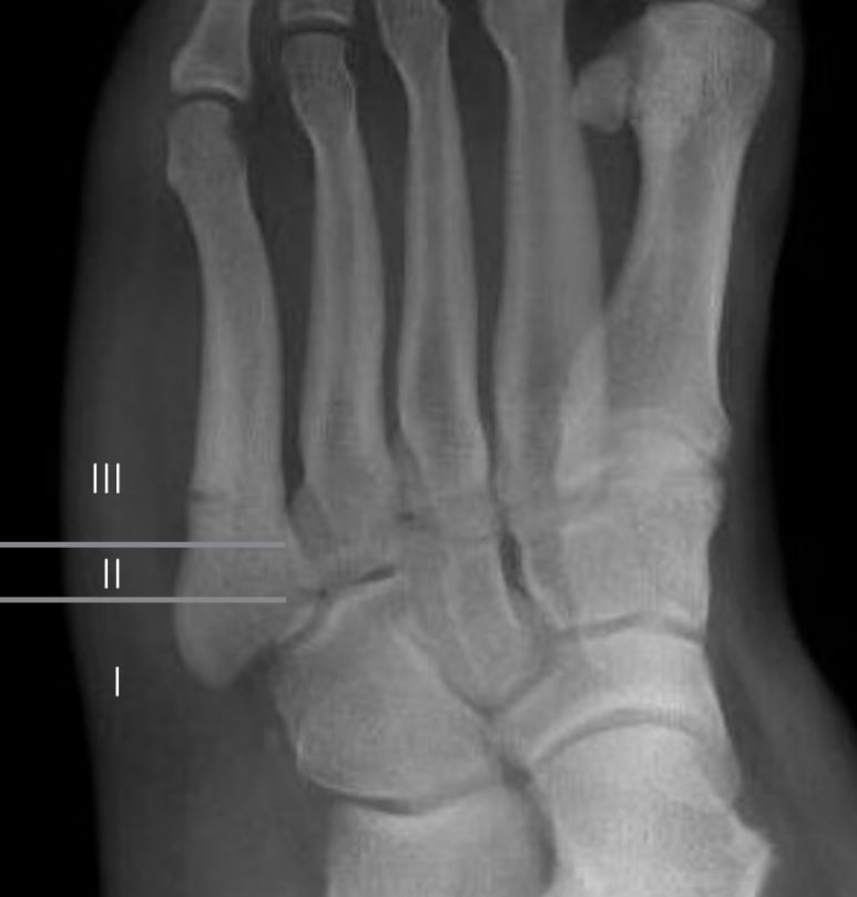
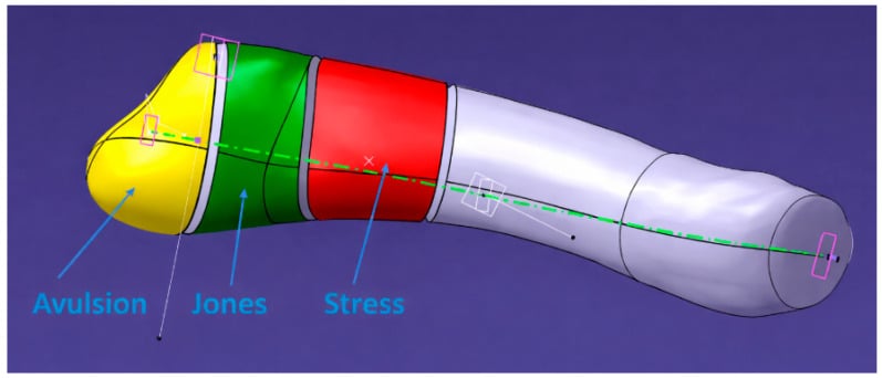
The watershed. Zone 2 and Zone 3 sit where the nutrient artery supply meets the periosteal supply, a relatively avascular region with poor healing potential. The other sites are supplied adequately, as the site table under Classification sets out.
What the nonunion figure really means. Attach the rate to a treatment, because it varies enormously with weight-bearing. In Torg's series the acute fractures managed in a non-weight-bearing toe-to-knee cast healed in 14 of 15 (93%) at a mean of 7 weeks, while those allowed to weight-bear united in only 4 of 10; in Japjec's athletes treated conservatively only 4 of 9 healed. The widely quoted "50% or more nonunion" therefore describes Zone 2-3 fractures that are weight-borne or inadequately immobilised, not the zone itself.
Why that changes nothing for the athlete. The clinical consequence is unchanged and arguably sharpened: an athlete will not accept three months non-weight-bearing with a one-in-two failure rate if they weight-bear, which is why fixation is the default in the competitive athlete. The honest statement to a non-athlete is that a strictly non-weight-bearing cast has a good chance of working.
Classification Systems
Three schemes are in use and they answer different questions: the site tells you the risk, the radiographic grade tells you how far the injury has progressed, and for the proximal fifth metatarsal Torg's types tell you whether the canal is sclerotic and needs grafting.
Site is the strongest predictor of outcome, and the blood supply is the reason.
- Blood supply
- Adequate nutrient artery supply
- Risk and nonunion rate
- Low; under 5% for the MT2 shaft
- Treatment
- Activity modification, CAM boot 4-6 weeks; excellent healing
- Blood supply
- Dual supply (medial and lateral plantar)
- Risk and nonunion rate
- Moderate; 10-15%
- Treatment
- NWB 6-8 weeks, consider bone stimulator; lower threshold for surgery because of its weight-bearing load
- Blood supply
- Periosteal supply adequate
- Risk and nonunion rate
- Low (the tuberosity avulsion is a different injury)
- Treatment
- Conservative treatment successful
- Blood supply
- Watershed zone, poor supply
- Risk and nonunion rate
- HIGH; 50%+
- Treatment
- Surgical fixation in athletes
- Blood supply
- Watershed zone, poor supply
- Risk and nonunion rate
- HIGH; 50%+
- Treatment
- Surgical fixation, bone graft if delayed
- Blood supply
- -
- Risk and nonunion rate
- High
- Treatment
- Comprehensive workup; address systemic causes (female athlete triad, nutrition, biomechanics)
Clinical Assessment
History. The onset is gradual and insidious, over weeks to months, in someone whose load has changed: repetitive loading, a training error, a recent increase in volume or intensity. Pain comes first with activity and progresses to pain at rest. Ask about the risk factors that will decide what happens after the fracture heals: the female athlete triad, nutrition and biomechanics.
Examination. Point tenderness over the specific metatarsal is the finding; swelling is localised and minimal in the early stages, and the neurovascular examination is usually intact. Two provocation tests help:
- Tuning fork test: vibration over the fracture site causes pain
- Hop test: a single-leg hop reproduces the pain
Fifth metatarsal Zone 2-3 stress fractures may present with minimal symptoms initially despite their nonunion risk. Any athlete with MT5 base pain and a training history should have early MRI to assess Zone 2-3 involvement. Delayed diagnosis leads to nonunion requiring bone graft.
Differential diagnosis. Forefoot pain has several sources, and point tenderness over bone is what separates the stress fracture from the rest.
- Key Features
- Point tenderness, insidious onset, training history
- Distinguishing Factor
- MRI shows fracture line and oedema
- Key Features
- Diffuse forefoot pain, no point tenderness
- Distinguishing Factor
- No fracture on imaging
- Key Features
- Interdigital pain, Mulder's click
- Distinguishing Factor
- Between metatarsals, not over bone
- Key Features
- MT2 head avascular necrosis
- Distinguishing Factor
- X-ray shows collapse, not stress fracture
Forefoot stress injury is not only metatarsal. A sesamoid stress fracture is a classic, easily missed cause of plantar first-ray pain in runners, dancers and sprinters, and a recognised high-risk, poor-healing site. The medial (tibial) sesamoid is more often affected because it bears more load; the pain is plantar under the first metatarsal head and worse on push-off and dorsiflexion of the hallux. The sesamoids have a tenuous, often single-vessel blood supply, so these fractures are prone to delayed union, nonunion and osteonecrosis despite the small bone.
The trap is the bipartite sesamoid, present in a notable minority and frequently bilateral: it has smooth, well-corticated, rounded margins, whereas a fracture has sharp, irregular, non-corticated edges, focal tenderness and marrow oedema on MRI. Get the contralateral film, and MRI or CT if unsure; bone scan is sensitive.
Treat non-operatively first: strict offloading in a stiff-soled shoe or boot with a dancer's pad to unload the sesamoid, then a graded return. For nonunion the options are partial sesamoidectomy or bone grafting. Avoid total excision of both sesamoids, or a tibial sesamoidectomy that destabilises the hallux, which causes a cock-up or hallux valgus or varus deformity.
Investigations
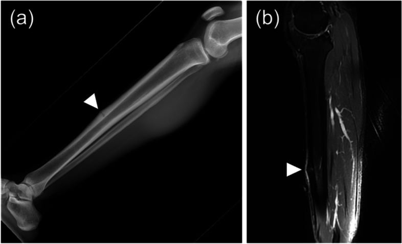
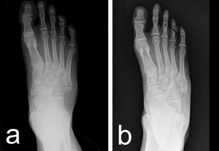
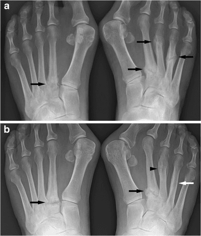
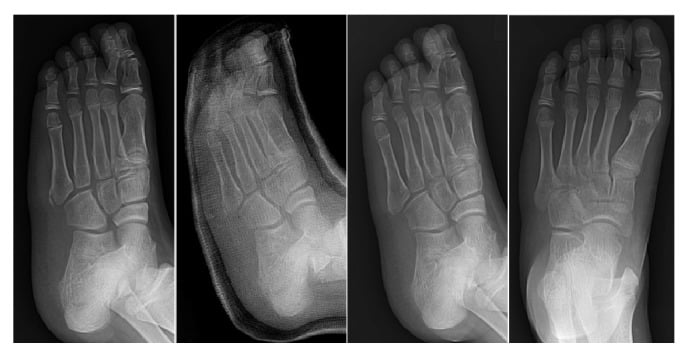
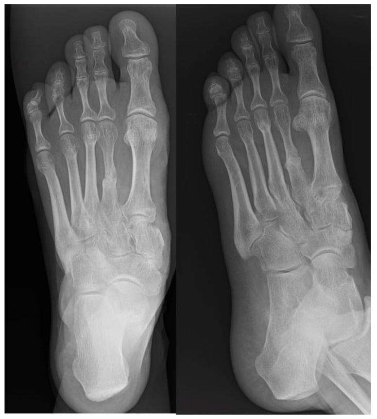
Radiographs first, knowing they lag. AP, lateral and oblique views of the foot are the first line, and the initial films are often negative: stress fractures take 2-3 weeks after symptom onset to show radiographic change. When change appears the sequence is predictable:
- Periosteal reaction at 2-3 weeks
- Fracture line at 3-4 weeks
- Callus at 4-6 weeks
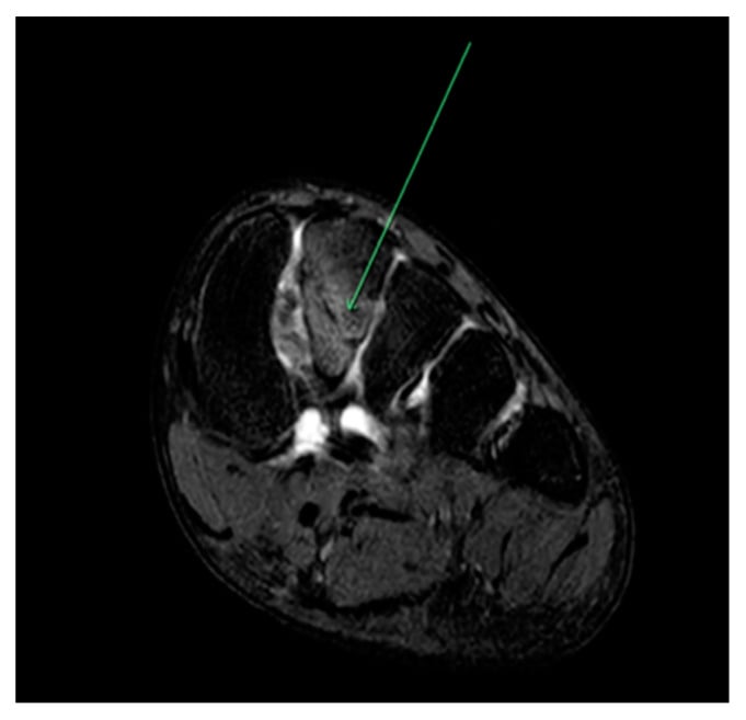
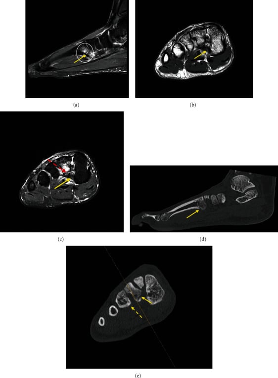
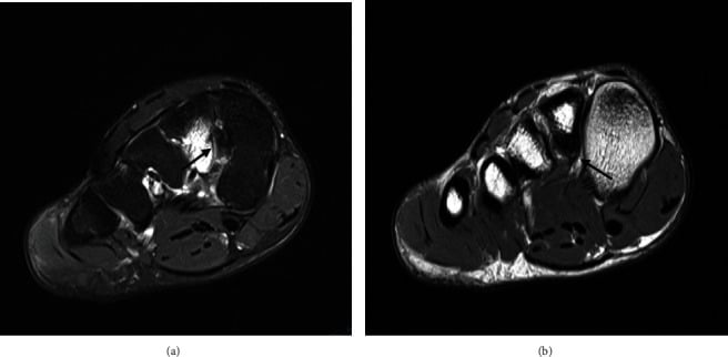
MRI is the gold standard for early diagnosis and shows changes within days of symptom onset: bone marrow oedema (T2 hyperintensity), a fracture line (a T1 hypointense line) and periosteal reaction. It is highly sensitive, shows the extent of the injury and guides treatment. If clinical suspicion is high, with point tenderness and a training history, order MRI immediately rather than waiting for the radiograph to change; early diagnosis allows prompt treatment and prevents progression to a complete fracture.
Bone scan shows increased uptake at the fracture site and is sensitive but less specific than MRI. Use it if MRI is unavailable; MRI is preferred for its specificity.
Management Algorithm
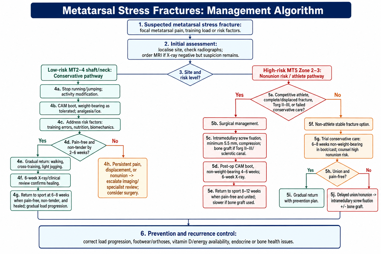
The decision. Localise the pain, image it, adding MRI when radiographs are negative but suspicion persists, and then the branch is risk by site. Low-risk MT2-4 shaft fractures are treated conservatively; high-risk MT5 Zone 2-3 fractures are fixed in the athlete. Whatever the site, the stress fracture is a symptom and not the disease, and the root causes must be addressed or it recurs.
Low-risk sites (MT2-4). The goal is pain-free healing with activity modification: stop running and jumping, a CAM boot with weight-bearing as tolerated, and correction of the training, nutritional and biomechanical causes. Return to sport is at 6-8 weeks once the fracture is pain-free; the week-by-week programme is under Rehabilitation. The success rate is over 95% with proper activity modification, and surgery is rarely needed unless the fracture is displaced or has gone to nonunion.
High-risk sites (MT5 Zone 2-3). The goal is to prevent nonunion and, in the athlete, to return to sport early. Surgery is indicated for:
- Athletes, competitive or professional
- Torg Type II-III (delayed union or nonunion)
- Displacement or a complete fracture
- Failed conservative treatment
The operation is intramedullary screw fixation, with bone graft for a Torg II-III sclerotic canal (technique below). Return to sport is at 8-10 weeks in athletes and 10-12 weeks if bone graft was used, once the patient is pain-free and the radiograph shows union.
The non-athlete. May attempt conservative treatment, 6-8 weeks non-weight-bearing, and must be counselled about the high nonunion risk and the need for eventual surgery if it fails. Whether that trial is honest depends on strict non-weight-bearing, for the reasons the Torg figures above give.
Risk factors, or it comes back. Four groups of causes, each with its fix:
- What goes wrong
- Sudden increase in volume or intensity; inadequate recovery between sessions; surface changes, hard to soft or vice versa; inappropriate or worn-out footwear
- Fix
- Gradual progression, periodisation
- What goes wrong
- Energy deficiency (inadequate caloric intake); amenorrhoea (hormonal disruption); low BMD (osteoporosis or osteopenia)
- Fix
- Nutrition counselling, endocrinology referral
- What goes wrong
- Foot structure (cavus, flatfoot); gait abnormalities (overpronation, supination); leg length discrepancy with compensatory loading
- Fix
- Orthotics, physiotherapy, gait analysis
- What goes wrong
- Inadequate calcium and vitamin D intake; insufficient protein for bone remodelling; caloric deficit and energy availability
- Fix
- Dietary assessment, supplementation
Stress fractures will recur if the underlying causes are not addressed. Every patient needs a comprehensive assessment of training, nutrition, biomechanics and, in females, menstrual function, with referral to sports medicine, nutrition and endocrinology as needed.
Surgical Technique
Intramedullary screw fixation of the MT5 Zone 2-3 stress fracture is the operation for the high-risk site in the athlete. The steps are few, and each has a way of going wrong.
Surgical Steps
Supine on a radiolucent table, the C-arm positioned for AP and lateral views, the limb free-draped so the base of the fifth metatarsal is accessible for screw insertion.
The tip of the fifth metatarsal tuberosity, the palpable prominence at the base of MT5. A small 1-2 cm longitudinal incision over it, protecting the sural nerve branches.
Under fluoroscopy, down the medullary canal and across the fracture until the far (distal) cortex is engaged; AP and lateral views confirm the position.
Minimum 5.5 mm diameter, and larger is better: studies show better outcomes with 5.5-6.5 mm screws than with 4.5 mm. Length is measured from the guidewire so that the screw engages the far cortex. Compression comes from a partially threaded screw, or a fully threaded screw with a compression technique. Fluoroscopy confirms compression and position before final tightening.
For Torg Type II-III, the sclerotic canal of delayed union or nonunion: curette the sclerotic bone, place iliac crest autograft around the fracture site, then fix with the screw. Allograft bone chips are the alternative if autograft is not wanted.
Subcutaneous and skin layers, a sterile non-adherent dressing, and a posterior splint for comfort, removed at 1-2 weeks.
What goes wrong. An undersized screw, under 5.5 mm, increases the failure risk. An entry point too medial risks injury to the peroneal tendons. A screw that does not engage the far cortex gives neither compression nor stability. And missed sclerosis is a Torg II-III fracture fixed without the bone graft it needs.
Complications
The complication that matters is the one the site predicts, and the table gives each its rate, its risk factors and its remedy.
- Incidence
- 50%+ (MT5 Zone 2-3 conservative)
- Risk Factors
- High-risk site, continued activity, Torg Type II-III
- Management
- Surgical fixation + bone graft
- Incidence
- 10-20% (all sites)
- Risk Factors
- Inadequate rest, poor nutrition, biomechanics
- Management
- Extended NWB, bone stimulator, address causes
- Incidence
- 20-30%
- Risk Factors
- Underlying causes not addressed
- Management
- Comprehensive risk factor management
- Incidence
- Rare (if displaced)
- Risk Factors
- Inadequate reduction, continued weight-bearing
- Management
- Osteotomy if symptomatic
- Incidence
- 5-10% (surgical)
- Risk Factors
- Undersized screw, poor technique
- Management
- Revision surgery if symptomatic
Postoperative Care and Rehabilitation
Two programmes, one for each pathway. The conservative programme advances on being pain-free, and pain returning stops it; the surgical programme advances on radiographic healing and then confirmed union, with pain and tenderness monitored along the way. In both, the 6-week radiograph is the checkpoint.
Rehabilitation Timeline
Complete rest from running and jumping, in a CAM boot with weight-bearing as tolerated. NSAIDs and ice for pain; swimming or cycling for cross-training if pain-free.
If pain-free, progress to walking without the boot and light activities of daily living, watching for any return of pain. Continue addressing the risk factors, training and nutrition.
Light jogging if pain-free, with a gradual increase in distance and intensity; stop if the pain returns. Radiograph at 6 weeks.
Criteria: pain-free, no tenderness, radiograph shows healing. Then a gradual, sport-specific return to full activity, maintaining the risk factor management.
Outcomes and Prognosis
- Conservative Success
- 95%+ (6-8 weeks)
- Surgical Success
- N/A (rarely needed)
- Return to Sport
- 6-8 weeks
- Conservative Success
- 85-90% (8-10 weeks)
- Surgical Success
- 95%+ (if needed)
- Return to Sport
- 8-10 weeks
- Conservative Success
- Under 50% (high nonunion)
- Surgical Success
- 90%+ (8-10 weeks)
- Return to Sport
- 8-10 weeks surgical
Site is the strongest predictor, as the table shows: MT2 excellent with conservative treatment, MT5 Zone 2-3 poor without surgery. Early diagnosis and treatment improve outcomes, addressing the risk factors prevents recurrence, and the athlete with a Zone 2-3 fracture should have early surgery for reliable healing and a timely return.
Guidelines, Registries & Global Practice
- Foot stress fractures: Among the commonest overuse bony injuries in runners, jumpers, dancers and military recruits worldwide
- Track and field: Overall stress fracture incidence ~21% over 12 months in competitive athletes; foot fractures cluster in sprint/jump events (Bennell et al, AJSM 1996)
- Military: Metatarsal stress fractures are a leading cause of recruit attrition during basic training globally
- Site pattern: MT2/MT3 shaft commonest overall; proximal MT5 (Zone 2-3) is uncommon but high-risk
- High-resource: Early MRI, CAM boot/orthoses, cannulated screw fixation, DEXA and sports-nutrition/endocrine pathways readily available
- Limited-resource: Diagnosis often clinical or delayed-radiograph based; activity modification and casting are mainstays; surgery reserved for clear high-risk/failed cases
- Athlete level: Elite/professional athletes are managed more aggressively (early fixation) to protect career timelines regardless of region
- Focus
- Proximal MT5 (Jones / Zone 2-3)
- Key Position
- Intramedullary screw fixation favoured in athletes for reliable union and earlier return to sport
- Focus
- Metatarsal & foot injuries
- Key Position
- Low-risk shaft fractures managed non-operatively; high-risk MT5 referred for surgical consideration
- Focus
- Bone stress injury & RED-S
- Key Position
- Risk-stratify by site (high vs low risk); screen and treat low energy availability / Triad as a root cause
- Focus
- Operative technique
- Key Position
- Canal-filling intramedullary screw with compression; graft sclerotic canals (Torg II-III)
Unlike arthroplasty, metatarsal stress fractures are not tracked in national joint registries. The best comparative evidence comes from systematic reviews and athlete cohorts (e.g. Mallee et al, BJSM 2014), which consistently show earlier return to sport with surgery for the proximal fifth metatarsal but at low overall evidence quality - so management is individualised by site, athlete level and resources rather than dictated by a single guideline.
- Document risk-factor assessment (training load, nutrition, energy availability, menstrual status, biomechanics)
- Counsel explicitly about the high nonunion rate of MT5 Zone 2-3 with conservative care and the surgical alternative
- Record the treatment rationale (site- and athlete-specific) and any multidisciplinary referrals (sports medicine, dietetics, endocrinology)
Controversies and Areas of Uncertainty
Screw size and type. Optimal diameter and design, solid or cannulated, partially or fully threaded, remain debated. The principle is a canal-filling screw with compression across the fracture: oversizing risks lateral cortex blow-out, undersizing risks refracture. No randomised trial defines a single best implant.
Surgery for non-athletes. Routine fixation of every proximal MT5 stress fracture in low-demand patients is not established. A conservative trial, non-weight-bearing, is reasonable, with surgery for delayed union, nonunion or those wanting a faster, more reliable return.
Biologic adjuncts. The role of bone stimulators, vitamin D optimisation, teriparatide and biologic augmentation is plausible but the evidence is limited and largely extrapolated; they should not replace addressing energy availability and mechanical load.
Return-to-sport criteria. There is no universally validated return-to-sport test battery. Radiographic union can lag clinical recovery, and re-fracture after early return, especially in cavovarus feet, drives interest in CT confirmation and gradual load progression.
A strong viva answer acknowledges that the "50% nonunion" figure for conservatively treated MT5 Zone 2-3 fractures comes from heterogeneous historical series, that the surgery-versus-conservative evidence is low quality (GRADE), and that the decision is individualised by site, Torg type, athlete level, foot alignment (cavovarus) and energy availability rather than a single rule.
MCQ Practice Points
Q: What is the most common site for metatarsal stress fractures? A: Second metatarsal (marching fracture) - most common site but has low nonunion risk with excellent prognosis using conservative treatment.
Q: Which metatarsal stress fracture location has the highest nonunion risk? A: Fifth metatarsal Zone 2-3 (metadiaphyseal junction and proximal diaphysis) - 50%+ nonunion rate with conservative treatment due to watershed blood supply. Athletes require surgical fixation.
Q: When do stress fractures become visible on X-ray after symptom onset? A: 2-3 weeks - X-rays are negative initially. MRI is gold standard for early diagnosis, showing changes within days of symptom onset.
Q: What is the minimum recommended screw diameter for MT5 Zone 2-3 stress fracture fixation? A: 5.5mm - Studies show better outcomes with larger screws (5.5-6.5mm) compared to 4.5mm. Screw size matters for reliable healing.
Q: What is the typical return to sport timeline for MT5 Zone 2-3 stress fractures treated surgically? A: 8-10 weeks - Surgical fixation allows reliable healing and earlier return compared to conservative treatment (which has 50%+ nonunion risk and 15+ weeks if it fails).
Q: What increases the risk of metatarsal stress fractures in female athletes? A: Female athlete triad / RED-S - low energy availability, menstrual dysfunction and low bone mineral density are the key female-specific risk factors (Bennell et al, AJSM 1996). Also training errors, nutrition, prior stress fracture, and biomechanical factors.
Exam Viva Scenarios
Practise clinical reasoning and management decisions out loud
“A 22-year-old female runner presents with 3 weeks of gradually worsening pain in her midfoot. Pain started after increasing her weekly mileage from 20 to 40 miles. Examination shows point tenderness over the second metatarsal shaft. X-rays are normal. What is your assessment and management?”
“A 19-year-old college basketball player presents with 6 weeks of pain at the base of his fifth metatarsal. He continued playing through the pain initially. Examination shows tenderness over MT5 base, pain with hop test. X-ray shows a fracture line at the metadiaphyseal junction extending into the 4-5 intermetatarsal articulation, with some medullary sclerosis. What is your assessment and management?”
“A 20-year-old female cross-country runner presents with her third metatarsal stress fracture in 18 months (previous MT2, MT3, now MT4). She has been compliant with activity modification each time. What is your approach?”
Key Anatomy
- MT2 = most common site (marching fracture) - low risk, excellent prognosis
- MT5 Zone 2-3 = HIGH RISK - watershed blood supply, 50%+ nonunion
- Watershed zone = metadiaphyseal junction where nutrient artery meets periosteal supply
- MT1 = moderate risk due to weight-bearing importance
Classification
- Site-based: MT2 (low), MT1 (moderate), MT5 Zone 2-3 (HIGH)
- Torg classification: Type I (acute), Type II (delayed with sclerosis), Type III (nonunion)
- Severity: Grade I (MRI only), Grade II (periosteal reaction), Grade III (fracture line), Grade IV (displaced)
- Zone classification (MT5): Zone 1 (tuberosity), Zone 2 (metaphyseal-diaphyseal), Zone 3 (proximal diaphysis)
Treatment Algorithm
- Low-risk (MT2-4): Activity modification, CAM boot 4-6 weeks, excellent prognosis
- High-risk (MT5 Zone 2-3): Surgical fixation (IM screw) in athletes, 50%+ nonunion with conservative
- Torg Type II-III: Bone graft + screw fixation for sclerotic canal
- Address risk factors: Training errors, nutrition, female athlete triad, biomechanics
Surgical Pearls
- Entry point: Tip of MT5 tuberosity (not too medial)
- Screw size: Minimum 5.5mm diameter (larger better outcomes)
- Technique: Engage far cortex for compression
- Bone graft: For Torg Type II-III (curettage + autograft)
Complications
- Nonunion: 50%+ risk MT5 Zone 2-3 with conservative treatment
- Delayed union: 10-20% all sites, address risk factors
- Recurrence: 20-30% if underlying causes not addressed
- Hardware issues: 5-10% with undersized screws or poor technique
Evidence Base and Key Trials
Torg Classification - Proximal Fifth Metatarsal Fractures
- Landmark series of 46 proximal fifth metatarsal fractures, mean follow-up 40 months
- Defined three radiographic types: acute (narrow line, no sclerosis), delayed union (widened line, medullary sclerosis), and nonunion (canal obliterated by sclerotic bone)
- 14 of 15 acute fractures treated with non-weight-bearing toe-to-knee cast healed in a mean of 7 weeks
- Only 4 of 10 acute fractures treated with weight-bearing methods united
- 19 of 20 fractures treated with medullary curettage and inlay bone graft healed
Lawrence & Botte Zone Classification of the Proximal Fifth Metatarsal
- Anatomical review separating three proximal fifth metatarsal fracture entities: tuberosity avulsion (Zone 1), Jones fracture at the metaphyseal-diaphyseal junction (Zone 2), and proximal diaphyseal stress fracture (Zone 3)
- Vascular study highlighted a watershed region at the metaphyseal-diaphyseal junction supplied by both the nutrient artery and metaphyseal vessels
- Diaphyseal stress fractures are frequently confused with Jones fractures, obscuring differences in prognosis and treatment
- Most acute fractures heal with immobilisation; displaced intra-articular fractures, delayed unions and nonunions usually need surgery
Risk Factors for Stress Fractures in Track-and-Field Athletes
- 12-month prospective cohort of 111 athletes (53 female, 58 male); stress fracture incidence 21.1%
- In men, none of the evaluated risk factors predicted stress fracture
- In women, lower bone density, a history of menstrual disturbance, less lower-limb lean mass, leg-length discrepancy and a lower-fat diet were significant risk factors
- Age at menarche and calf girth were the best independent predictors in women, correctly classifying 80%
Incidence and Distribution of Stress Fractures in Athletes
- Same prospective cohort: 20 athletes sustained 26 stress fractures (21.1%), rate 0.70 per 1000 training hours
- No significant difference in stress fracture rate between men and women
- Sprints, hurdles and jumps were associated with more foot fractures; middle/long-distance running with more long-bone and pelvic fractures
- Tibia (46%), navicular (15%) and fibula (12%) were the commonest overall sites