Plasma Cell Neoplasm | Lytic Bone Disease | CRAB Criteria
- CRAB criteria define symptomatic myeloma requiring treatment - must have end-organ damage
- Lytic lesions show NO blastic response - purely osteolytic, unlike metastases which may show healing
- Bisphosphonates are mandatory for all patients with bone disease to prevent skeletal events
- Pathological fractures common in vertebrae, ribs, and long bones - prophylactic fixation for impending fractures
- Diagnosis requires serum protein electrophoresis (SPEP), urine protein, and bone marrow biopsy showing over 10% plasma cells
- “Purely lytic lesions with NO sclerotic response differentiates myeloma from metastatic disease
- “Whole-body MRI or PET-CT more sensitive than skeletal survey for detecting bone involvement
- “Impending pathological fracture needs prophylactic fixation - use intramedullary nails for long bones
- “Spinal cord compression is an emergency - dexamethasone, radiotherapy, and consider surgery
Overview and Epidemiology
Multiple myeloma is a malignant neoplasm of plasma cells: a clonal proliferation in the bone marrow that produces a monoclonal immunoglobulin (M-protein) and damages end organs. It accounts for approximately 1-2% of all cancers and is the most common primary malignancy of bone in adults over 40 years of age.
Why it matters to an orthopaedic surgeon. Skeletal involvement occurs in 90% of patients and is often the presenting feature: pathological fractures, severe bone pain or spinal cord compression. The fractures are common and require internal fixation (arthroplasty is avoided at the spine). The surgeon has to recognise the characteristic punched-out lytic lesion without a sclerotic response, know when an operation is indicated, and understand that bisphosphonate therapy is critical to preventing skeletal complications.
Who. The median age at diagnosis is 65 years; only 2% of cases occur under 40. There is a slight male predominance (1.4:1), the incidence is two-fold higher in African populations, and it runs at 4-6 per 100,000 per year.
Where. In descending order of frequency; the distal skeleton is rarely involved:
- Vertebral column - 70%, the most common site
- Ribs - 50%
- Skull - 40%, the classic "punched-out" lesions
- Pelvis - 30%
- Proximal long bones - 25%, femur and humerus
The precursor spectrum. Myeloma sits at the end of a continuum, and where the patient sits on it decides whether anything is treated.
- MGUS (monoclonal gammopathy of undetermined significance): M-protein under 30 g/L, bone marrow plasma cells under 10%, no CRAB features. It progresses to myeloma at 1% per year and requires annual monitoring.
- Smouldering myeloma: M-protein of 30 g/L or higher or bone marrow plasma cells of 10-60%, with no CRAB features, which is the key distinction. It progresses to symptomatic myeloma at 10% per year in the first 5 years and is observed, not treated.
- Symptomatic myeloma: one or more CRAB criteria, or a myeloma-defining biomarker (the SLiM criteria, set out with the CRAB criteria below). It requires chemotherapy and supportive care.
Pathophysiology and Skeletal Mechanisms
The clone produces excessive amounts of M-protein, detectable in serum and urine, and the skeletal disease comes from a profound disruption of normal bone remodelling: resorption is driven up while formation is shut down.
Osteoclast activation. Myeloma cells secrete RANKL (receptor activator of nuclear factor kappa-B ligand), which dramatically increases osteoclast activity, drives bone resorption and creates the lytic lesions. Decreased OPG (osteoprotegerin) removes the natural brake on osteoclast function.
Osteoblast inhibition. Myeloma cells produce DKK1 and sclerostin, which inhibit the Wnt signalling pathway essential for osteoblast function. Osteoblasts cannot form new bone, which is why myeloma lesions show no sclerotic or healing response.
Cytokine dysregulation. IL-6, IL-1 and TNF-alpha are overproduced, promoting myeloma cell growth and survival while further stimulating bone resorption. The same cytokines contribute to systemic symptoms such as fatigue and weight loss.
Angiogenesis. VEGF production by myeloma cells promotes new blood vessel formation in the bone marrow microenvironment, supporting tumour growth and creating the vascular network seen on MRI.
The absence of any sclerotic or blastic response in a myeloma bone lesion is pathognomonic and distinguishes myeloma from metastatic carcinoma, because DKK1 and sclerostin from myeloma cells completely suppress osteoblast differentiation and function. Even with successful chemotherapy, lesions rarely show healing or sclerosis, so any sclerotic change in a presumed myeloma lesion should prompt reconsideration of the diagnosis: healing or sclerotic lesions suggest metastases instead.
Classification and Staging
Myeloma is described in three ways: by clinical stage, by the immunoglobulin it secretes, and by prognostic risk.
By clinical stage. Smouldering (asymptomatic) or symptomatic, as set out in the precursor spectrum above.
By immunoglobulin type.
- IgG myeloma - most common, 50-55%
- IgA myeloma - 20-25%
- Light chain only (Bence Jones) - 15-20%
- IgD, IgE or non-secretory - rare
By prognostic risk. Standard-risk or high-risk cytogenetics, defined below.
International Staging System (ISS)
- Criteria
- Beta-2 microglobulin under 3.5 mg/L AND albumin 35 g/L or higher
- Median Survival
- 62 months
- Frequency
- 30% of patients
- Criteria
- Neither Stage I nor Stage III
- Median Survival
- 44 months
- Frequency
- 40% of patients
- Criteria
- Beta-2 microglobulin 5.5 mg/L or higher
- Median Survival
- 29 months
- Frequency
- 30% of patients
Revised International Staging System (R-ISS)
The R-ISS adds LDH and high-risk cytogenetics to the ISS stage for better prognostication, and it is the preferred current system. Its survival figures are in the Prognosis section.
- Criteria
- ISS Stage I AND standard-risk cytogenetics AND normal LDH
- Clinical Implication
- Excellent prognosis - may defer treatment in smouldering myeloma
- Criteria
- Not R-ISS I or III
- Clinical Implication
- Intermediate prognosis - standard treatment approach
- Criteria
- ISS Stage III AND (high-risk cytogenetics OR elevated LDH)
- Clinical Implication
- Poor prognosis - consider novel agents and early transplant
High-risk cytogenetics, detected by FISH on bone marrow plasma cells:
- del(17p) - TP53 deletion - worst prognosis
- t(4;14) - FGFR3/MMSET translocation - high risk
- t(14;16) - MAF translocation - high risk
- Gain 1q - chromosome 1q gain/amplification - adverse
- del(13) - chromosome 13 deletion - adverse when detected by conventional cytogenetics
Unlike other haematological malignancies, myeloma plasma cells often have a low mitotic index, which makes conventional karyotyping difficult. FISH (fluorescence in situ hybridisation) is essential to detect the high-risk translocations and deletions. Presence of del(17p) or t(4;14) indicates aggressive disease requiring intensive treatment.
Clinical Presentation and CRAB Criteria
Most patients present with symptoms of skeletal involvement or of the systemic effects of plasma cell proliferation.
Skeletal symptoms. Persistent bone pain, often in the back or chest, is the usual complaint. Pathological fractures follow minimal trauma, vertebral compression fractures cause height loss and can compress a nerve root to produce radiculopathy, and spinal cord compression is the emergency presentation.
Systemic symptoms. Fatigue and weakness from anaemia, recurrent infections from hypogammaglobulinaemia, weight loss from the cachexia of tumour burden and, rarely, a bleeding tendency from hyperviscosity syndrome.
Renal symptoms. Renal insufficiency from light chain cast nephropathy, dehydration induced by hypercalcaemia, AL amyloid deposition in 10-15%, and tubular dysfunction (Fanconi syndrome).
CRAB criteria define symptomatic myeloma requiring treatment. The presence of ANY ONE CRAB feature (or myeloma-defining biomarker) mandates initiation of chemotherapy. Without CRAB features, patients have smouldering myeloma and should NOT be treated.
- Definition
- Serum calcium over 2.75 mmol/L (11 mg/dL), or corrected calcium over 2.75 mmol/L, or ionised calcium over 1.30 mmol/L
- Pathophysiology
- Osteoclast-mediated bone resorption releases calcium; renal insufficiency impairs calcium excretion
- Management
- Aggressive IV hydration (3-4L per day), bisphosphonates (zoledronic acid), calcitonin if severe, treat underlying myeloma
- Definition
- Creatinine clearance under 40 mL/min or creatinine over 173 micromol/L (over 2 mg/dL)
- Pathophysiology
- Light chain cast nephropathy (myeloma kidney), hypercalcaemia, dehydration, nephrotoxic drugs
- Management
- Hydration, treat hypercalcaemia, avoid NSAIDs and contrast, dialysis if needed, chemotherapy to reduce light chains
- Definition
- Haemoglobin under 100 g/L (under 10 g/dL) or over 20 g/L below normal
- Pathophysiology
- Bone marrow infiltration by plasma cells suppresses normal haematopoiesis; renal insufficiency decreases EPO
- Management
- Transfusion if symptomatic, erythropoietin, treat underlying myeloma to restore marrow function
- Definition
- One or more osteolytic lesions on skeletal survey, CT, or PET-CT
- Pathophysiology
- RANKL-mediated osteoclast activation plus DKK1/sclerostin-mediated osteoblast suppression creates purely lytic lesions
- Management
- Bisphosphonates (zoledronic acid or pamidronate monthly), fixation for fractures/impending fractures, vertebroplasty for painful VCFs
Myeloma-defining biomarkers (SLiM). Any one of these defines symptomatic myeloma even in the absence of CRAB features, and allows earlier treatment of high-risk patients before end-organ damage occurs:
- Sixty per cent or more clonal plasma cells on bone marrow biopsy
- Light chain ratio of 100 or higher (involved to uninvolved serum free light chain)
- MRI with more than one focal lesion at least 5 mm in size
The differential. Metastatic disease and osteoporosis are the two to separate from myeloma, and the serum protein and the marrow do it.
- Multiple Myeloma
- Purely lytic, punched-out, NO sclerosis
- Metastatic Disease
- Mixed lytic-blastic or purely blastic
- Osteoporosis
- Diffuse osteopenia, no focal lesions
- Multiple Myeloma
- M-protein spike on SPEP
- Metastatic Disease
- Normal protein electrophoresis
- Osteoporosis
- Normal protein electrophoresis
- Multiple Myeloma
- Over 10% clonal plasma cells
- Metastatic Disease
- Metastatic carcinoma cells
- Osteoporosis
- Normal marrow
- Multiple Myeloma
- Vertebrae, ribs, proximal femur/humerus
- Metastatic Disease
- Vertebrae, femur, pelvis
- Osteoporosis
- Vertebrae, distal radius, hip
- Multiple Myeloma
- Chemotherapy plus bisphosphonates
- Metastatic Disease
- Treat primary cancer plus targeted therapy
- Osteoporosis
- Bisphosphonates, calcium, vitamin D
Hyperviscosity Syndrome
Hyperviscosity is the rare cause of the bleeding tendency above, and it earns its own section because, unlike most myeloma complications, it is a treatable emergency with a specific intervention.
Mechanism. A high circulating monoclonal protein raises serum viscosity and impairs the microcirculation. It is more likely with IgA, which polymerises, and with IgM (Waldenstrom macroglobulinaemia) than with typical IgG myeloma, but is possible at very high paraprotein levels.
The triad. Suspect it in a patient with a very high paraprotein who has:
- Mucosal or spontaneous bleeding (epistaxis, gum bleeding)
- Visual disturbance (blurring; engorged "sausage-shaped" retinal veins and haemorrhages on fundoscopy)
- Neurological symptoms (headache, dizziness, confusion, drowsiness; rarely seizure or stroke)
Volume overload may cause dyspnoea.
Diagnosis. Measured serum viscosity, together with fundoscopy. Symptoms usually appear above about 4 centipoise; normal relative viscosity is roughly 1.4 to 1.8.
Treatment. Plasmapheresis (plasma exchange) removes the paraprotein rapidly and gives immediate symptom relief; systemic anti-myeloma therapy alongside it gives durable control. Avoid red-cell transfusion before plasmapheresis, as it can further raise viscosity.
Myeloma Kidney: Cast Nephropathy and Acute Kidney Injury
Renal impairment is the R of CRAB. It deserves its own section because the injury is potentially reversible if treated early, and because it directly shapes perioperative care.
Cast nephropathy. Filtered monoclonal free light chains precipitate with Tamm-Horsfall protein (uromodulin) in the distal tubules and form obstructing casts. This "myeloma kidney" is the commonest cause of renal failure in myeloma; light-chain deposition disease, AL amyloidosis, hypercalcaemic nephropathy and dehydration also contribute.
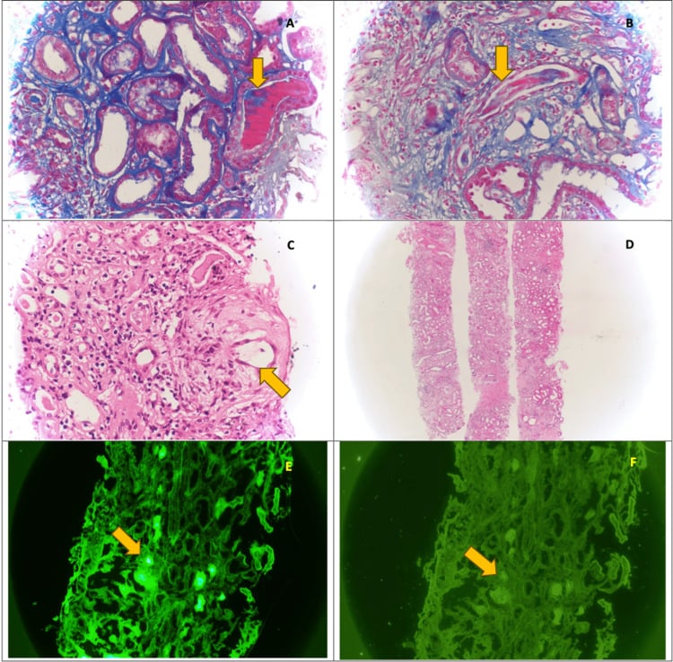
Precipitants. Hypercalcaemia, dehydration and nephrotoxins must be stopped or avoided: especially NSAIDs and iodinated contrast, and also aminoglycosides.
Management. Aggressive hydration, correction of hypercalcaemia and withdrawal of nephrotoxins, with dialysis if required. The key step is to reduce the toxic light chains rapidly with bortezomib-based anti-myeloma therapy. Extracorporeal light-chain removal (plasma exchange or high-cut-off haemodialysis) has not shown a clear renal-recovery benefit and is not standard.
What it means for the surgeon. Avoid iodinated contrast and NSAIDs, maintain hydration, correct hypercalcaemia, and adjust drug and anaesthetic dosing. For bone protection in renal impairment, denosumab is preferred over a bisphosphonate.
Investigations
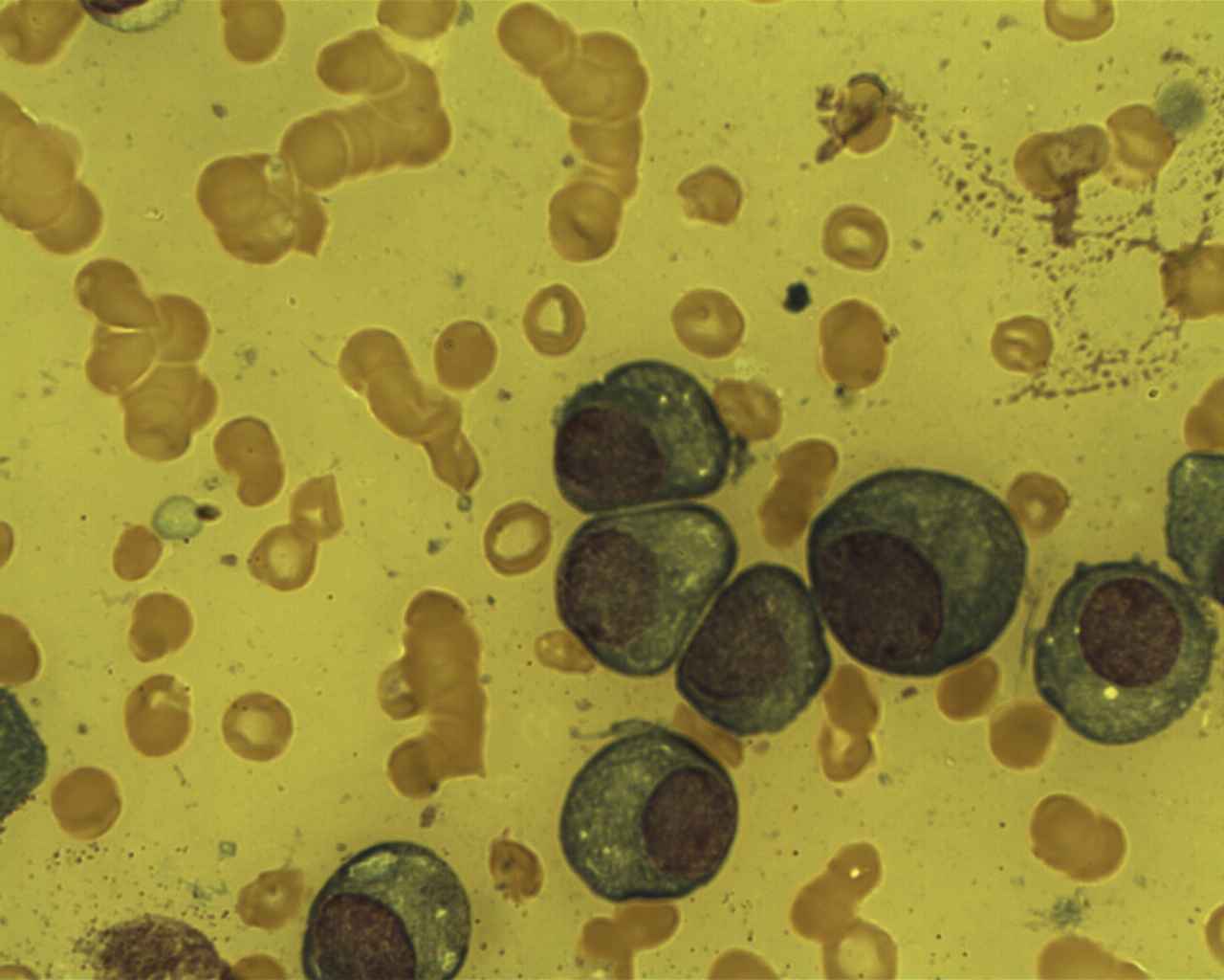
Laboratory tests. The diagnosis rests on finding the paraprotein and the clone. Serum and urine electrophoresis and the free light chain assay find the protein; the bone marrow biopsy proves the clone; the blood count, renal function and calcium record the CRAB damage; and beta-2 microglobulin, albumin and LDH feed the staging systems.
- Purpose
- Detect and quantify M-protein
- Typical Finding in Myeloma
- Monoclonal spike in gamma region (IgG 50-55%, IgA 20-25%)
- Clinical Significance
- Diagnostic - quantifies disease burden; M-protein level correlates with tumour mass
- Purpose
- Detect Bence Jones protein
- Typical Finding in Myeloma
- Monoclonal light chains (kappa or lambda)
- Clinical Significance
- Present in 75% - indicates light chain production; nephrotoxic
- Purpose
- Quantify free light chains
- Typical Finding in Myeloma
- Elevated involved FLC; abnormal kappa/lambda ratio
- Clinical Significance
- More sensitive than UPEP; useful for monitoring non-secretory myeloma
- Purpose
- Confirm clonal plasma cells
- Typical Finding in Myeloma
- Over 10% clonal plasma cells; often 30-90%
- Clinical Significance
- Diagnostic gold standard - required for diagnosis
- Purpose
- Assess cytopenias
- Typical Finding in Myeloma
- Anaemia common (Hb under 100 g/L); leukopenia and thrombocytopenia in advanced disease
- Clinical Significance
- Monitors CRAB criteria (anaemia); assesses bone marrow reserve
- Purpose
- Assess renal impairment
- Typical Finding in Myeloma
- Elevated creatinine over 173 micromol/L in 25%
- Clinical Significance
- Monitors CRAB criteria (renal); impacts chemotherapy dosing
- Purpose
- Detect hypercalcaemia
- Typical Finding in Myeloma
- Elevated over 2.75 mmol/L in 20-30%
- Clinical Significance
- Monitors CRAB criteria (calcium); emergency if severe
- Purpose
- Prognostic marker
- Typical Finding in Myeloma
- Elevated in advanced disease
- Clinical Significance
- Part of ISS staging - higher levels = worse prognosis
- Purpose
- Prognostic marker
- Typical Finding in Myeloma
- Low in advanced disease
- Clinical Significance
- Part of ISS staging - lower levels = worse prognosis
- Purpose
- Tumour burden marker
- Typical Finding in Myeloma
- Elevated in high tumour burden
- Clinical Significance
- Part of revised ISS (R-ISS); indicates aggressive disease
Imaging. The modalities differ mainly in how early they see marrow disease, and the skeletal survey sees it last.
- Sensitivity
- 40-50% (low)
- Advantages
- Widely available; low cost; traditional standard
- Disadvantages
- Misses early lesions; requires 30-50% bone loss to visualise; radiation exposure
- Clinical Use
- Initial screening; shows classic punched-out lesions; useful for fracture assessment
- Sensitivity
- 90% (very high)
- Advantages
- Most sensitive for bone marrow involvement; no radiation; detects early focal lesions
- Disadvantages
- Expensive; time-consuming; not widely available; claustrophobia
- Clinical Use
- Preferred first-line imaging; detects disease before skeletal survey positive
- Sensitivity
- 85-90% (high)
- Advantages
- Detects metabolically active disease; whole-body assessment; useful for monitoring response
- Disadvantages
- Radiation exposure; expensive; false negatives in low-grade disease
- Clinical Use
- Alternative to MRI; excellent for assessing treatment response and detecting extramedullary disease
- Sensitivity
- 70-80% (moderate-high)
- Advantages
- Better than X-ray; detects smaller lesions; fast acquisition
- Disadvantages
- Radiation exposure; less sensitive than MRI/PET-CT for marrow disease
- Clinical Use
- Alternative when MRI unavailable; good for cortical bone assessment and surgical planning
Why not a bone scan. Technetium-99m bone scans rely on osteoblastic activity to show uptake. Because myeloma lesions are purely osteolytic with suppressed osteoblast function, the scan is typically negative or shows decreased uptake ("cold spots"), the opposite of metastatic disease, which usually shows "hot spots" of increased uptake.
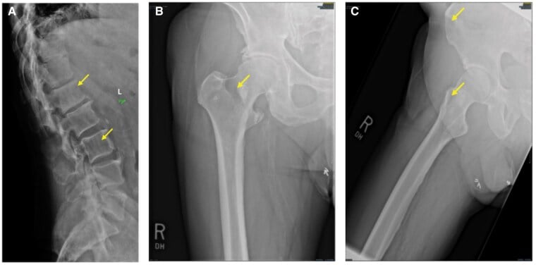
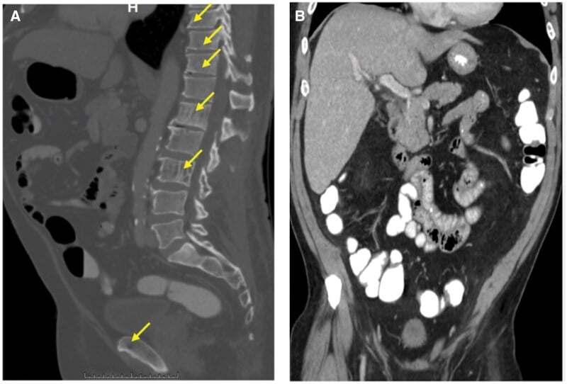
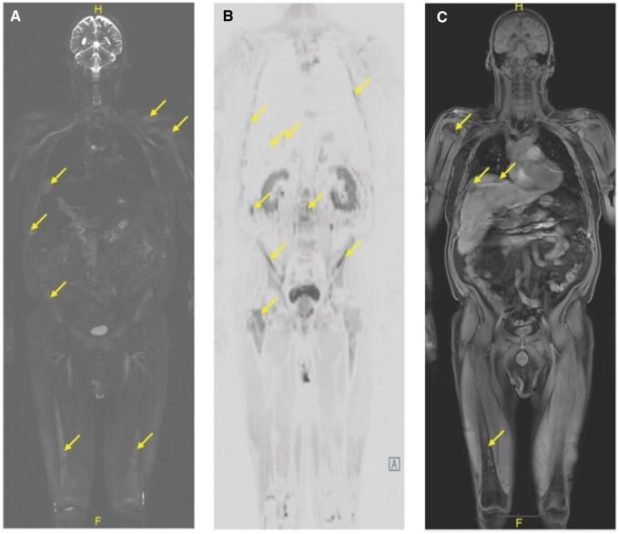
Radiographic features. Myeloma is characteristically polyostotic, and each region has its own signature.
Skull. "Punched-out" lesions: multiple well-defined, round lytic lesions with sharp margins and no sclerotic rim, the classic "moth-eaten" or "Swiss cheese" appearance, most visible on the lateral skull radiograph.
Spine. Vertebral compression fractures, often at multiple levels, on a background of diffuse osteopenia. Vertebral body collapse creates the "coin-on-edge" appearance, and the posterior elements are usually spared, unlike metastases.
Pelvis. Multiple lytic lesions of the ilium, pubis and ischium, which may cause pathological fractures, and pelvic insufficiency fractures in osteopenic bone.
Long bones. Involvement is preferentially proximal, in the femur and humerus (red marrow sites), with endosteal scalloping. Cortical destruction over 50% carries a risk of pathological fracture.
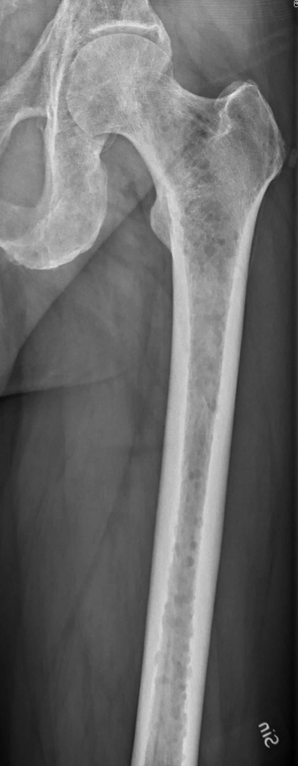
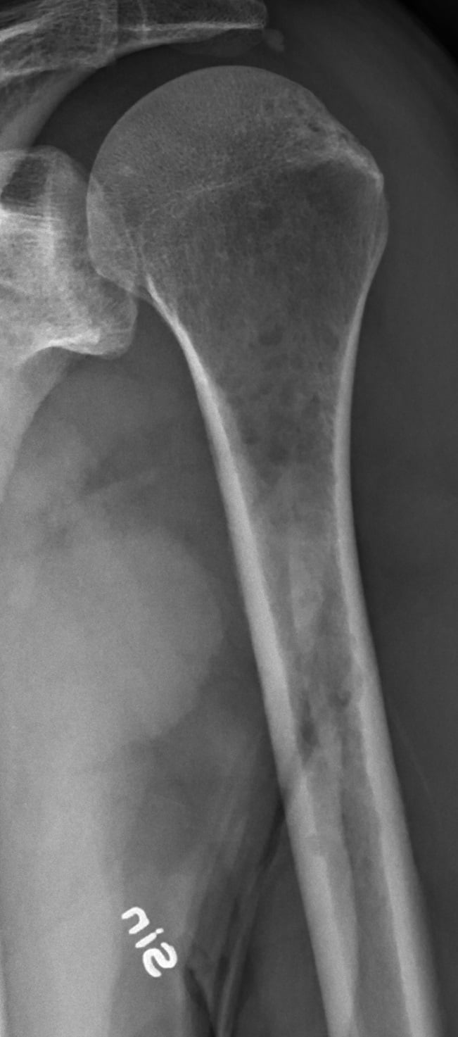
Diagnostic criteria (IMWG 2014). The diagnosis of multiple myeloma requires both:
- Clonal bone marrow plasma cells of 10% or more, or a biopsy-proven plasmacytoma, PLUS
- One or more of: any CRAB feature, or any myeloma-defining (SLiM) biomarker
Management Algorithm
Myeloma is managed jointly by haematology and orthopaedics, and the pathway below runs both threads: systemic therapy chosen by disease status and transplant eligibility, bone protection for everyone with bone disease, and a surgical decision for each skeletal complication.
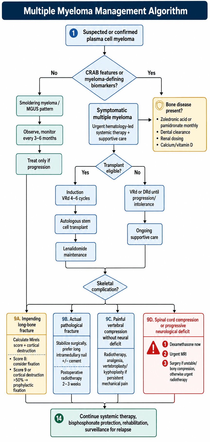
Smouldering myeloma. No CRAB criteria and no myeloma-defining biomarkers: monitor closely every 3-6 months with repeat SPEP, free light chains and imaging. Do not treat; observation was superior to early treatment in trials, and treatment begins only on progression to symptomatic myeloma.
Newly diagnosed, transplant eligible. Induction chemotherapy with bortezomib, lenalidomide and dexamethasone (VRd) for 4-6 cycles, then autologous stem cell transplant (high-dose melphalan followed by stem cell rescue), then lenalidomide maintenance continued until progression, with monthly zoledronic acid or pamidronate. Median progression-free survival is 50 months or more.
Newly diagnosed, transplant ineligible. VRd, or daratumumab, lenalidomide and dexamethasone (DRd), continued until disease progression or intolerance, with monthly zoledronic acid or pamidronate. Median overall survival is 4-5 years.
Relapsed or refractory disease. Second-line agents (carfilzomib, ixazomib, daratumumab, elotuzumab, pomalidomide), CAR T-cell therapy (ide-cel, cilta-cel) for heavily pretreated patients, clinical trials, and palliation and supportive care.
- Agents
- Bortezomib, carfilzomib, ixazomib
- Mechanism and Role
- Inhibit protein degradation, causing myeloma cell apoptosis; backbone of most regimens
- Side Effects
- Peripheral neuropathy, thrombocytopenia
- Agents
- Lenalidomide, pomalidomide, thalidomide
- Mechanism and Role
- Immune modulation and anti-angiogenic effects; highly effective in combination regimens
- Side Effects
- Thrombosis (require anticoagulation), neuropathy, teratogenicity
- Agents
- Daratumumab, isatuximab, elotuzumab
- Mechanism and Role
- Target CD38 or SLAMF7 on myeloma cells; dramatic responses in combination therapy
- Side Effects
- Infusion reactions, infections, cytopenias
- Agents
- Dexamethasone, prednisone
- Mechanism and Role
- Direct anti-myeloma effect and anti-inflammatory; used in all regimens
- Side Effects
- Hyperglycaemia, insomnia, avascular necrosis, infections
Bisphosphonate Therapy - Essential for Skeletal Protection
Who. All patients with myeloma bone disease should receive a bisphosphonate. Hold it before surgery.
How big the effect is. Quote it accurately, because the figure in wide circulation is the wrong one. The "40%" comes from Berenson's 1998 pamidronate trial (reference 13), which reported 1.3 skeletal events per year on pamidronate against 2.2 on placebo: a 41% reduction in event rate, in stage III myeloma with at least one lytic lesion, on a single agent. The pooled evidence is the Cochrane network meta-analysis of 24 randomised trials in 7,293 participants, which found a 26% relative reduction in skeletal-related events (RR 0.74, 95% CI 0.63-0.88) and the same 26% in pathological vertebral fractures (RR 0.74, 95% CI 0.62-0.89), both moderate-quality. Use 26 per cent: it is the meta-analytic estimate across the whole class, and Berenson's larger figure is one trial, one drug, one selected population, and a rate rather than a risk.
Two negatives matter as much to a surgeon. There was no reduction in non-vertebral fractures (RR 1.03, 95% CI 0.68-1.56): the drug does not protect the femur, and it is never a reason to defer fixing an impending fracture. And the pooled overall survival effect was not significant (HR 0.90, 95% CI 0.76-1.07); the survival advantage attributed to zoledronate rests on MRC Myeloma IX and on indirect network comparison, and Cochrane's own authors call for head-to-head trials to settle it. Berenson found no overall survival difference either.
Which agent, and for how long. MRC Myeloma IX continued bisphosphonate at least until progression and the Cochrane search closed before denosumab, so neither addresses duration or agent choice in a patient who now survives many years. The IMWG Bone Working Group's 2021 recommendations (reference 18) do, using GRADE:
- Zoledronic acid is the preferred agent in newly diagnosed myeloma, with or without bone disease.
- There is a stopping rule. Once the patient reaches a very good partial response or better, after at least 12 months of monthly zoledronic acid, the treating physician can consider reducing the frequency or discontinuing. Note the hedge: this is consensus, not a prospective comparison against continued dosing.
- Denosumab can be considered, particularly in renal impairment, where it is not renally cleared, and might prolong progression-free survival in transplant-eligible patients with bone disease. It is easy to start and hard to stop: discontinuation causes a rebound rise in bone turnover with rapid bone loss and new vertebral fractures, so plan the exit before the first dose.
- Surgery is reserved for prevention and restoration of long-bone pathological fracture, vertebral column instability, and cord compression caused by bone fragments in the canal; soft tumour in this exquisitely radiosensitive disease will often answer to radiotherapy alone.
- Dosing
- 4mg IV over 15 minutes every 4 weeks
- Advantages
- Most potent bisphosphonate; convenient monthly dosing
- Precautions
- Requires renal dose adjustment if CrCl under 60; risk of ONJ; hold before dental procedures
- Dosing
- 90mg IV over 2-4 hours every 4 weeks
- Advantages
- Alternative if renal impairment
- Precautions
- Longer infusion time; less potent than zoledronic acid; risk of ONJ
- Dosing
- 120mg SubQ every 4 weeks
- Advantages
- Can use in renal failure; no dose adjustment needed
- Precautions
- Higher risk of hypocalcaemia; ensure calcium/vitamin D supplementation; risk of ONJ
Duration. Continue monthly for the first 2 years, then consider reducing to every 3 months if a complete response is achieved.
Osteonecrosis of the jaw. The risk rises with the duration of therapy (a cumulative effect), and prevention is a protocol:
- Dental examination and clearance before starting bisphosphonates
- Maintain excellent oral hygiene
- Avoid invasive dental procedures while on therapy
- If dental surgery is required, hold bisphosphonates for 2-3 months before and after
Orthopaedic Management and Surgical Indications
Myeloma is a systemic disease requiring systemic treatment. Surgery is palliative and aims to:
- Stabilise actual or impending pathological fractures to restore function
- Decompress neural structures in spinal cord compression
- Provide pain relief through stabilisation or vertebroplasty/kyphoplasty
- Improve quality of life by restoring mobility and independence
Surgery alone NEVER cures myeloma - chemotherapy is essential for disease control.
Impending pathological fracture. The Mirels score adds four variables and the total decides who is fixed before the bone breaks.
- 1 Point
- Upper limb
- 2 Points
- Lower limb
- 3 Points
- Peritrochanteric
- 1 Point
- Mild
- 2 Points
- Moderate
- 3 Points
- Functional (severe)
- 1 Point
- Blastic
- 2 Points
- Mixed
- 3 Points
- Lytic
- 1 Point
- Less than 1/3 diameter
- 2 Points
- 1/3 to 2/3 diameter
- 3 Points
- Over 2/3 diameter
Interpretation.
- Score ≤7: low fracture risk - observation, bisphosphonates, radiotherapy if painful
- Score 8: intermediate risk - consider prophylactic fixation
- Score ≥9: high fracture risk - prophylactic fixation indicated
Mirels scoring was developed for metastatic disease, but myeloma lesions have NO sclerotic response and may be at higher fracture risk than the score suggests. Cortical destruction over 50% or lesion over 3cm in long bone should prompt strong consideration of prophylactic fixation regardless of score.
Surgical Technique for Long Bone Fractures
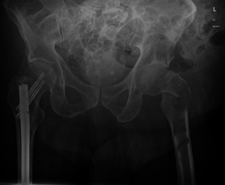
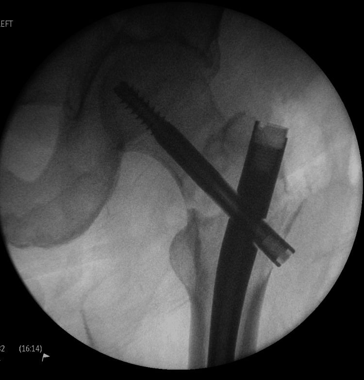
The device. An intramedullary nail is preferred over plate fixation. It is load sharing rather than load bearing, it protects the entire bone including skip lesions, it has a lower reoperation rate when the disease progresses, and it allows early weight-bearing.
The length of fixation. Protect the entire bone at risk, assume the disease may progress, bridge all lytic lesions, and use locked interlocking screws.
Cement. PMMA is indicated for large segmental defects, periarticular fractures and as an adjunct to an intramedullary nail; it gives immediate pain relief and stability.
Adjuvant radiotherapy. Consider postoperative radiotherapy for local disease control and pain, at 2-3 weeks after surgery to allow wound healing, with a typical dose of 20-30 Gy in 5-10 fractions.
The femur. In the proximal femur (intertrochanteric or subtrochanteric), a long cephalomedullary nail (a long Gamma nail or trochanteric femoral nail) protects the whole femur down to the supracondylar region; consider cemented hip arthroplasty if the femoral head or neck is extensively involved. In the shaft, a long antegrade nail from subtrochanteric to supracondylar region, reamed if cortical destruction is significant and statically locked proximally and distally. In the distal femur, a retrograde nail or a lateral locked plate with cement augmentation of the screw holes; consider distal femoral replacement if metaphyseal involvement is extensive.
The humerus. In the proximal humerus, an antegrade humeral nail or a shoulder hemiarthroplasty or reverse arthroplasty, with arthroplasty preferred if the humeral head is extensively destroyed or the patient is elderly. In the shaft, an antegrade nail is preferred, with plate fixation the alternative if there is concern for the radial nerve or the lesion is very distal; protect the full length of the diaphysis.
Spinal Involvement and Cord Compression
Spinal cord compression is an orthopaedic and oncological EMERGENCY. Prognosis depends on neurological status at treatment initiation - patients who lose ambulation rarely regain it. Immediate dexamethasone 10mg IV, MRI whole spine, and urgent oncology consultation are mandatory.
Presentation. Back pain (95%) is often the first symptom; lower-limb motor weakness (75%), sensory changes (50%, numbness and paraesthesiae) and bladder or bowel dysfunction (40%, a late finding with a poor prognosis) follow. Cauda equina syndrome declares itself with saddle anaesthesia and urinary retention.
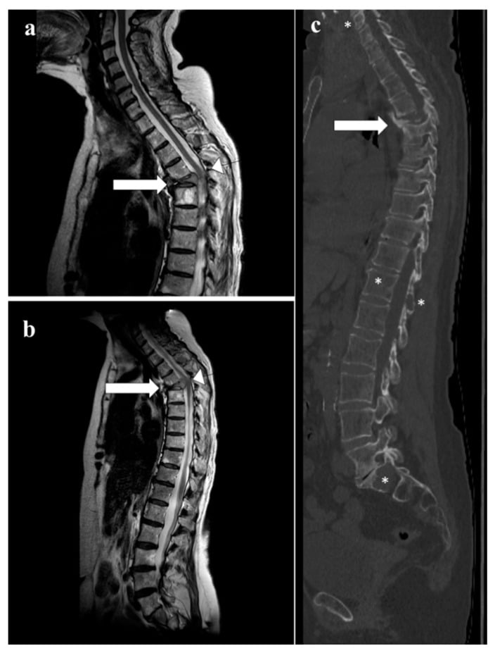
Imaging. MRI of the whole spine is the gold standard: it identifies the level or levels of compression, assesses spinal stability, and finds the multiple sites that 30% of patients have. The STIR sequence is best for oedema and disease.
- Spinal Stability
- Stable spine
- Treatment
- Dexamethasone + radiotherapy + bisphosphonates + chemotherapy
- Prognosis for Ambulation
- Over 90% maintain ambulation
- Spinal Stability
- Unstable spine
- Treatment
- Posterior stabilisation + decompression, then radiotherapy
- Prognosis for Ambulation
- Over 90% maintain ambulation
- Spinal Stability
- Stable or unstable
- Treatment
- Dexamethasone + URGENT radiotherapy OR surgery if unstable + chemotherapy
- Prognosis for Ambulation
- 60-80% maintain/regain ambulation
- Spinal Stability
- Any stability
- Treatment
- Dexamethasone + EMERGENCY surgery (decompression + stabilisation) + radiotherapy
- Prognosis for Ambulation
- 40-60% regain ambulation
- Spinal Stability
- Any stability
- Treatment
- Dexamethasone + radiotherapy (surgery unlikely to help) + chemotherapy
- Prognosis for Ambulation
- Under 20% regain ambulation
Vertebroplasty and Kyphoplasty
A painful vertebral compression fracture without neurology is treated with radiotherapy and analgesia, and with vertebroplasty or kyphoplasty if mechanical pain persists. Cement can give rapid pain relief and mechanical support to a selected painful compression fracture, but it does not treat neural compression or gross instability, and it needs a posterior wall intact enough to control the leakage risk.
Indications.
- Painful vertebral compression fractures
- Failed conservative management (analgesia, bracing)
- No spinal cord compression
- Fracture under 3 months old (better outcomes)
- Vertebral body height over 33% preserved
Contraindications.
- Spinal cord compression
- Posterior vertebral wall disruption
- Infection (osteomyelitis, discitis)
- Uncorrectable coagulopathy
- Extensive vertebral collapse (under 33% height)
- Vertebroplasty
- Direct PMMA injection into vertebral body
- Kyphoplasty
- Balloon inflation to create cavity, then PMMA injection
- Vertebroplasty
- Minimal (10-20%)
- Kyphoplasty
- Moderate (30-40%)
- Vertebroplasty
- 10-15% (higher)
- Kyphoplasty
- 5-7% (lower)
- Vertebroplasty
- 75-85% achieve significant relief
- Kyphoplasty
- 80-90% achieve significant relief
- Vertebroplasty
- Lower
- Kyphoplasty
- Higher (balloon equipment)
- Vertebroplasty
- 30-45 minutes
- Kyphoplasty
- 60-90 minutes
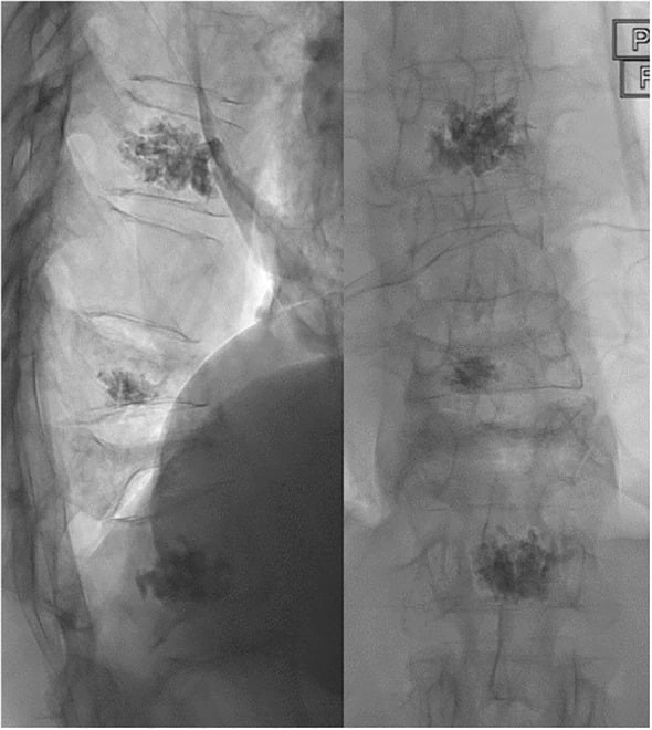
Outcomes. 75-90% of patients achieve significant pain relief within 24-48 hours, with reduced analgesic requirements and improved mobility, and the relief is sustained in 80% at 1 year. Cement leakage is usually asymptomatic and rarely causes neural compression (under 1%).
Prognosis and Survival
Survival in myeloma has improved dramatically over the past two decades with the introduction of novel agents (proteasome inhibitors, immunomodulatory drugs, monoclonal antibodies) and autologous stem cell transplantation. The factors below separate the patients who do well from those who do not.
- Favourable Prognosis
- Under 65 years
- Adverse Prognosis
- Over 75 years
- Favourable Prognosis
- Stage I (beta-2M under 3.5, albumin 35 or higher)
- Adverse Prognosis
- Stage III (beta-2M over 5.5)
- Favourable Prognosis
- Standard-risk: t(11;14), hyperdiploidy
- Adverse Prognosis
- High-risk: del(17p), t(4;14), t(14;16), gain 1q
- Favourable Prognosis
- Normal
- Adverse Prognosis
- Elevated
- Favourable Prognosis
- Complete response or better
- Adverse Prognosis
- Stable disease or progressive disease
- Favourable Prognosis
- Creatinine under 173 micromol/L
- Adverse Prognosis
- Creatinine over 173 micromol/L or dialysis-dependent
- Favourable Prognosis
- ECOG 0-1 (fully active)
- Adverse Prognosis
- ECOG 3-4 (limited self-care)
- Favourable Prognosis
- No fractures, limited lytic lesions
- Adverse Prognosis
- Multiple pathological fractures, extensive lytic disease
Survival by R-ISS stage (contemporary data).
- 5-Year OS
- 82%
- Median PFS
- 66 months
- Median OS
- Not reached (over 10 years)
- 5-Year OS
- 62%
- Median PFS
- 42 months
- Median OS
- 83 months
- 5-Year OS
- 40%
- Median PFS
- 29 months
- Median OS
- 43 months
OS = Overall Survival; PFS = Progression-Free Survival
MRC Myeloma IX: Zoledronic Acid vs Clodronic Acid
VISTA Trial: Bortezomib in Newly Diagnosed Myeloma
Bisphosphonates in Multiple Myeloma: Cochrane Network Meta-Analysis
IMWG Updated Diagnostic Criteria for Multiple Myeloma
Revised International Staging System (R-ISS)
Guidelines, Registries & Global Practice
Global Epidemiology
According to the Global Burden of Disease Study 2016 (Cowan et al., JAMA Oncology 2018), there were approximately 138,500 incident cases of multiple myeloma worldwide in 2016, with an age-standardised incidence rate of 2.1 per 100,000 persons. Incident cases rose by 126% between 1990 and 2016, driven by population growth and ageing. Incidence is highly variable geographically, being highest in Australasia, North America and Western Europe, and substantially higher (approximately two-fold) in populations of African ancestry. Access to autologous stem-cell transplantation and to novel agents (lenalidomide, bortezomib) is concentrated in high-income regions and remains very limited in sub-Saharan Africa and parts of the Middle East.
- Finding
- Approximately 138,500 new cases; age-standardised rate 2.1 per 100,000
- Source / Evidence
- GBD 2016 (Cowan, JAMA Oncol 2018, PMID 29800065)
- Finding
- 126% rise in incident cases (largest increase in low and middle SDI countries)
- Source / Evidence
- GBD 2016 (PMID 29800065)
- Finding
- Australasia, North America, Western Europe; two-fold higher in African-ancestry populations
- Source / Evidence
- GBD 2016 (PMID 29800065)
- Finding
- Routine in high-income regions; under 5% utilisation in Africa/East Mediterranean
- Source / Evidence
- WBMT/GBD analysis (Cowan, Biol Blood Marrow Transplant 2020, PMID 32846200)
Guideline Comparison Across Jurisdictions
- Diagnostic Framework
- CRAB plus SLiM biomarkers (clonal plasma cells 60% or more, FLC ratio 100 or higher, more than 1 focal MRI lesion)
- First-line Imaging
- Whole-body low-dose CT, whole-body MRI or PET-CT preferred over skeletal survey
- Bone-Directed Therapy
- Zoledronic acid or pamidronate for all with bone disease; denosumab if renal impairment
- Evidence Level
- Level I (Rajkumar, Lancet Oncol 2014, PMID 25439696)
- Diagnostic Framework
- Adopts IMWG criteria
- First-line Imaging
- Whole-body MRI first-line; whole-body low-dose CT if MRI unsuitable; avoid isolated skeletal survey
- Bone-Directed Therapy
- Zoledronic acid offered to all (survival and SRE benefit); bisphosphonate continued
- Evidence Level
- Level I (informed by MRC Myeloma IX, PMID 21131037)
- Diagnostic Framework
- IMWG criteria; R-ISS for staging
- First-line Imaging
- Whole-body low-dose CT or PET-CT; MRI for suspected cord compression or smouldering disease
- Bone-Directed Therapy
- Zoledronic acid preferred; denosumab non-inferior alternative, useful in renal impairment
- Evidence Level
- Level I (Dimopoulos, Ann Oncol 2021)
- Diagnostic Framework
- IMWG criteria; R-ISS staging
- First-line Imaging
- Whole-body low-dose CT, PET-CT or MRI; skeletal survey only if advanced imaging unavailable
- Bone-Directed Therapy
- Bisphosphonate (zoledronic acid/pamidronate) or denosumab for all with bone disease
- Evidence Level
- Level I
There is strong international consensus across IMWG, NICE, EHA-ESMO and NCCN: diagnosis follows the CRAB-plus-SLiM framework, cross-sectional imaging (whole-body low-dose CT, whole-body MRI or PET-CT) has replaced the plain skeletal survey, and a bisphosphonate (preferably zoledronic acid) is offered to every patient with bone disease. The principal practice variation is access-driven, not evidence-driven: transplant and novel-agent availability diverge sharply between high-income and low/middle-income countries.
Registry Evidence
Large national and international registries (the US SEER programme, the European EBMT/WBMT transplant registries and the Australian Myeloma and Related Diseases Registry) document a substantial improvement in survival over the past two decades, coinciding with the introduction of proteasome inhibitors, immunomodulatory drugs, monoclonal antibodies and routine autologous transplantation. Registry data also confirm marked global disparities in access to autologous transplantation, with utilisation below 5% of incident cases in Africa and the East Mediterranean compared with over 20% in North America and Europe (Cowan et al., Biol Blood Marrow Transplant 2020).
Supportive Care and Prophylaxis
International consensus (IMWG, NCCN, EHA-ESMO) supports the following supportive-care measures for patients on multi-agent myeloma therapy:
- Pneumocystis jirovecii prophylaxis: trimethoprim-sulfamethoxazole for patients on high-dose corticosteroids or bortezomib-based regimens
- Herpes-zoster prophylaxis: aciclovir or valaciclovir during and after bortezomib-based therapy (reactivation risk)
- Venous thromboembolism prophylaxis during immunomodulatory therapy (lenalidomide, pomalidomide, thalidomide): aspirin for standard-risk patients; low-molecular-weight heparin or a direct oral anticoagulant (e.g. apixaban, rivaroxaban) for high-risk patients
MCQ Practice Points
Q: What is the characteristic radiographic appearance of multiple myeloma bone lesions?
A: Punched-out lytic lesions without surrounding sclerosis or periosteal reaction. Most common in axial skeleton: spine, skull, pelvis, ribs, proximal femur/humerus. No bone scan uptake (purely osteolytic with suppressed osteoblasts) - use skeletal survey or whole-body MRI instead. Lesions represent replaced marrow.
Q: What laboratory findings are diagnostic for multiple myeloma?
A: CRAB criteria: Calcium elevation, Renal insufficiency, Anemia, Bone lesions. M-spike (monoclonal protein) on serum protein electrophoresis. Bence Jones proteinuria (light chains). Bone marrow with greater than 10% plasma cells. Rouleaux formation on blood smear. ESR markedly elevated. Normal ALP (osteoblasts suppressed).
Q: Why is the alkaline phosphatase (ALP) typically normal in multiple myeloma?
A: Myeloma cells produce osteoclast-activating factors (RANKL, IL-6, MIP-1α) causing bone resorption WITHOUT compensatory bone formation. Osteoblast activity suppressed by Dickkopf-1 (DKK1). Hence ALP (marker of osteoblastic activity) remains normal despite extensive bone destruction. Distinguishes from metastatic bone disease.
Q: What is the surgical approach to pathological fractures in multiple myeloma?
A: Prophylactic fixation for impending fractures (Mirels score ≥9). Internal fixation + cement augmentation for actual fractures. Avoid intramedullary devices alone in spine - tumor extends into canal. Spine: Vertebroplasty/kyphoplasty for compression fractures, decompression + stabilization for cord compression. Radiation post-operatively.
Q: What is the difference between multiple myeloma and solitary plasmacytoma?
A: Solitary plasmacytoma: Single bone or soft tissue lesion, normal bone marrow (less than 10% plasma cells), absent/minimal M-protein, no other CRAB features. Better prognosis - treat with radiation ± surgery. ~50% progress to multiple myeloma within 10 years. Multiple myeloma requires systemic chemotherapy and may require autologous stem cell transplant.
Exam Viva Scenarios
Practise clinical reasoning and management decisions out loud
“A 68-year-old man presents with 3 months of progressive back pain. Plain radiographs show multiple lytic lesions in the thoracolumbar spine with compression fractures at T8 and L2. How would you approach this patient?”
“A 72-year-old woman with known multiple myeloma on chemotherapy presents with acute onset right thigh pain after a fall at home. X-ray shows a complete subtrochanteric femur fracture through a 5cm lytic lesion. Her oncologist asks your advice on management. What would you recommend?”
“You are called to the emergency department at 2 AM. A 65-year-old man with newly diagnosed multiple myeloma (started chemotherapy 2 weeks ago) presents with 24 hours of progressive lower limb weakness and urinary retention. On examination, he has grade 3/5 power in both lower limbs, a sensory level at T10, and absent ankle reflexes. Walk me through your emergency management.”
Key Definition
- Plasma cell neoplasm with clonal bone marrow plasma cells over 10% PLUS CRAB criteria or myeloma-defining biomarkers
- Most common primary bone malignancy in adults over 40 years
- Median age 65 years; 90% have skeletal involvement
CRAB Criteria (Must Know)
- C - Calcium elevated over 2.75 mmol/L
- R - Renal insufficiency (creatinine over 173 micromol/L)
- A - Anemia (Hb under 100 g/L)
- B - Bone lesions (one or more lytic lesions)
- ANY ONE = symptomatic myeloma requiring treatment
Pathognomonic Radiology
- Purely LYTIC lesions with NO sclerotic response (vs metastases)
- Punched-out lesions in skull - classic appearance
- Vertebral compression fractures (70% involve spine)
- Whole-body MRI or PET-CT more sensitive than skeletal survey
- Bone scan NOT useful (no osteoblastic activity)
Diagnostic Triad
- 1. SPEP - monoclonal protein spike (IgG 50-55%, IgA 20-25%)
- 2. Urine protein electrophoresis - Bence Jones protein (75%)
- 3. Bone marrow biopsy - over 10% clonal plasma cells
- PLUS: Serum free light chains, calcium, renal function, imaging
Staging (R-ISS Preferred)
- R-ISS I: ISS I + standard cytogenetics + normal LDH (82% 5yr survival)
- R-ISS II: Not I or III (62% 5yr survival)
- R-ISS III: ISS III + high-risk cytogenetics OR elevated LDH (40% 5yr survival)
- High-risk cytogenetics: del(17p), t(4;14), t(14;16), gain 1q
Medical Treatment
- Transplant eligible: VRd induction → ASCT → lenalidomide maintenance
- Transplant ineligible: VRd or DRd until progression
- ALL with bone disease: Bisphosphonates (zoledronic acid 4mg IV monthly)
- Bisphosphonates reduce skeletal-related events and vertebral fractures by 26% (RR 0.74 each, Cochrane) - but NOT non-vertebral fractures (RR 1.03), so they do not protect the femur
- Beware ONJ - dental clearance before starting bisphosphonates
Surgical Indications
- Pathological fractures: Intramedullary nailing preferred (long nail, protect full bone)
- Impending fractures: Mirels over 8 or cortical destruction over 50%
- Spinal cord compression: Emergency decompression + stabilization if unstable spine
- Vertebroplasty/kyphoplasty: Painful VCFs, 75-90% pain relief
- Cement augmentation: Immediate stability for large defects
Surgical Principles
- Surgery is PALLIATIVE - chemotherapy is definitive treatment
- Intramedullary nail over plate (load-sharing, protects full bone, early weight-bearing)
- LONG fixation - protect entire bone (skip lesions and progression risk)
- Adjuvant radiotherapy: 20-30 Gy starting 2-3 weeks post-op
- Weight-bearing as tolerated - goal is quality of life
Spinal Cord Compression (Emergency)
- Dexamethasone 10mg IV IMMEDIATELY (within 30 min)
- MRI whole spine urgently (within 1 hour)
- Ambulatory at presentation = 60-80% preserve function
- Paraplegic over 48hrs = under 20% recover ambulation
- Surgery if unstable spine or bony compression; RT if soft tissue mass
Complications to Know
- SREs (skeletal-related events): fracture, RT, surgery, cord compression - 60-70% experience
- Hypercalcemia: IV hydration + bisphosphonates (response 48-72hrs)
- Renal failure: Light chain cast nephropathy - hydration, avoid NSAIDs, treat myeloma
- Infections: Hypogammaglobulinemia - leading cause of death early
- ONJ from bisphosphonates: 1-10% incidence, increases with duration
Prognosis
- Median survival: 8-10 years for standard-risk with modern therapy (was 3 years in 2000)
- R-ISS I: Median OS over 10 years (82% at 5 years)
- R-ISS III: Median OS 43 months (40% at 5 years)
- Causes of death: Progressive myeloma (40-50%), infection (25-30%), renal failure (10-15%)
Exam Day Pearls
- No blastic response = pathognomonic for myeloma (vs metastases which heal)
- CRAB backwards and forwards - defines symptomatic disease
- Smoldering myeloma (no CRAB) = DO NOT TREAT, only observe
- Bisphosphonates mandatory for ALL with bone disease
- Spinal cord compression = dexamethasone within 30 min, MRI within 1 hour, treatment within 24 hours
- Intramedullary nail over plate, long fixation over short, cement augmentation for large defects
References
-
Kyle RA, Gertz MA, Witzig TE, et al. Review of 1027 patients with newly diagnosed multiple myeloma. Mayo Clin Proc. 2003;78(1):21-33.
-
Rajkumar SV, Dimopoulos MA, Palumbo A, et al. International Myeloma Working Group updated criteria for the diagnosis of multiple myeloma. Lancet Oncol. 2014;15(12):e538-e548.
-
Terpos E, Ntanasis-Stathopoulos I, Gavriatopoulou M, Dimopoulos MA. Pathogenesis of bone disease in multiple myeloma: from bench to bedside. Blood Cancer J. 2018;8(1):7.
-
Roodman GD. Pathogenesis of myeloma bone disease. Leukemia. 2009;23(3):435-441.
-
Palumbo A, Avet-Loiseau H, Oliva S, et al. Revised International Staging System for Multiple Myeloma: A Report From International Myeloma Working Group. J Clin Oncol. 2015;33(26):2863-2869.
-
Hillengass J, Usmani S, Rajkumar SV, et al. International myeloma working group consensus recommendations on imaging in monoclonal plasma cell disorders. Lancet Oncol. 2019;20(6):e302-e312.
-
Dimopoulos MA, Moreau P, Terpos E, et al. Multiple myeloma: EHA-ESMO Clinical Practice Guidelines for diagnosis, treatment and follow-up. Ann Oncol. 2021;32(3):309-322.
-
Moreau P, Kumar SK, San Miguel J, et al. Treatment of relapsed and refractory multiple myeloma: recommendations from the International Myeloma Working Group. Lancet Oncol. 2021;22(3):e105-e118.
-
Mhaskar R, Kumar A, Miladinovic B, Djulbegovic B. Bisphosphonates in multiple myeloma: an updated network meta-analysis. Cochrane Database Syst Rev. 2017;12(12):CD003188.
-
Terpos E, Kleber M, Engelhardt M, et al. European Myeloma Network guidelines for the management of multiple myeloma-related complications. Haematologica. 2015;100(10):1254-1266.
-
Mirels H. Metastatic disease in long bones: A proposed scoring system for diagnosing impending pathologic fractures. Clin Orthop Relat Res. 1989;(249):256-264.
-
Patchell RA, Tibbs PA, Regine WF, et al. Direct decompressive surgical resection in the treatment of spinal cord compression caused by metastatic cancer: a randomised trial. Lancet. 2005;366(9486):643-648.
-
Berenson JR, Lichtenstein A, Porter L, et al. Long-term pamidronate treatment of advanced multiple myeloma patients reduces skeletal events. J Clin Oncol. 1998;16(2):593-602.
-
Morgan GJ, Davies FE, Gregory WM, et al. First-line treatment with zoledronic acid as compared with clodronic acid in multiple myeloma (MRC Myeloma IX): a randomised controlled trial. Lancet. 2010;376(9757):1989-1999.
-
Kumar SK, Dispenzieri A, Lacy MQ, et al. Continued improvement in survival in multiple myeloma: changes in early mortality and outcomes in older patients. Leukemia. 2014;28(5):1122-1128.
-
Rajkumar SV. Multiple myeloma: 2020 update on diagnosis, risk-stratification and management. Am J Hematol. 2020;95(5):548-567.
-
San Miguel JF, Schlag R, Khuageva NK, et al. Bortezomib plus melphalan and prednisone for initial treatment of multiple myeloma. N Engl J Med. 2008;359(9):906-917. PMID 18753647. doi:10.1056/NEJMoa0801479
-
Terpos E, Zamagni E, Lentzsch S, et al; Bone Working Group of the International Myeloma Working Group. Treatment of multiple myeloma-related bone disease: recommendations from the Bone Working Group of the International Myeloma Working Group. Lancet Oncol. 2021;22(3):e119-e130. PMID 33545067. doi:10.1016/S1470-2045(20)30559-3
Citation note. Reference 17 (the VISTA trial) was carded in the Evidence Base but was absent from this list; it has been added. Two figures on this page were also reconciled against their sources: the frequently quoted "40% reduction in skeletal events" traces to Berenson (reference 13), whose result is 1.3 versus 2.2 skeletal events per year, a reduction in event rate in a single pamidronate trial - whereas the pooled Cochrane estimate across 24 trials (reference 9) is a 26% relative risk reduction (RR 0.74), which is the figure now used throughout. The same review's finding of no benefit for non-vertebral fractures (RR 1.03) has been added wherever bisphosphonate efficacy is stated.