Strain Classification | Satellite Cells | Regeneration vs Fibrosis | Return to Sport
- Satellite cells are muscle stem cells essential for regeneration
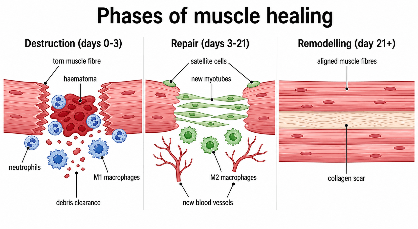
- Three healing phases: Destruction (0-3 days), Repair (3-21 days), Remodelling (21+ days)
- Myotendinous junction is most common injury site
- Early mobilisation promotes regeneration over fibrosis
- Fibrosis occurs when regenerative capacity is overwhelmed
- “Satellite cells express Pax7 and are located beneath basal lamina
- “Type IIb fibres are most susceptible to strain injury
- “Eccentric contractions cause most muscle injuries
- “NSAIDs may impair early healing but reduce fibrosis
Overview
Skeletal muscle injury is among the most common presentations in sports medicine and trauma, and skeletal muscle is one of the few adult tissues with a genuine capacity for scar-free regeneration, through a dedicated stem-cell population. Why injured muscle sometimes regenerates fully and sometimes heals by fibrosis is a core basic-science theme of the examination, and a favourite vehicle for probing tissue healing principles that generalise to tendon, ligament and bone.
The thread through the topic. The regeneration-versus-fibrosis balance explains re-injury, chronic stiffness and the rationale for early controlled loading. It links the basic science (satellite cells, myogenic regulatory factors, macrophage polarisation) to the clinical management (classification, POLICE, eccentric rehabilitation, return to sport), and the candidate is expected to move between the two.
Muscle Structure and Biology
The tissue. Skeletal muscle constitutes approximately 40% of body mass and is a highly organised tissue capable of both force generation and regeneration. It is built as a hierarchy:
- Muscle fibre (myofibre): a multinucleated syncytium, 10-100 μm in diameter
- Myofibril: the contractile unit, containing sarcomeres
- Sarcomere: Z-line to Z-line, containing actin and myosin
- Fascicle: a bundle of fibres surrounded by perimysium
- Muscle belly: multiple fascicles within the epimysium
Fibre types. Fibre types differ in metabolism, contraction speed and fatigue resistance, and with them in susceptibility to injury.
- Metabolism
- Oxidative
- Contraction
- Slow
- Fatigue Resistance
- High
- Injury Risk
- Lower
- Metabolism
- Oxidative-Glycolytic
- Contraction
- Fast
- Fatigue Resistance
- Moderate
- Injury Risk
- Moderate
- Metabolism
- Glycolytic
- Contraction
- Fast
- Fatigue Resistance
- Low
- Injury Risk
- Highest
Type IIb fibres are the most susceptible to strain because they generate high forces rapidly but fatigue quickly. Their low oxidative capacity means they rely on anaerobic metabolism and are more prone to metabolic failure during sustained eccentric loading.
Satellite cells. These are the muscle stem cells, and they sit in a niche between the sarcolemma and the basal lamina. In the quiescent state they express Pax7 and remain dormant; injury releases HGF and FGF, which activate them and trigger proliferation, and the differentiating cells then express the myogenic regulatory factors in sequence: MyoD, Myf5, myogenin and MRF4. Desmin is an early marker of myogenic commitment.
Self-renewal. Division is asymmetric: some daughter cells differentiate, others replenish the pool, so the population is a self-renewing reserve maintained through life. Satellite cells are essential for postnatal muscle growth and regeneration. Without them, damaged muscle cannot regenerate and heals only by fibrosis.
Mechanisms of Muscle Injury
Strain. The most common mechanism, accounting for over 90% of sports-related muscle injuries: injury from excessive tensile force during muscle contraction. Eccentric contraction (active lengthening) carries the highest risk; the force exceeds the tensile strength at the myotendinous junction, and fast-twitch fibres fail first. The high-risk situations are:
- Late swing phase of running (hamstrings)
- Kicking (rectus femoris)
- Push-off (gastrocnemius)
Risk factors for strain. The recognised predisposing factors:
- Previous injury: scar tissue reduces compliance
- Muscle imbalance: weak hamstrings relative to quadriceps
- Fatigue: reduced force absorption capacity
- Poor flexibility: reduced extensibility
- Inadequate warm-up
FASTFAST TWITCH - Injury Risk Factors
Hook:FAST twitch fibres fail first during eccentric loading
Contusion. Direct blunt trauma, and the second most common muscle injury in athletes. Compression crushes the fibres and an intramuscular haematoma forms; the damage is more localised than a strain. The common sites are the quadriceps (contact sports), the biceps (falls) and the calf (direct kicks). Severity is classified by active range of motion:
- Mild: greater than 90 degrees
- Moderate: 45-90 degrees
- Severe: less than 45 degrees
The complications to anticipate after a contusion are myositis ossificans (heterotopic ossification) and, rarely but seriously, compartment syndrome.
Laceration. Direct cutting or tearing of muscle by sharp or penetrating trauma, with complete fibre disruption in the injury zone; neurovascular structures may be involved. Surgical repair may be indicated, nerve regeneration is needed if a nerve is transected, and the fibrosis potential is higher. Prognosis depends on the extent of injury, nerve involvement worsens it, and there may be a permanent functional deficit. Lacerations are less common in sport but important in trauma.
The myotendinous junction. The MTJ is the most common site of strain, and Grade III injuries often avulse here. It is the transition from compliant muscle to stiff tendon, so stress concentrates at it; all contractile force passes through it; and it is a watershed region with limited blood supply. Its interdigitating membrane folds increase the surface area 10-20 fold for force transmission, yet the junction remains vulnerable during eccentric loading and is the weak link in the muscle-tendon unit.
Classification
Clinical grading. The Grade I-III system grades a strain by the proportion of fibres disrupted and the function lost. It is simple but imprecise, with poor prognostic value.
- Grade I (Mild)
- Less than 5%
- Grade II (Moderate)
- 5-50%
- Grade III (Severe)
- More than 50% or complete
- Grade I (Mild)
- Localised tenderness
- Grade II (Moderate)
- Palpable defect, weakness
- Grade III (Severe)
- Complete loss of function
- Grade I (Mild)
- Minimal
- Grade II (Moderate)
- Moderate
- Grade III (Severe)
- Significant with haematoma
- Grade I (Mild)
- Able
- Grade II (Moderate)
- Antalgic gait
- Grade III (Severe)
- Unable
- Grade I (Mild)
- 1-2 weeks
- Grade II (Moderate)
- 3-6 weeks
- Grade III (Severe)
- 3-6 months
- Grade I (Mild)
- Often normal
- Grade II (Moderate)
- Partial tear on MRI
- Grade III (Severe)
- Complete disruption
- Grade I (Mild)
- RICE, early ROM
- Grade II (Moderate)
- Protected rehab
- Grade III (Severe)
- Surgery consideration
Imaging-based classification. Elite sports medicine now uses imaging-based classifications that separate functional disorders from true structural tears and locate the injury.
Munich consensus (Mueller-Wohlfahrt 2013). Functional disorders, with no macroscopic fibre tear, are separated from structural injuries with one.
- Type
- Type 1: overexertion-related
- Description
- 1a fatigue-induced; 1b delayed-onset muscle soreness (DOMS)
- Type
- Type 2: neuromuscular
- Description
- 2a spine-related; 2b muscle-related (neuromuscular control)
- Type
- Type 3: partial tear
- Description
- 3a minor (less than a fascicle/bundle); 3b moderate partial tear
- Type
- Type 4: (sub)total tear / avulsion
- Description
- Subtotal or complete muscle tear or tendinous avulsion
British Athletics Muscle Injury Classification (BAMIC, Pollock 2014). Grades 0-4 for size are combined with a site letter: a is myofascial, b musculotendinous (at the MTJ), c intratendinous (extending into the tendon). Grade 0 is MRI-negative (0a focal, 0b DOMS); grades 1-4 mark increasing extent; grade 4 is a complete tear.
In BAMIC, the "c" (intratendinous) injuries carry the worst prognosis and the longest return-to-play, even for a given grade, because the tendon is the slow-healing, load-bearing element. Site (especially intratendinous involvement) predicts return-to-play time better than size alone, which is why modern classification records the site, not just the percentage of fibres torn: a key advance over the Grade I-III system.
Phases of Muscle Healing
Healing runs through three overlapping phases, and the timing of each is what the rehabilitation programme is built around.
Destruction (days 0-3). Fibres necrose and rupture and a haematoma forms. Inflammatory cells infiltrate, neutrophils first, peaking at 24 hours, then macrophages; the pro-inflammatory M1 macrophages dominate and phagocytose the necrotic debris. Growth factors are released (HGF, FGF, IGF-1), and satellite cells are activated but not yet proliferating.
Repair (days 3-21). Satellite cells proliferate and differentiate, and the myoblasts fuse into myotubes, visible by day 5, with myoblast proliferation peaking at days 5-7; the new fibres express embryonic myosin. The anti-inflammatory M2 macrophages now dominate, angiogenesis revascularises the zone, and a connective tissue scaffold of collagen III is laid down.
Remodelling (day 21 onwards). Myofibres mature and hypertrophy, express the mature myosin isoforms, and the neuromuscular junction is re-established. Collagen III is replaced by collagen I, the scar remodels and the fibres align with stress, so tensile strength increases progressively. Complete remodelling may take 6-12 months, and some scar tissue may persist permanently.
Molecular control. The factors driving each phase, and the one that tips healing toward scar:
- Key Factors
- TNF-α, IL-1β
- Role
- Pro-inflammatory signalling
- Key Factors
- HGF
- Role
- Satellite cell activation
- Key Factors
- FGF, IGF-1
- Role
- Myoblast proliferation
- Key Factors
- Myostatin
- Role
- Negative regulator (inhibits growth)
- Key Factors
- MyoD, Myogenin
- Role
- Myogenic differentiation
- Key Factors
- TGF-β
- Role
- Fibrosis (if excessive)
- Key Factors
- Mechanical loading
- Role
- Fibre alignment, hypertrophy
The transition from pro-inflammatory M1 macrophages to anti-inflammatory M2 macrophages around day 3-4 is critical for successful regeneration. M1 macrophages clear debris but also release factors that can impair regeneration if prolonged. M2 macrophages promote myoblast differentiation and angiogenesis.
Regeneration versus Fibrosis
The competition. Muscle healing is a competition between regeneration (restoration of functional muscle) and fibrosis (scar formation), and the two progress in parallel. Understanding the factors that tilt the balance is essential.
Factors favouring regeneration:
- Satellite cell availability and activation
- Adequate blood supply
- Early controlled mobilisation
- Preserved basal lamina scaffold
- Limited injury extent
- Young age
Factors favouring fibrosis:
- Satellite cell depletion
- Poor vascularity
- Prolonged immobilisation
- Extensive basal lamina disruption
- Large injury gap
- Repeated injury to the same area
- Advanced age
- Regeneration
- Satellite cells, myoblasts
- Fibrosis
- Fibroblasts
- Regeneration
- New muscle fibres
- Fibrosis
- Collagen scar
- Regeneration
- Contractile, normal
- Fibrosis
- Non-contractile, stiff
- Regeneration
- Normal capillary bed
- Fibrosis
- Reduced vessels
- Regeneration
- MyoD, IGF-1
- Fibrosis
- TGF-β, CTGF
- Regeneration
- Weeks to months
- Fibrosis
- Forms within weeks
TGF-β, the fibrosis switch. Transforming growth factor beta plays the central role in deciding between regeneration and fibrosis. At physiological levels it promotes matrix production for the scaffold; at excessive levels it induces fibroblast proliferation and collagen deposition. It is a therapeutic target: TGF-β inhibition reduces fibrosis in animal models.
What this means clinically. Severe injuries with large gaps tend toward fibrosis, and repeated injuries to the same location create progressively more scar. Complete ruptures may require surgical approximation to allow regeneration.
Clinical Assessment
History. The answers that classify the injury and shape the plan:
- Mechanism of injury (eccentric loading, direct trauma)
- Precise location of pain
- Immediate versus delayed onset
- Audible pop or tearing sensation
- Functional limitations
- Previous injury to the same muscle
Examination. The findings by grade:
- Grade I
- Localised tenderness
- Grade II
- Diffuse tenderness
- Grade III
- Over defect
- Grade I
- No
- Grade II
- May be present
- Grade III
- Yes (palpable gap)
- Grade I
- Delayed, minimal
- Grade II
- Moderate
- Grade III
- Extensive, early
- Grade I
- Nearly full
- Grade II
- Reduced, painful
- Grade III
- Absent or minimal
- Grade I
- Full but painful
- Grade II
- Reduced
- Grade III
- Unable
- Grade I
- Minor limitation
- Grade II
- Moderate limitation
- Grade III
- Unable to function
Ultrasound. The first-line imaging for acute injuries: dynamic assessment is possible and it identifies haematoma and fibre disruption, but it is operator dependent.
MRI. The gold standard for characterising the injury. It grades the oedema and fibre disruption, identifies extent and location, and is useful for surgical planning in Grade III. MRI grades on the same cross-sectional thresholds as the clinical system: Grade I is oedema without fibre disruption, Grade II partial fibre disruption, and Grade III complete disruption.
Differential diagnosis. The "felt a pop, sudden pain" presentation is not always a simple strain. The alternatives must be excluded, especially in skeletally immature or older patients.
- Distinguishing features
- Eccentric mechanism, tender at myotendinous junction, pain on resisted contraction
- Key investigation
- MRI / ultrasound shows feathery oedema at MTJ
- Distinguishing features
- Palpable gap, marked weakness, ecchymosis tracking distally
- Key investigation
- MRI shows tendon discontinuity and retraction
- Distinguishing features
- Skeletally immature, ischial or AIIS pain, bony fragment
- Key investigation
- Plain radiograph shows displaced apophysis
- Distinguishing features
- Calf swelling, no clear mechanism, risk factors present
- Key investigation
- Doppler ultrasound; do not aggressively mobilise
- Distinguishing features
- Pain out of proportion, pain on passive stretch, tense compartment
- Key investigation
- Clinical diagnosis; compartment pressures if unclear
- Distinguishing features
- Diffuse, bilateral, peaks 24-72h after unaccustomed exercise
- Key investigation
- Clinical; self-limiting, no focal defect
- Distinguishing features
- Firm, enlarging mass weeks after contusion, restricted ROM
- Key investigation
- Radiograph/CT shows peripheral zonal ossification
Treatment Principles
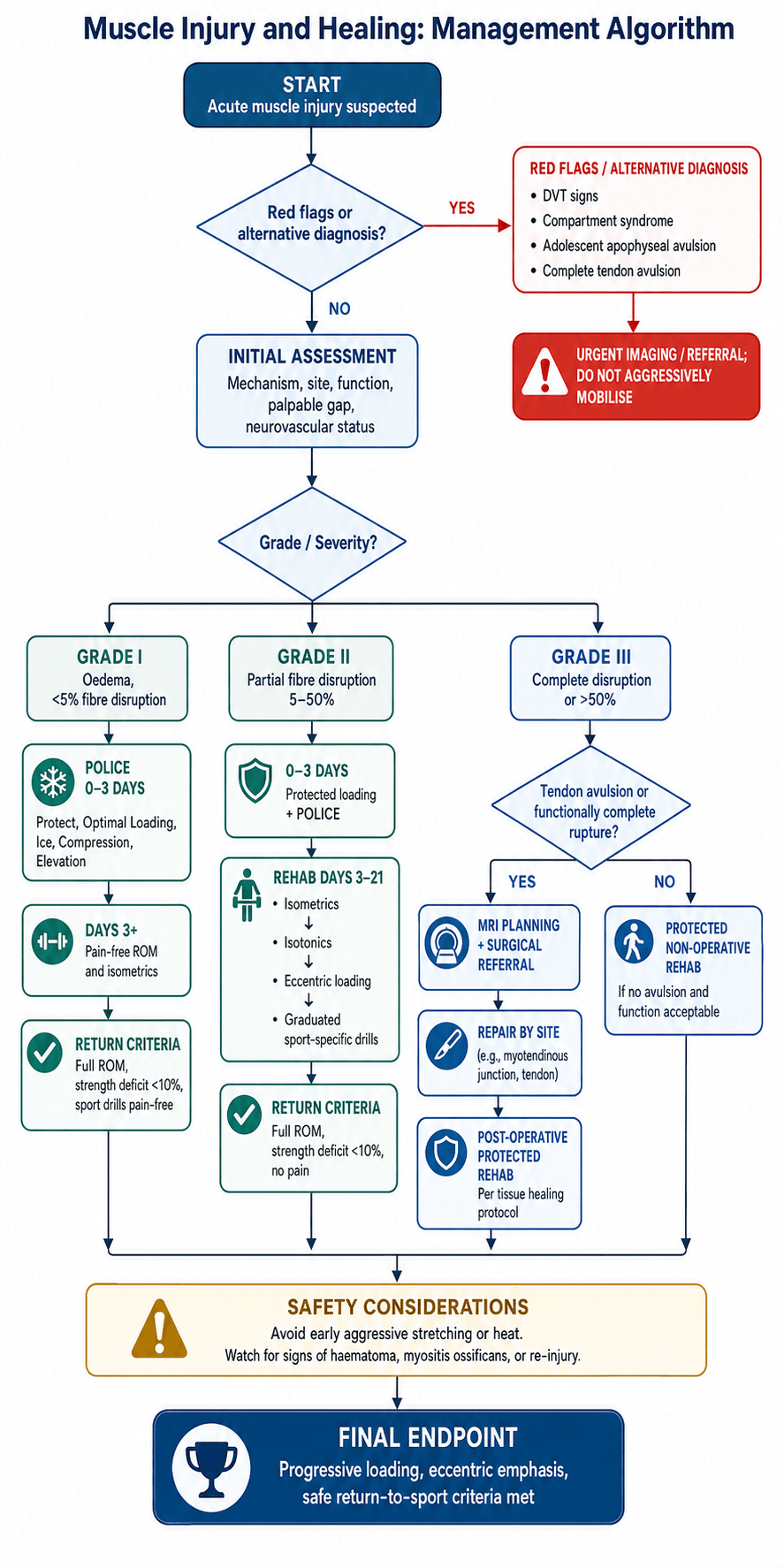
Acute phase (days 0-3). The modern approach replaces RICE with POLICE: Protection, Optimal Loading, Ice, Compression, Elevation. The change is Rest becoming Optimal Loading, because complete rest is not recommended.
- Protection: avoid aggravating activities, use crutches if weight bearing is painful, and apply compression bandaging
- Optimal loading: early protected movement within pain limits, with isometric contractions when comfortable
- Ice: 15-20 minutes every 2-3 hours, reducing metabolic demand and limiting secondary hypoxic injury
- Compression: reduces haematoma expansion and limits oedema formation
- Elevation: reduces venous pressure and promotes lymphatic drainage
This is the balance described in the Järvinen review in the evidence base: brief protection while the scar gains strength, then early controlled mobilisation, is superior to either prolonged rest or immediate aggressive loading.
Repair phase (days 3-21). The goals are to restore range of motion, load progressively, maintain cardiovascular fitness and prevent muscle atrophy. The exercise progression:
- Isometrics (days 3-7): pain-free submaximal contractions, at multiple angles if there is no pain
- Isotonics (days 7-14): concentric before eccentric; light resistance, high repetitions
- Eccentric loading (days 14-21): critical for tendon and MTJ remodelling; progress gradually
Why early mobilisation works. It promotes satellite cell activation, aligns the regenerating fibres with stress, reduces excessive scar formation and maintains neuromuscular function. Studies show early mobilisation improves tensile strength and reduces time to return to sport.
Remodelling phase (day 21 onwards). Functional rehabilitation has three strands. Strength training uses progressive resistance with an eccentric emphasis and sport-specific loading; flexibility work combines static stretching, dynamic stretching before activity and PNF techniques; proprioception is rebuilt with balance training, perturbation exercises and sport-specific drills.
Return to sport. The criteria are functional rather than time-based:
- Full pain-free range of motion
- Strength equal to the uninjured side (less than 10% deficit)
- No pain with sport-specific activities
- Successful completion of a graduated return protocol
Premature return to sport is a leading cause of re-injury.
Surgery (Grade III injuries). Operative repair is reserved for the complete avulsions and ruptures:
- Surgery Indications
- Complete avulsion from ischium
- Technique
- Suture anchor repair
- Surgery Indications
- Complete distal rupture
- Technique
- End-to-end repair
- Surgery Indications
- Complete rupture off humerus
- Technique
- Suture anchor reattachment
- Surgery Indications
- Complete rupture (see dedicated topic)
- Technique
- End-to-end repair or augmentation
Pharmacological Considerations
NSAIDs. Their role is controversial.
- Evidence
- Effective in acute phase
- Evidence
- Reduces early inflammation
- Evidence
- May delay satellite cell activation
- Evidence
- May decrease scar formation
The current recommendation is to limit NSAIDs to the first 48-72 hours if needed for pain, avoid prolonged use during the repair phase, and prefer paracetamol for ongoing analgesia.
Corticosteroids. Generally contraindicated, with a risk of delayed healing and of tendon rupture at the MTJ. They may be considered for specific indications, such as a severe contusion with compartment concerns.
Platelet-rich plasma. There is a theoretical benefit from its growth factors, but the clinical evidence is mixed and it is not currently standard of care. It may have a role in chronic non-healing injuries.
Emerging therapies. Most remain experimental:
- Growth factor therapy (IGF-1, HGF)
- Anti-TGF-β agents (reduce fibrosis)
- Stem cell therapies
- Gene therapy approaches
Complications
Myositis ossificans. Heterotopic bone forms within the muscle, most commonly after a contusion. The risk factors are aggressive early treatment, repeat trauma and haematoma aspiration; prevention is to avoid aggressive stretching and heat in the early phase. Treatment is observation, with excision after maturation at 6-12 months.
Compartment syndrome. Rare but serious, usually after a severe contusion or crush injury, and it requires urgent fasciotomy.
Chronic muscle dysfunction. Persistent weakness, reduced flexibility and susceptibility to re-injury, which may result from excessive fibrosis.
Re-injury. The most common complication. Previous injury is the strongest risk factor, and re-injury usually occurs in the same location; prevention is complete rehabilitation before return to sport.
Five Ps (often late signs):
- Pain out of proportion
- Pain with passive stretch
- Paraesthesias
- Pallor
- Pulselessness (very late)
Early sign: Increasing analgesic requirements
Maintain a high index of suspicion after crush injuries or severe contusions.
Guidelines, Registries and Global Practice
Global epidemiology.
- Muscle injuries account for roughly 30% of all time-loss injuries in professional football, with thigh injuries alone representing about 25% of injuries in the UEFA Elite League cohort.
- Indirect (strain) injuries vastly outnumber direct (contusion) injuries (88% vs 12% of thigh injuries) and cause markedly longer absence (mean 18.5 vs 7 days).
- The hamstrings are the single most commonly injured muscle group in sprinting sports; rectus femoris and gastrocnemius follow. Recurrence rates of 12-30% make muscle injury one of the costliest problems in elite sport.
Side-by-side guidance and classification systems.
- Focus
- Terminology and classification
- Key recommendation
- Distinguishes functional disorders (types 1-2) from structural injuries (types 3-4); standardises language
- Focus
- MRI-based grading
- Key recommendation
- Grades 0-4 plus a/b/c by site (myofascial, musculotendinous, intratendinous); intratendinous (c) injuries have worse prognosis
- Focus
- Clinical plus imaging
- Key recommendation
- Combines mechanism and imaging to guide prognosis
- Focus
- Rehabilitation principles
- Key recommendation
- Early controlled loading and progressive eccentric exercise; functional return-to-sport criteria over fixed timelines
- Focus
- Tissue healing principles
- Key recommendation
- Protect, then progressively load; surgery reserved for complete avulsions/ruptures
Registry and high-level data: Unlike arthroplasty, muscle injuries are not tracked in implant registries; the most authoritative population-level data come from prospective surveillance cohorts such as the UEFA Elite Club Injury Study, which functions as a de facto registry for elite-football soft-tissue injury and underpins much of the global epidemiology above.
High-resource versus limited-resource practice variation.
- High-resource settings: Early MRI for grading and prognosis, individualised eccentric rehabilitation with objective strength/return-to-sport testing, and access to physiotherapy-led graded loading.
- Limited-resource settings: Clinical grading (Grade I-III) without routine MRI, with management centred on the universally available and evidence-supported core: relative rest, protection, progressive loading and graded return. The key message is that the highest-value interventions (early controlled loading and progressive eccentric rehabilitation) require no expensive technology.
Controversies and Areas of Uncertainty
Muscle-injury management contains several genuinely unresolved questions that examiners use to test depth of understanding. Acknowledging uncertainty, rather than overstating dogma, is a marker of consultant-level judgement.
NSAIDs: harm or help? Animal data suggest NSAIDs blunt the inflammatory signalling and satellite-cell activation needed for regeneration, yet human clinical trials are inconsistent and most show no clear functional harm with short courses. The pragmatic consensus of short-term analgesia in the acute phase and paracetamol thereafter is opinion-led, not high-level evidence.
PRP and biologics. Despite biological plausibility, the best evidence (Pas/Reurink 2015 meta-analysis) shows no benefit of PRP for acute hamstring injury. PRP, stem-cell and growth-factor therapies remain experimental for muscle, and routine clinical use is not supported.
Which classification? Imaging-based systems (Munich, British Athletics, ISMuLT) add detail and prognostic discrimination to the simple clinical grades, but they are less universally adopted and add cost. No single system is the agreed global standard.
Timing and dose of loading. Early controlled loading is favoured over immobilisation, but the optimal timing, intensity and progression of eccentric loading, and the precise return-to-sport thresholds, remain debated. Most protocols are extrapolated from small studies and expert consensus.
MCQ Practice Points
Q: Which marker identifies a quiescent satellite cell? A: Pax7. Quiescent satellite cells reside beneath the basal lamina and express the transcription factor Pax7. On activation they upregulate MyoD and Myf5, then myogenin and MRF4 as they differentiate into myoblasts. A cell co-expressing Pax7 but not MyoD is quiescent; loss of Pax7 with gain of myogenin signals terminal differentiation.
Q: Which fibre type and contraction mode carry the highest strain risk? A: Fast-twitch Type IIb fibres during eccentric (active lengthening) contraction. Biarticular muscles with a high proportion of Type IIb fibres (hamstrings, rectus femoris, gastrocnemius) fail most often, typically at or near the myotendinous junction during the late swing phase of sprinting.
Q: What single cellular event best distinguishes regeneration from fibrosis? A: Satellite cell activation and myotube formation versus fibroblast-driven collagen deposition. When satellite cells are available and the basal lamina scaffold is preserved, new contractile fibres form. When satellite capacity is overwhelmed or the scaffold is destroyed, TGF-beta1-driven fibroblast activity lays down non-contractile collagen scar.
Q: For an acute hamstring strain, what intervention has the strongest evidence? A: Progressive lengthening (eccentric) rehabilitation exercises, not platelet-rich plasma. Meta-analysis (Pas/Reurink 2015) showed lengthening rehabilitation significantly shortened return to play (HR 3.22) while PRP showed no benefit over control.
Q: When does the macrophage phenotype switch occur and why does it matter? A: Around day 3-4, pro-inflammatory M1 macrophages give way to anti-inflammatory M2 macrophages. M1 cells phagocytose necrotic debris; M2 cells promote myoblast differentiation and angiogenesis. A failed or prolonged M1 phase impairs regeneration and favours fibrosis.
Rapid-fire high-yield facts:
- Satellite cells express Pax7 (quiescent) and MyoD (activated), located between sarcolemma and basal lamina
- Type IIb fast-twitch fibres are most susceptible to strain
- Myotendinous junction is the most common strain site
- Phases: Destruction (days 0-3), Repair (days 3-21), Remodeling (day 21 onwards)
- M1 macrophages are pro-inflammatory; M2 macrophages promote repair
- TGF-beta drives fibrosis; early controlled mobilization favours regeneration
- Myositis ossificans is the classic complication of quadriceps contusion
Exam Viva Scenarios
Practise clinical reasoning and management decisions out loud
“A basic science examiner asks you to describe the phases of muscle healing following a Grade II hamstring strain.”
“A 25-year-old footballer presents with acute posterior thigh pain after sprinting. He felt a pop and has weakness with knee flexion. How do you classify and manage this injury?”
“Explain the role of satellite cells in muscle regeneration and what happens if they are depleted.”
Key Anatomy
- Satellite cells between sarcolemma and basal lamina
- Pax7+ quiescent, MyoD+ activated
- Type IIb fast-twitch most vulnerable
- MTJ is most common injury site
Healing Phases
- Destruction: Days 0-3, necrosis, M1 macrophages
- Repair: Days 3-21, satellite activation, M2 macrophages
- Remodeling: Day 21+, fiber maturation, collagen conversion
Strain Classification
- Grade I: Less than 5% fibers, return 1-2 weeks
- Grade II: 5-50%, partial tear, return 3-6 weeks
- Grade III: More than 50% or complete, return 3-6 months
Treatment Principles
- POLICE not RICE (Optimal Loading)
- Early mobilization promotes regeneration
- Progress: Isometrics to Isotonics to Eccentrics
- Return when less than 10% strength deficit
Regeneration vs Fibrosis
- Satellite cells = regeneration
- TGF-β excess = fibrosis
- Early mobilization favors regeneration
- Repeated injury increases scar
Complications
- Myositis ossificans after contusion
- Re-injury is most common complication
- Compartment syndrome rare but serious
- Previous injury is biggest risk factor
Evidence Base
Muscle injuries: biology and treatment
- Foundational narrative review defining the three-phase model: destruction, repair, remodeling
- Satellite cells (located beneath the basal lamina) are essential for true regeneration
- Brief immobilization to allow scar to gain strength, then early controlled mobilization, is superior to either prolonged rest or immediate aggressive loading
- Fibrosis (granulation tissue and scar) and regeneration progress in parallel and compete
Muscle injuries and repair: current trends in research
- TGF-beta1 is a central driver of post-injury fibrosis in skeletal muscle
- Antifibrotic agents (e.g. decorin, suramin, gamma-interferon) reduced scar in animal models
- Growth factors (IGF-1, bFGF, NGF) enhanced myoblast proliferation and muscle regeneration in vivo
- Combined antifibrosis plus growth-factor strategies improved functional recovery experimentally
Inflammatory processes in muscle injury and repair
- Neutrophils invade rapidly after injury and can promote further muscle damage via free radicals
- Pro-inflammatory (M1) macrophages clear debris; anti-inflammatory (M2) macrophages support repair, regeneration and growth
- Macrophage actions are coordinated with satellite cell activation, proliferation and differentiation
- Muscle-derived nitric oxide modulates inflammatory cell invasion, protecting healthy fibres