Skeletal muscle structure, contraction mechanisms, fiber types, and motor unit physiology
Critical Must-Knows
- Sliding filament theory: actin-myosin cross-bridge cycling
- Calcium from sarcoplasmic reticulum (NOT extracellular like cardiac)
- A-band constant during contraction; I-band and H-zone shorten
- Motor unit recruitment follows size principle (small to large)
- Force-length relationship: optimal at resting muscle length
Clinical Pearls
- "Draw sarcomere structure: A-band, I-band, H-zone, M-line, Z-lines
- "Know fiber type distribution: Soleus 80% Type I, Gastrocnemius 50/50
- "Training shifts IIx to IIa but NOT Type II to Type I
- "Immobilization: 30% strength loss in 2 weeks, contracture formation
Clinical Imaging
Imaging Gallery

![(A) Epi-illumination of a rat skinned myocyte treated with anti-α-actinin antibody-QDs excited by blue light during ADP-SPOC (see [25] for composition of the ADP-SPOC solution). Note the clear striati](/_next/image?url=%2Fimages%2Ftopics%2Fmuscle-physiology%2Fweb-sourced%2F2-muscle-physiology.png&w=1920&q=85)
![(A) Top, Epi-illumination image of an intact cardiomyocyte treated with anti-α1B-adrenergic receptor antibody (sc-27136, Santa Cruz, CA; see [38]) QDs at 3 nM in oxygenated Ca2+-free-HEPES-Tyrode's so](/_next/image?url=%2Fimages%2Ftopics%2Fmuscle-physiology%2Fweb-sourced%2F3-muscle-physiology.png&w=1920&q=85)
Clinical Warning
Examiner Focus: Sliding Filament Theory
Viva Trap: Fiber Type Distribution
Common Error: Calcium Source
High-Yield Concept: Length-Tension
SO FAT GASMuscle Fiber Types
Hook:Type I fibers are SLOW but can go FAR (marathon runners), Type IIx fibers provide quick GAS but run out fast (sprinters)
ACh DART Catches SR CalciumExcitation-Contraction Coupling Steps
Hook:Imagine throwing a DART at the muscle membrane that pierces through to CATCH calcium stored inside
A Always Stays, I and H DisappearSarcomere Bands During Contraction
Hook:A is for ANCHOR - the A-band is anchored and doesn't change size. I and H are the SPACES that close up
ABCD - Attach, Bend, Come off, DoneCross-Bridge Cycle Four Steps
Hook:The cross-bridge cycle is as simple as ABCD - just remember ATP is needed to Come off (detach), not to Attach
Muscle Fiber Ultrastructure
Hierarchical Organization
Skeletal muscle exhibits a highly organized structural hierarchy essential for coordinated force generation:
Macroscopic to Microscopic Levels:
- Whole muscle: Ensheathed by epimysium (dense irregular connective tissue)
- Fascicles: Bundles of 10-100 fibers wrapped by perimysium containing neurovascular structures
- Muscle fibers (cells): Individual multinucleated cells (10-100 micrometers diameter, up to 30 cm length) surrounded by endomysium
- Myofibrils: Cylindrical organelles (1-2 micrometers diameter) comprising 80% of fiber volume
- Sarcomeres: Fundamental contractile unit (2.5 micrometers at rest) arranged in series within myofibrils
Sarcomere Architecture
The sarcomere extends from one Z-line to the next and contains precisely arranged contractile proteins:
Thick Filaments (15 nm diameter):
- Primarily myosin II (molecular weight 500 kDa)
- Each myosin molecule has 2 heavy chains forming tail and 2 head regions
- Myosin heads contain actin-binding site and ATP-binding site
- Arranged in bipolar fashion with heads projecting outward
- Located in A-band (1.6 micrometers length remains constant during contraction)
Thin Filaments (7 nm diameter):
- F-actin: double-stranded helix of G-actin monomers (375 monomers per filament)
- Tropomyosin: coiled-coil protein lying in actin grooves, blocking myosin-binding sites at rest
- Troponin complex: heterotrimeric regulatory protein every 7 actin monomers
- TnT: binds tropomyosin
- TnI: inhibitory subunit blocking actin-myosin interaction
- TnC: calcium-binding subunit (4 binding sites, 2 high-affinity structural, 2 low-affinity regulatory)
- Anchored at Z-line via alpha-actinin
Sarcomere Bands and Zones:
- A-band: Contains entire thick filament length (appears dark on EM, anisotropic to polarized light)
- I-band: Thin filaments only, no overlap with thick filaments (appears light, isotropic)
- H-zone: Central region of A-band containing only thick filament tails (no actin overlap)
- M-line: Central anchoring proteins (myomesin, M-protein) connecting thick filaments
- Z-line (Z-disc): Anchoring point for thin filaments via alpha-actinin
Key Point
During contraction, the I-band and H-zone decrease in width while the A-band remains constant. This demonstrates the sliding filament mechanism where thin filaments slide past thick filaments rather than filaments themselves shortening. The Z-lines move closer together, reducing sarcomere length from 2.5 micrometers to approximately 1.5 micrometers at maximal contraction.
Membrane Systems and Calcium Handling
Transverse Tubules (T-tubules):
- Invaginations of sarcolemma penetrating deep into fiber
- Located at A-I junction in mammalian muscle (2 per sarcomere)
- Contain voltage-gated L-type calcium channels (dihydropyridine receptors, DHPRs)
- Rapidly conduct action potentials to fiber interior
- Critical for synchronous activation of all myofibrils
Sarcoplasmic Reticulum (SR):
- Specialized smooth endoplasmic reticulum surrounding each myofibril
- Terminal cisternae: expanded SR regions flanking T-tubules forming triads
- Contains ryanodine receptors (RyR1) - calcium release channels
- SR lumen calcium concentration: 1-2 mM (10,000x higher than cytoplasm)
- SERCA pumps (SR Ca2+-ATPase) actively sequester calcium during relaxation
- Calsequestrin: calcium-binding protein in SR lumen (stores 80% of SR calcium)
Neuromuscular Junction:
- Specialized synapse between motor neuron terminal and muscle fiber
- Motor end plate: highly folded sarcolemma increasing surface area
- Acetylcholine receptors concentrated at junctional folds (10,000 per micrometer squared)
- Acetylcholinesterase in synaptic cleft rapidly hydrolyzes ACh (ensures brief signal)
Sliding Filament Theory and Contraction Mechanism
Cross-Bridge Cycle
The molecular basis of muscle contraction involves cyclical interactions between myosin heads and actin binding sites, powered by ATP hydrolysis. Each cycle produces approximately 10 nm of filament sliding and generates 3-4 pN of force.
Four-Step Cross-Bridge Cycle:
-
ATP Binding and Myosin Detachment
- ATP binds to myosin head causing conformational change
- Myosin-actin affinity decreases dramatically (1000-fold)
- Cross-bridge detaches from actin
- In rigor mortis (no ATP), myosin remains bound creating muscle stiffness
-
ATP Hydrolysis and Myosin Cocking
- Myosin ATPase hydrolyzes ATP to ADP plus Pi (both remain bound)
- Energy from hydrolysis cocks myosin head into high-energy conformation
- Myosin head rotates 90 degrees to "ready" position
- This step stores elastic energy in myosin neck region
-
Cross-Bridge Formation (Power Stroke)
- When calcium levels are high, tropomyosin moves, exposing actin binding sites
- Cocked myosin head binds strongly to actin
- Pi release triggers power stroke
- Myosin head rotates, pulling actin filament toward M-line
- Generates approximately 10 nm of sliding motion
-
ADP Release and Force Maintenance
- ADP released from myosin completing power stroke
- Myosin remains tightly bound to actin until next ATP binds
- Multiple asynchronous cross-bridges maintain steady force
- Cycle repeats if calcium and ATP remain available
Energetics:
- One ATP consumed per cross-bridge cycle
- Approximately 50% efficiency converting chemical to mechanical energy
- Resting muscle ATP concentration: 5 mM (sufficient for 2-3 seconds maximal contraction)
- Phosphocreatine provides rapid ATP regeneration (30 mM concentration)
- Glycolysis and oxidative phosphorylation sustain prolonged activity
Key Point
The rate-limiting step for shortening velocity is ADP release, which is faster in Type II fibers than Type I fibers. This explains why Type II fibers can contract 3-5 times faster. Myosin heavy chain isoforms (MHC I, MHC IIa, MHC IIx) have different ATPase rates determining fiber type contractile speed.
Excitation-Contraction Coupling
This process links electrical excitation of the sarcolemma to mechanical contraction of myofibrils:
Step-by-Step Sequence:
-
Neuromuscular Transmission (1 ms)
- Motor neuron action potential reaches axon terminal
- Voltage-gated calcium channels open, calcium influx
- Synaptic vesicles fuse, releasing ACh into synaptic cleft (200-300 vesicles)
- Each vesicle contains 5,000-10,000 ACh molecules (quantum)
-
Muscle Fiber Depolarization (2-3 ms)
- ACh binds nicotinic receptors on motor end plate
- Sodium influx creates end-plate potential (50-70 mV)
- Exceeds threshold, triggering action potential in adjacent sarcolemma
- Action potential propagates bidirectionally along fiber at 3-5 m/s
-
T-tubule Conduction (1-2 ms)
- Action potential rapidly conducted into T-tubules
- Reaches A-I junctions throughout fiber cross-section
- Ensures synchronous activation of all myofibrils
- Voltage-gated DHPR (L-type calcium channels) detect depolarization
-
Calcium-Induced Calcium Release (5-10 ms)
- DHPR conformational change mechanically coupled to RyR1
- RyR1 calcium release channels open in SR membrane
- SR calcium floods into cytoplasm (100-fold increase to 10 micrometers)
- Unlike cardiac muscle, this is mechanical coupling NOT calcium-induced
-
Thin Filament Activation (10-20 ms)
- Calcium binds to TnC (2 calcium ions per troponin)
- Troponin complex undergoes conformational change
- Tropomyosin shifts 25 Angstrom deeper into actin groove
- Myosin-binding sites on actin exposed
- Cross-bridge cycling initiated
-
Relaxation Phase (50-100 ms)
- Sarcolemma repolarizes, DHPR returns to resting conformation
- RyR1 channels close
- SERCA pumps actively transport calcium back into SR
- Requires ATP (1 ATP per 2 calcium ions)
- Cytoplasmic calcium decreases below 0.1 micrometers
- Calcium dissociates from TnC
- Tropomyosin returns to blocking position
- Myosin-actin interactions cease
Clinical Relevance:
- Malignant hyperthermia: RyR1 mutation causing uncontrolled calcium release
- Central core disease: RyR1 mutations causing structural abnormalities and weakness
- Periodic paralysis: mutations in DHPR or sodium channels causing episodic weakness
- Myasthenia gravis: antibodies against ACh receptors reducing end-plate potentials
Muscle Fiber Types and Motor Units
Fiber Type Classification
Skeletal muscle fibers are classified based on contractile speed and metabolic profile:
Muscle Fiber Type Characteristics
| feature | typeI | typeIIa | typeIIx |
|---|---|---|---|
| Myosin ATPase Activity | Low (slow) | High (fast) | Highest (very fast) |
| Contraction Speed | Slow (110 ms) | Fast (50 ms) | Very fast (40 ms) |
| Fatigue Resistance | Very high | Intermediate | Low (rapid fatigue) |
| Mitochondrial Density | Very high | High | Low |
| Capillary Density | High | Intermediate | Low |
| Myoglobin Content | High (red muscle) | Intermediate | Low (white muscle) |
| Primary Metabolism | Oxidative | Oxidative-glycolytic | Glycolytic |
| Glycogen Stores | Low | Intermediate | High |
| Motor Neuron Size | Small alpha neurons | Larger alpha neurons | Largest alpha neurons |
| Fiber Diameter | Small (50 micrometers) | Intermediate (60 micrometers) | Large (80 micrometers) |
| Force Production | Low per fiber | Intermediate | High per fiber |
| Example Muscles | Soleus (80%), paravertebrals | Gastrocnemius, deltoid | Extraocular, hand intrinsics |
Fiber Type Distribution Patterns:
- Most human muscles contain mixed fiber types (40-60% Type I, 30-50% Type IIa, 5-10% Type IIx)
- Postural muscles: predominantly Type I (soleus 80%, paravertebrals 70%)
- Phasic muscles: higher Type II (gastrocnemius 50/50, vastus lateralis 45/55)
- Within muscle: superficial regions often have more Type II than deep regions
- Individual variation: genetic factors determine baseline distribution
Fiber Type Plasticity:
- Type IIx to Type IIa transitions occur with training or detraining
- Endurance training: shifts IIx to IIa, increases oxidative capacity of Type II
- Strength training: hypertrophy of Type II fibers, minimal type conversion
- Denervation: shift toward faster phenotype (Type I to Type II characteristics)
- Cross-innervation experiments: motor neuron determines fiber type (neural control)
- Type I to Type II transformation extremely rare in adults (thyroid hormone can induce)
Key Point
Rotator cuff tears demonstrate fiber type changes: chronic tears show increased Type II fibers and fatty infiltration. Goutallier grading on MRI correlates with irreversible muscle changes. This explains why massive chronic tears have poor surgical outcomes - the muscle has undergone irreversible transformation.
Motor Unit Organization
A motor unit consists of one alpha motor neuron and all muscle fibers it innervates.
Motor Unit Characteristics:
- Innervation ratio: varies from 10:1 (extraocular muscles) to 2000:1 (gastrocnemius)
- Fine motor control: small motor units (finger muscles: 100:1)
- Gross movement: large motor units (back muscles: 1000-2000:1)
- All fibers in a motor unit are the same type (Type I or Type II)
- Fiber territory: distributed over 5-10 mm diameter region (interspersed with other motor units)
Size Principle of Motor Unit Recruitment: Henneman's size principle describes orderly recruitment based on motor neuron size:
-
Low-Force Tasks: Small Type I motor units recruited first
- Small motor neurons have lower activation threshold
- Higher input resistance amplifies synaptic input
- Provides fine gradation of force at low levels
-
Moderate-Force Tasks: Type IIa motor units added
- Larger motor neurons require greater synaptic drive
- Increases force production while maintaining some fatigue resistance
-
High-Force Tasks: Type IIx motor units recruited last
- Largest motor neurons, highest threshold
- Maximal force production but rapid fatigue
- Only recruited for ballistic movements or maximal efforts
Rate Coding:
- Once recruited, motor units increase firing frequency to generate more force
- Initial firing rate: 8-12 Hz (unfused tetanus, visible twitches)
- Maximal firing rate: 50-60 Hz (fused tetanus, smooth contraction)
- Rate coding contributes 50% of force modulation in intermediate force ranges
Clinical Implications:
- Denervation: loss of motor units, remaining units reinnervate orphaned fibers (enlarged motor units)
- EMG shows increased motor unit amplitude and duration with reinnervation
- Primary muscle disease: motor units normal but reduced force per fiber
- Upper motor neuron lesions: impaired recruitment, firing rate abnormalities (spasticity)
Force Generation and Biomechanics
Length-Tension Relationship
Force generation depends critically on sarcomere length due to actin-myosin filament overlap:
Zones of the Length-Tension Curve:
-
Over-Stretched Position (sarcomere length greater than 3.6 micrometers)
- Minimal actin-myosin overlap
- Few cross-bridges can form
- Force production reduced to 0-20% maximum
- Clinical example: over-lengthened muscle after nerve palsy
-
Descending Limb (3.6-2.5 micrometers)
- Progressive increase in filament overlap
- Linear relationship between length and force
- Each 0.1 micrometer decrease adds approximately 10% more cross-bridges
- Passive elastic elements contribute minimal force
-
Plateau Region (2.5-2.0 micrometers)
- Optimal actin-myosin overlap throughout entire thick filament
- Maximum active force generation (100%)
- Corresponds to resting muscle length in situ
- Most stable operating range for physiological function
-
Ascending Limb (2.0-1.5 micrometers)
- Actin filaments begin overlapping from opposite ends
- Interference reduces available binding sites
- Thick filament compression against Z-lines
- Force decreases to 60-70% at 1.5 micrometers
-
Extreme Shortening (less than 1.5 micrometers)
- Severe actin overlap and structural distortion
- Force production drops below 50%
- Rarely achieved in vivo except in specialized muscles
Passive Tension Component:
- At lengths beyond resting, elastic elements (titin, connective tissue) generate passive force
- Titin: giant protein (3-4 MDa) spanning from Z-line to M-line
- Acts as molecular spring resisting over-stretch
- Total force = active (cross-bridges) plus passive (elastic) components
Clinical Applications:
- Joint immobilization in shortened position: sarcomeres adapt by reducing number in series
- Results in shortened optimal length and contracture formation
- Immobilization in lengthened position: sarcomeres added in series
- Tendon transfers: muscle set to appropriate tension intraoperatively for optimal length-tension
- Achilles tendon lengthening: must preserve sufficient overlap for push-off strength
Force-Velocity Relationship
Shortening velocity inversely relates to force production:
Concentric Contractions (Muscle Shortening):
- Maximum velocity (Vmax) occurs at zero load
- Type I fibers: Vmax = 4-5 fiber lengths/second
- Type IIx fibers: Vmax = 15-20 fiber lengths/second
- As load increases, velocity decreases hyperbolically
- At maximum isometric force, velocity = 0
- Power (force times velocity) peaks at approximately 30% Vmax and 30% maximum force
Isometric Contractions:
- Velocity = 0, force determined by activation level and length
- Reference point for force-velocity curve
- Maximum tetanic force defined as F0 (100%)
Eccentric Contractions (Muscle Lengthening):
- Force production exceeds isometric maximum (up to 150% F0)
- Mechanisms:
- Cross-bridges forcibly detached while bound (energy absorbed)
- Titin and elastic elements stretched, contributing passive force
- Some cross-bridges remain attached longer during forced lengthening
- Lower metabolic cost per unit force (ATP consumed only during attachment)
- Muscle damage occurs more readily (delayed-onset muscle soreness)
- Training effect: rapid strength gains, protective adaptation to eccentric stress
Key Point
Eccentric contractions generate higher forces with lower metabolic cost, making them efficient for deceleration and shock absorption. However, unaccustomed eccentric exercise causes Z-line streaming, membrane disruption, and delayed-onset muscle soreness (DOMS) peaking at 24-72 hours. This explains post-operative pain patterns and guides rehabilitation progression.
Muscle Architecture and Force Production
Muscle architecture determines functional capacity:
Architectural Parameters:
-
Physiological Cross-Sectional Area (PCSA)
- Total cross-sectional area of all fibers perpendicular to fiber direction
- PCSA = (muscle mass times cosine pennation angle) divided by (fiber length times muscle density)
- Directly proportional to maximum force (specific tension: 20-30 N/cm squared)
- Example: gastrocnemius PCSA 50 cm squared, maximum force 1000-1500 N
-
Fiber Length
- Determines maximum shortening distance and velocity
- Longer fibers = greater excursion and higher Vmax
- Sarcomeres in series multiply shortening distance
- Example: sartorius (long fibers) vs. soleus (short fibers)
-
Pennation Angle
- Angle between fiber direction and muscle's line of action
- Ranges from 0 degrees (fusiform) to 30 degrees (unipennate) to 45 degrees (multipennate)
- Packing more fibers (higher PCSA) but reducing effective force transmission
- Force along tendon = fiber force times cosine (pennation angle)
- Example: deltoid pennation 20 degrees, force reduction 6% but allows 3x more fibers
Muscle Architectural Types:
-
Fusiform (parallel fibers): sartorius, biceps brachii
- Long excursion, high velocity, lower force
-
Unipennate: extensor digitorum longus, vastus lateralis
- Fibers on one side of central tendon
- Intermediate force and excursion
-
Bipennate: rectus femoris, flexor hallucis longus
- Fibers on both sides of central tendon
- Higher force, moderate excursion
-
Multipennate: deltoid, subscapularis
- Multiple pennation planes
- Highest force production, shortest excursion
Force Transmission:
- Longitudinal transmission: via tendons to skeleton
- Lateral transmission: through endomysium/perimysium to adjacent structures (30% of force)
- Costameres: link sarcolemma to extracellular matrix
- Dystrophin-glycoprotein complex: critical for membrane stability
- Muscular dystrophies: defects in force transmission proteins causing progressive weakness
Muscle Adaptation and Pathophysiology
Training Adaptations
Endurance Training:
- Mitochondrial biogenesis (PGC-1alpha upregulation): 50-100% increase in mitochondrial density
- Capillary angiogenesis: 20-30% increase in capillary:fiber ratio
- Oxidative enzyme upregulation (citrate synthase, succinate dehydrogenase)
- Fiber type shift: IIx to IIa conversion (more fatigue-resistant)
- Myoglobin content increases 80%
- Glycogen storage capacity increases
- Improved lactate clearance
- Minimal hypertrophy (fiber size increases less than 10%)
Resistance Training:
- Muscle hypertrophy: 20-50% fiber cross-sectional area increase in 12 weeks
- Type II fibers hypertrophy more than Type I (especially IIx)
- Satellite cell activation and myonuclear addition
- Protein synthesis exceeds breakdown (mTOR pathway activation)
- Neural adaptations (first 4-6 weeks):
- Increased motor unit recruitment
- Improved firing rate synchronization
- Reduced antagonist co-contraction
- Tendon stiffness increases (force transmission efficiency)
- Minimal metabolic adaptations
Eccentric Training:
- Rapid strength gains (20% in 4 weeks)
- Addition of sarcomeres in series (shifts optimal length)
- Protective "repeated bout effect" - reduced DOMS with subsequent sessions
- Z-line remodeling and cytoskeletal reinforcement
- Desmin and dystrophin upregulation
- Useful for tendinopathy rehabilitation (Alfredson protocol for Achilles)
Immobilization and Denervation
Immobilization Effects (Time Course):
Week 1:
- Protein synthesis decreases 50%
- Type I fibers preferentially affected initially
- Mitochondrial enzyme activity decreases
- 3-5% strength loss
Week 2:
- 20-30% strength loss
- Muscle atrophy 10-15% (fiber cross-sectional area)
- Shift toward Type II fiber characteristics
- Collagen deposition begins in endomysium
Week 4-6:
- 30-40% strength loss
- Sarcomere number adaptation:
- Shortened position: sarcomeres in series decrease (contracture develops)
- Lengthened position: sarcomeres added in series
- Joint stiffness from capsular contracture and muscle shortening
- Type I fiber atrophy up to 30%
Month 3 plus:
- Fatty infiltration begins (adipogenic differentiation of muscle progenitors)
- Fibrosis replaces functional tissue
- Contracture formation resistant to stretching
- Strength loss plateaus at 50-60% if immobilization continues
Recovery from Immobilization:
- Strength returns faster than muscle mass (neural adaptations)
- Complete recovery takes 2-3x immobilization duration
- Contractures may be permanent if immobilization exceeds 12 weeks
- Early mobilization critical for preventing irreversible changes
Denervation Effects:
Acute Phase (0-4 weeks):
- Muscle fiber membrane instability (fibrillation potentials on EMG)
- Acetylcholine receptors spread beyond motor end plate
- Fiber atrophy begins (10% loss in first month)
- Preserved fiber type characteristics initially
Subacute Phase (1-6 months):
- Progressive atrophy (50% fiber size reduction by 6 months)
- Reinnervation potential if nerve regenerates (1 mm/day)
- Surviving motor neurons sprout collaterals to reinnervate orphaned fibers
- Motor unit territory expansion (giant motor units on EMG)
- Fiber type grouping (all fibers in a region become same type)
Chronic Phase (beyond 6 months):
- Irreversible fatty infiltration and fibrosis
- Motor end plates degenerate
- Muscle loses capacity for reinnervation (point of no return 12-18 months)
- Goutallier grading on MRI:
- Grade 0: Normal muscle
- Grade 1: Some fatty streaks
- Grade 2: Less than 50% fat
- Grade 3: Equal muscle and fat
- Grade 4: More fat than muscle
- Grade 3-4 indicates poor surgical prognosis for rotator cuff repair
Key Point
The "point of no return" for muscle reinnervation is approximately 12-18 months after complete denervation. Beyond this, motor end plates are lost, and fatty infiltration/fibrosis is irreversible. This timeline guides surgical decision-making for nerve repairs and tendon transfers. Nerve repairs have best outcomes if performed within 6 months of injury.
Muscle Injury and Regeneration
Strain Injury Mechanisms:
- Most common during eccentric contractions at long muscle length
- Myotendinous junction: weakest structural point (75% of strains)
- Grade I: micro-tears, less than 5% fibers, 1-2 weeks recovery
- Grade II: partial tear, 5-50% fibers, 3-6 weeks recovery
- Grade III: complete tear, surgical consideration, 3 plus months recovery
Regeneration Process:
-
Destruction Phase (1-3 days):
- Membrane disruption, calcium influx, protein degradation
- Neutrophil infiltration, inflammatory cytokines
- Satellite cell activation (normally quiescent between basal lamina and sarcolemma)
-
Repair Phase (3-14 days):
- Satellite cells proliferate and differentiate into myoblasts
- Myoblasts fuse forming myotubes
- Macrophages clear debris, release growth factors (IGF-1)
- Angiogenesis and neural sprouting
-
Remodeling Phase (2-6 weeks):
- Myotube maturation and sarcomere organization
- Connective tissue scar formation
- Functional strength returns to 80-90%
- Residual scar remains potential weak point
Factors Impairing Regeneration:
- Age: satellite cell number and function decline with age
- Corticosteroids: inhibit satellite cell proliferation
- NSAIDs: controversial, may impair early inflammation but long-term effect minimal
- Excessive fibrosis: TGF-beta pathway overactivation
- Severe injury: complete disruption of basal lamina scaffold prevents organized regeneration
Clinical Decision Scenarios
Use these scenarios to practise clinical reasoning and management decisions
"A 45-year-old marathon runner presents with progressive fatigue and reduced endurance over 6 months. Muscle biopsy shows increased proportion of Type IIx fibers compared to Type I. Explain the normal fiber type composition in endurance athletes and what this finding might suggest."
"You're performing a rotator cuff repair on a 65-year-old with a chronic massive tear. MRI shows Goutallier Grade 3 fatty infiltration of the supraspinatus. Explain the pathophysiology of fatty infiltration and why this affects surgical prognosis."
"A medical student asks you to explain why muscle can generate more force during eccentric contractions than concentric contractions. Explain the mechanisms and clinical relevance."
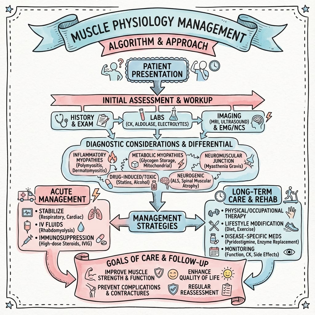
Management Algorithm
Clinical summary
Sarcomere Structure
- •Z-line to Z-line = 2.5 micrometers at rest
- •A-band: thick filaments (constant 1.6 micrometers)
- •I-band: thin filaments only (decreases with contraction)
- •H-zone: myosin only (decreases with contraction)
- •M-line: thick filament anchoring
- •Troponin complex: TnC (calcium binding), TnI (inhibitory), TnT (tropomyosin binding)
- •During contraction: I-band and H-zone shorten, A-band stays constant
Excitation-Contraction Coupling
- •ACh to end-plate depolarization to action potential to T-tubule conduction
- •DHPR conformational change to RyR1 opens to SR calcium release (100x increase to 10 micrometers)
- •Calcium binds TnC to tropomyosin shifts to myosin-actin binding to cross-bridge cycle
- •Relaxation: SERCA pumps return calcium to SR (1 ATP per 2 calcium ions)
- •Malignant hyperthermia = RyR1 mutation
Cross-Bridge Cycle
- •Step 1: ATP binds, myosin detaches
- •Step 2: ATP hydrolysis, myosin cocks (high energy)
- •Step 3: Myosin binds actin, Pi release, power stroke (10 nm sliding)
- •Step 4: ADP release, ready for next ATP
- •Rigor mortis = no ATP, myosin stays bound
- •Rate-limiting step = ADP release (faster in Type II)
Fiber Types Quick Reference
- •Type I (SO): slow, oxidative, fatigue-resistant, high mitochondria, small diameter, red
- •Type IIa (FOG): fast, oxidative-glycolytic, intermediate fatigue resistance
- •Type IIx (FG): very fast, glycolytic, rapid fatigue, low mitochondria, white
- •Soleus 80% Type I; Gastrocnemius 50/50
- •Training: IIx to IIa (reversible); Type I to Type II does NOT occur
Size Principle
- •Henneman's size principle: orderly recruitment based on motor neuron size
- •Small Type I units recruited first (low threshold, fine control)
- •Type IIa added for moderate force
- •Type IIx recruited last for maximal force (high threshold, rapid fatigue)
- •Rate coding: increase firing frequency 8-12 Hz (unfused) to 50-60 Hz (fused tetanus)
Length-Tension Relationship
- •Optimal force at 2.0-2.5 micrometers (resting length), maximum actin-myosin overlap
- •Overstretched (greater than 3.6 micrometers): minimal overlap, low force
- •Shortened (less than 1.5 micrometers): actin interference, reduced force
- •Passive tension from titin (Z-line to M-line molecular spring)
- •Immobilization: sarcomeres adapt (fewer in series if shortened, more if lengthened)
Force-Velocity Relationship
- •Concentric: inverse relationship, Vmax at zero load, velocity = 0 at maximum force
- •Type I Vmax = 4-5 lengths per second, Type IIx = 15-20 lengths per second
- •Power peaks at 30% Vmax
- •Eccentric: force up to 150% maximum isometric force, lower ATP cost
- •Eccentric mechanisms: forced cross-bridge detachment plus titin stretch plus prolonged attachment
- •Causes DOMS (24-72 hours)
Muscle Architecture
- •PCSA (physiological cross-sectional area) proportional to maximum force (20-30 N per cm squared)
- •Fiber length proportional to excursion and velocity
- •Pennation angle: allows more fibers but reduces effective force (force times cosine angle)
- •Fusiform (sartorius): long excursion
- •Multipennate (deltoid): high force, short excursion
Training Adaptations
- •Endurance: mitochondria plus 50-100%, capillaries plus 20-30%, IIx to IIa shift, minimal hypertrophy
- •Resistance: fiber hypertrophy plus 20-50% (Type II more), satellite cell activation, neural adaptations (first 4-6 weeks)
- •Eccentric: rapid strength gains, sarcomeres added in series, repeated bout effect (protective adaptation)
Immobilization Timeline
- •Week 1: protein synthesis decreased 50%, 3-5% strength loss
- •Week 2: 20-30% strength loss, 10-15% atrophy
- •Week 4-6: 30-40% strength loss, sarcomere adaptation, contractures develop
- •Month 3 plus: fatty infiltration, fibrosis, 50-60% strength loss
- •Recovery takes 2-3x immobilization duration
Denervation Effects
- •Acute (0-4 weeks): fibrillations on EMG, ACh receptor spread
- •Subacute (1-6 months): 50% atrophy, reinnervation possible (nerve regenerates 1 mm per day), motor unit sprouting, fiber type grouping
- •Chronic (beyond 6 months): fatty infiltration, fibrosis, motor end plate loss
- •Point of no return = 12-18 months
Goutallier Grading (MRI Fatty Infiltration)
- •Grade 0: normal
- •Grade 1: some fatty streaks
- •Grade 2: less than 50% fat
- •Grade 3: equal muscle and fat (poor prognosis)
- •Grade 4: more fat than muscle (very poor prognosis)
- •Grade 3-4 indicates irreversible changes, re-tear rate greater than 50% after rotator cuff repair
- •Acute repairs 90% success versus chronic massive tears 50%
Fiber Type Distribution in Human Muscles
- Landmark autopsy study quantifying fiber type composition across 36 human muscles. Found wide between-muscle variability: soleus approximately 84% Type I (slow oxidative), tibialis anterior approximately 73%, with limb muscles such as triceps and biceps brachii closer to a mixed composition. Substantial individual variation. Postural/antigravity muscles are predominantly Type I; most limb muscles are mixed. Provides the baseline reference data still cited for muscle-specific fiber typing and biopsy interpretation.
Henneman Size Principle of Motor Unit Recruitment
- Seminal work establishing that motor units are recruited in order of increasing size (small to large) during voluntary contractions. Demonstrated that small motor neurons innervating Type I fibers have higher input resistance and lower activation thresholds, recruited first for fine motor control. Large motor neurons innervating Type II fibers recruited only for high-force demands. This orderly recruitment pattern optimizes efficiency and force gradation. Has become fundamental principle in motor control understanding, rehabilitation design, and interpretation of EMG studies.
Myosin Heavy Chain IIX Overshoot with Training and Detraining
- Vastus lateralis MHC isoforms were measured in sedentary men before 3 months of heavy resistance training and after 3 months of detraining. Resistance training reduced MHC IIX from 9.3% to 2.0% with a reciprocal rise in MHC IIA, confirming the physiological IIX-to-IIA shift. Critically, detraining drove MHC IIX to 17.2%, markedly ABOVE pre-training values (the IIX overshoot). This establishes that IIX-IIA transitions are activity-regulated and reversible, and that complete Type I to Type II conversion does not occur with normal training. Underpins the principle that detraining/disuse pushes muscle toward a faster, more fatigable phenotype.
Goutallier Classification for Rotator Cuff Fatty Infiltration
- Original CT-based five-stage grading of rotator cuff fatty degeneration in 63 patients scheduled for cuff repair: Grade 0 normal, Grade 1 some fatty streaks, Grade 2 fat less than muscle, Grade 3 equal fat and muscle, Grade 4 fat more than muscle. Fatty degeneration worsened with time and correlated with functional impairment of external rotation. After effective repair, moderate supraspinatus degeneration regressed in only a minority and infraspinatus degeneration never regressed (often progressed). Supraspinatus repair recurrence was 25%, and infraspinatus degeneration strongly predicted poorer outcome. Conclusion: operate before irreversible muscular degeneration develops. Foundational prognostic classification in cuff surgery.
Fatty Degeneration of Cuff Muscles: CT versus MRI Grading
- Forty-one shoulders were graded for fatty degeneration on both CT and MRI. Interobserver reproducibility was good to excellent for each modality, validating the translation of Goutallier grading to MRI. CT-MRI agreement was only fair to moderate even when simplified to a three-grade scale, so grading should be reported per-modality. Degree of fatty degeneration correlated significantly with muscle atrophy on parasagittal MRI. This work underpins the now-standard MRI-based Goutallier/Fuchs grading used clinically for cuff prognosis.
Discovery of the Skeletal Muscle Satellite Cell
- Classic electron-microscopy report describing a distinct mononuclear cell wedged between the basal lamina and sarcolemma of the muscle fiber, named the satellite cell. Mauro hypothesized it was a dormant myoblast retained for growth and repair. This single observation founded the field of muscle stem-cell biology: satellite cells are now established as the resident myogenic progenitors that activate, proliferate, and fuse to regenerate injured fibers, and whose decline with age and pathological adipogenic switching underlie impaired regeneration and fatty infiltration.
Malignant Hyperthermia: RYR1 Pharmacogenetics and Dantrolene
- Comprehensive review of malignant hyperthermia (MH), a pharmacogenetic skeletal-muscle disorder triggered by volatile anaesthetics and succinylcholine via uncontrolled myoplasmic calcium release. Reaction incidence ranges from about 1:10,000 to 1:250,000 anaesthetics, with genetic susceptibility possibly as common as 1:400. Inheritance is autosomal dominant; most cases involve RYR1 variants on chromosome 19q13.1 (over 400 variants described, at least 34 causal), with a minority in CACNA1S. Diagnosis rests on the in-vitro contracture (caffeine-halothane) test plus DNA testing. Dantrolene is the specific antagonist and has reduced mortality from approximately 80% historically to under 5%.
Differential Diagnosis of the Weak or Wasted Muscle
Muscle weakness/wasting is a common viva stem. The key is to localise the lesion (upper motor neuron, lower motor neuron, neuromuscular junction, or muscle) using the pattern of weakness, reflexes, EMG, and CK.
Localising the Cause of Weakness and Wasting
| feature | neurogenic | myopathic | nmj | umn |
|---|---|---|---|---|
| Distribution | Myotomal / nerve territory, often distal | Symmetrical, proximal (limb-girdle) | Fatigable, ocular/bulbar onset | Pyramidal pattern, anti-gravity weakness |
| Wasting | Marked, early | Present, proportional to weakness | Minimal (until severe/chronic) | Disuse only (late) |
| Tone / Reflexes | Reduced tone, absent/reduced reflexes | Normal or reduced reflexes | Normal reflexes | Increased tone, hyper-reflexia, clonus |
| Fasciculations | Present (denervation) | Absent | Absent | Absent |
| Serum CK | Normal or mildly raised | Often markedly raised | Normal | Normal |
| EMG | Fibrillations, large polyphasic long-duration units | Small short-duration low-amplitude units, early recruitment | Decrement on repetitive stimulation (myasthenia) | Reduced recruitment, normal unit morphology |
| Examples | Peripheral nerve injury, radiculopathy, motor neuron disease | Muscular dystrophy, inflammatory myopathy, disuse atrophy | Myasthenia gravis, Lambert-Eaton | Stroke, spinal cord injury, cerebral palsy |
Controversies and Areas of Uncertainty
-
Reversibility of fatty infiltration after cuff repair. Goutallier and most series show fatty degeneration does not regress and often progresses despite anatomic healing, yet some report stabilisation or modest improvement after early, successful repair. There is no consensus on a precise Goutallier threshold above which repair is futile; Grade 3-4 is widely treated as a relative contraindication, but biology, tear size, and patient demand all modify the decision.
-
Type I to Type II conversion. Adult human muscle shows robust IIX-IIA interconversion with training and detraining (Andersen and Aagaard), but whether true slow-to-fast (Type I to Type II) conversion ever occurs physiologically remains debated; cross-innervation and chronic stimulation studies suggest the neural firing pattern is the dominant determinant, with hybrid fibers as transitional states.
-
The denervation "point of no return". The commonly quoted 12-18 month window for irreversible motor end-plate loss is derived largely from animal and observational human data; outcomes are highly variable, and some nerve transfers succeed beyond this window. Timing remains individualised.
-
NSAIDs and muscle healing. Short-term NSAIDs may blunt the early inflammatory/satellite-cell response in animal models, but clinical significance in humans is uncertain and the analgesic and anti-heterotopic-ossification benefits often dominate practice.
-
Eccentric loading for tendinopathy. Eccentric protocols (e.g. Alfredson for Achilles) are well established, but head-to-head trials increasingly show heavy slow resistance and combined loading are at least equivalent, so "eccentric-only" dogma is being revised.
Guidelines, Registries & Global Practice
Muscle physiology is a basic-science topic, so formal disease-specific society guidelines are limited; the most relevant guidance concerns conditions where muscle physiology drives clinical decisions (rotator cuff disease, malignant hyperthermia, denervation/nerve repair).
Global epidemiology and relevance
- Skeletal muscle is approximately 40% of body mass and is the largest protein reservoir; sarcopenia (age-related muscle loss) affects an estimated 10-16% of older adults worldwide and is a growing global burden.
- Malignant hyperthermia susceptibility may be present in up to roughly 1 in 400 individuals genetically, though clinical reactions are rare (about 1:10,000 to 1:250,000 anaesthetics).
Side-by-side guidance
Selected Society Positions Relevant to Applied Muscle Physiology
| feature | aaos | ukEurope | global |
|---|---|---|---|
| Rotator cuff fatty infiltration | AAOS cuff guidance: advanced atrophy/fatty infiltration predicts poorer repair outcomes; shared decision-making | BESS/BOA (UK) and ESSKA (Europe): use Goutallier/Fuchs MRI grading to guide reparability and timing | Consistent global theme: repair earlier, before Grade 3-4 change |
| Malignant hyperthermia | MHAUS (US): dantrolene immediately available wherever volatile agents/succinylcholine used | AAGBI/EMHG (UK/Europe): identical core principle; structured crisis algorithm and referral for IVCT/genetic testing | Universal: stop trigger, dantrolene, cool, treat hyperkalaemia/acidosis |
| Nerve repair timing (denervation) | Repair/reconstruct early; reinnervation declines with prolonged denervation | BOA/BSSH (UK): early exploration of high-energy/sharp nerve injuries; nerve transfers for proximal injuries | Limited-resource settings: pragmatic delayed primary repair, prioritise high-yield transfers |
Registry and high- vs limited-resource notes
- No registry tracks muscle physiology per se, but arthroplasty/shoulder registries (NJR-UK, AOANJRR-Australia, AJRR-US) capture outcomes of procedures (e.g. reverse shoulder arthroplasty) chosen when muscle is irreparably degenerated.
- In high-resource settings, MRI Goutallier grading, EMG/nerve conduction studies, and IVCT/genetic MH testing are routinely available. In limited-resource settings, decisions rely more on clinical pattern, ultrasound, and serum CK; dantrolene stocking and MH testing may be unavailable, shifting practice toward total intravenous anaesthesia to avoid triggers.
Summary
Muscle physiology encompasses the structural, biochemical, and biomechanical principles underlying skeletal muscle function. Understanding sarcomere architecture, excitation-contraction coupling mechanisms, fiber type characteristics, motor unit organization, and force generation relationships is essential for orthopaedic practice.
The sliding filament theory explains contraction at the molecular level through ATP-dependent cross-bridge cycling between actin and myosin filaments, regulated by calcium-mediated troponin-tropomyosin interactions. Fiber type diversity (Type I slow oxidative, Type IIa fast oxidative-glycolytic, Type IIx fast glycolytic) provides functional specialization for different muscular demands, with recruitment following Henneman's size principle.
Force production depends on length-tension relationships (optimal at resting length), force-velocity relationships (inverse during concentric contractions, enhanced during eccentric contractions), and architectural parameters (PCSA, fiber length, pennation angle). Muscle adapts to training stimuli through metabolic, structural, and neural mechanisms, while immobilization and denervation cause progressive atrophy, fatty infiltration, and potentially irreversible functional loss.
Clinical applications include understanding tendon transfer biomechanics, rotator cuff tear prognosis based on fatty infiltration grading, rehabilitation protocol design incorporating fiber type and motor unit recruitment principles, and recognition of time-sensitive intervention windows for denervation injuries. These fundamental concepts underpin evidence-based orthopaedic decision-making across trauma, reconstructive, and sports medicine specialties.