Surgical Emergency | Pain Out of Proportion | Early Debridement Saves Lives
- Pain out of proportion to clinical findings is the classic early sign - do not dismiss this
- Hard signs: Crepitus, skin necrosis, bullae, 'dishwater' pus - immediate surgery
- Soft signs: Disproportionate pain, rapidly spreading erythema, systemic toxicity - high suspicion
- LRINEC score ≥6: Intermediate risk; ≥8 strongly predictive - do not rely on score alone
- Finger test: Incision, lack of bleeding, 'dishwater' pus, fascial necrosis confirms diagnosis
- Time is tissue: Delay to debridement is the key modifiable driver of mortality (non-survivors waited ~90h vs ~25h for survivors)
- “Pain out of proportion + systemic toxicity = necrotizing fasciitis until proven otherwise
- “Type I (polymicrobial): Diabetics, post-op, perineum (Fournier's) - mixed organisms
- “Type II (monomicrobial): GAS in healthy adults - TOXIC SHOCK SYNDROME
- “Clindamycin is essential - inhibits toxin production (Eagle effect)
- “Surgery is diagnosis AND treatment - CT/MRI delays cost lives
Overview and Epidemiology
Necrotising fasciitis is a rapidly progressive, life-threatening soft tissue infection in which the fascia and subcutaneous tissue necrose while the overlying skin (at first) and the underlying muscle are relatively spared. It is a surgical emergency, and early recognition and aggressive debridement are the only interventions that improve survival. [1]
What it is. A necrotising soft tissue infection (NSTI) of the fascial planes. The infection spreads along the relatively avascular fascia, thrombosing the perforating vessels as it goes, and the skin necrosis is secondary. Unlike cellulitis, it travels in the deep fascial planes, which is why the surface examination is misleadingly benign. [2]
How common. Incidence is 0.4-0.9 per 100,000 per year [3] and has risen 2-4 fold over the past 20 years. Men outnumber women 2-3:1, and the age distribution is bimodal, peaking in neonates and in adults over 50. Overall mortality is 20-40%; the figures by timing of treatment and by organism are in the Prognosis section.
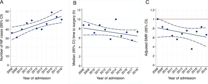
Who gets it. Most cases occur in diabetics and the immunocompromised, though Type II GAS disease can occur in healthy adults:
- Diabetes mellitus - the major risk factor: impaired immunity and vascular disease
- Immunocompromise - HIV, chemotherapy, steroids, transplantation
- Alcohol excess - liver disease, malnutrition, poor wound care
- Breaks in the skin - surgical wounds, injection sites, trauma
- Extremes of age - the very young and the elderly
- Tissue ischaemia - peripheral vascular disease, vasculitis, pressure sores
- Intravenous drug use - contaminated needles, injection-site infection
- Chronic illness - renal failure, malignancy, malnutrition
History. Joseph Jones, a Confederate surgeon, first described the disease in 1871 during the American Civil War, and Wilson coined the term "necrotising fasciitis" in 1952. It was previously known as "hospital gangrene" and "streptococcal gangrene", and it remains associated with military trauma, natural disasters and epidemics of intravenous drug use.
Necrotising fasciitis is an orthopaedic oral examination favourite because it tests five things at once:
- Recognition of a surgical emergency from clinical signs
- Understanding of soft tissue anatomy and fascial planes
- Knowledge of microbiology and antibiotic selection
- Surgical decision-making: when to operate, when to return
- The ethical aspects of life-threatening disease and limb sacrifice
The examiner wants to hear: "Pain out of proportion = NF until proven otherwise. When in doubt, cut it out."
Pathophysiology
Why it spreads along the fascia. The fascia is relatively avascular, so few immune cells reach the infection. Bacterial enzymes (hyaluronidase, lipases, collagenases) digest the connective tissue, and thrombosis of the perforating vessels starves the skin above, which is the ischaemia behind pain out of proportion to what is visible. Toxin production drives the systemic toxicity and shock. Because necrosis advances along the plane so quickly, a deceptively small area of involvement at presentation may become extensive within hours if it is not debrided.
Type I, polymicrobial. Aerobes and anaerobes infect synergistically: the aerobes consume oxygen, the anaerobes thrive in the hypoxia that results and produce gas (crepitus), and enzymes from several organisms accelerate the destruction. It typically arises in compromised tissue: diabetes, peripheral vascular disease, a surgical wound.
Type II, monomicrobial group A Streptococcus. GAS carries several virulence factors:
- Streptolysin O and S - pore-forming toxins that lyse cells
- Streptococcal pyrogenic exotoxins (SPE-A, B, C) - superantigens that trigger massive cytokine release
- M-protein - antiphagocytic, promotes adherence
- Streptokinase - activates plasminogen and promotes spread
Streptococcal toxic shock syndrome (STSS). A superantigen activates 20-30% of T-cells, against 0.01% in a normal antigen response, and the cytokine storm that follows is STSS.
Type III, clostridial gas gangrene. Clostridium perfringens produces alpha-toxin, a lecithinase that lyses cell membranes and causes massive tissue necrosis. Gas (CO2, H2) forms rapidly and gives the crepitus, and the course is so fast that it can be fatal within hours.
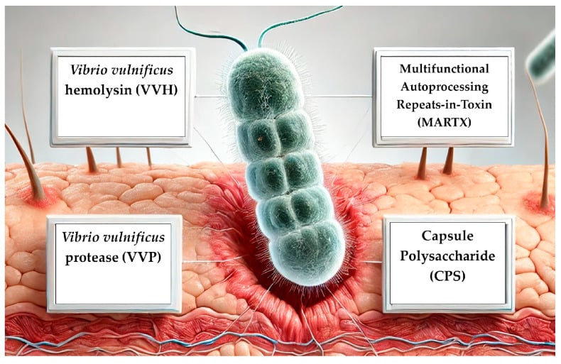
Vibrio vulnificus. A Gram-negative rod of warm seawater that enters through minor wounds while handling seafood or swimming, and produces cytolysins and proteases. It is particularly severe in patients with liver disease or haemochromatosis, iron overload feeding bacterial growth, and septicaemic infection carries a mortality of 50-60%.
The two common types differ in who they affect, how they present and how they are treated:
- Type I (Polymicrobial)
- Mixed aerobic + anaerobic (Streptococcus, Enterococcus, E. coli, Bacteroides, Peptostreptococcus)
- Type II (Monomicrobial)
- Single organism: GAS (Strep pyogenes), S. aureus (MRSA), less commonly Clostridium
- Type I (Polymicrobial)
- Elderly, diabetics, immunocompromised, post-surgical, perineal (Fournier's)
- Type II (Monomicrobial)
- Previously healthy adults, children, minor trauma or NSAID use
- Type I (Polymicrobial)
- Surgery, perianal abscess, decubitus ulcer, diabetic foot
- Type II (Monomicrobial)
- Minor skin break, insect bite, blunt trauma, varicella (children)
- Type I (Polymicrobial)
- Common (anaerobic organisms)
- Type II (Monomicrobial)
- Less common (unless clostridial superinfection)
- Type I (Polymicrobial)
- Less common
- Type II (Monomicrobial)
- Very common with GAS - high mortality
- Type I (Polymicrobial)
- Broad-spectrum: Piperacillin-tazobactam + Clindamycin ± Vancomycin
- Type II (Monomicrobial)
- Penicillin + Clindamycin (GAS); Vancomycin + Clindamycin (MRSA)
- Type I (Polymicrobial)
- 15-30%
- Type II (Monomicrobial)
- 30-50% (higher with TSS)
Clinical Presentation
Early necrotising fasciitis mimics cellulitis. The diagnosis lies in the discordance between what the patient reports, which is severe, and what you can see, which is relatively little.
History
Pain out of proportion is the cardinal feature. The pain seems excessive for the appearance, extends beyond the visible erythema and responds poorly to analgesia. It may paradoxically improve as the nerves themselves necrose, which is an ominous sign rather than a reassuring one.
Rapid progression. "It wasn't like this four hours ago": erythema spreading despite antibiotics, blisters appearing over hours.
Systemic symptoms. Fever, rigors and sweating; nausea, vomiting and diarrhoea; confusion or altered consciousness; and the patient who tells you they feel they are going to die.
Precipitants. Ask about:
- Minor trauma - a scratch, insect bite or abrasion
- Recent surgery - abdominal, perineal or orthopaedic
- Injection - intravenous drug use or an intramuscular injection
- Perianal abscess or pilonidal disease
- Childbirth or episiotomy
- Varicella in children
- NSAID use, which may mask the early symptoms (below)
The risk-factor lists name NSAIDs and varicella, but the reason they matter is a recurring viva discussion point that is worth holding clearly:
- NSAIDs mask the diagnosis. By blunting fever, pain and the inflammatory response, NSAIDs can make early necrotising fasciitis look like a settling cellulitis - delaying recognition and surgery (the one modifiable mortality driver). A child or young adult on regular NSAIDs whose "cellulitis" is not improving deserves heightened, not lowered, suspicion.
- The proposed biological harm (controversial). Beyond masking, NSAIDs have been hypothesised to worsen invasive group A streptococcal disease - by impairing neutrophil function and possibly up-regulating cytokine/superantigen responses. The evidence is observational and confounded (sicker children get more analgesia), so causation is unproven, but the association is strong enough that avoiding NSAIDs in suspected invasive GAS infection is widely advised.
- Varicella in children is the classic precipitant for paediatric Type II (GAS) necrotising fasciitis - the chickenpox lesions are the skin breach. A febrile, disproportionately painful, rapidly worsening lesion in a child with varicella is necrotising fasciitis until proven otherwise.
Physical Examination
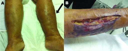
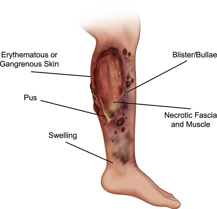
Hard signs are 100% specific. Any one of them mandates immediate surgery:
- Crepitus - gas in the tissues, felt as a crackling, best at the periphery of the infection; may be absent in Type II (GAS)
- Skin necrosis - dusky, purplish, non-blanching skin, with loss of sensation as the cutaneous nerves die
- Bullae - haemorrhagic bullae are the more ominous, serous ones still concerning; both mean the dermis has separated from the epidermis through vascular thrombosis
- Dishwater discharge - thin, grey, foul-smelling fluid, "washing-up water", seen where the skin breaks down spontaneously or on aspiration
- Visible fascial necrosis - grey, non-viable fascia seen through a break in the skin
Soft signs demand high clinical suspicion, and several together should prompt surgical exploration:
- Pain out of proportion, as in the history, with pain extending beyond the erythema
- Rapidly spreading erythema - mark the border with a pen and look again in 1-2 hours; spread despite intravenous antibiotics
- Systemic toxicity - heart rate above 100, systolic pressure below 90, temperature above 38.5°C or below 36°C, altered mental state
- Failure to respond to antibiotics - no improvement, or worsening, after 24-48 hours of appropriate intravenous antibiotics; this should prompt urgent surgical review
- Skin changes beyond cellulitis - woody induration, oedema beyond the erythema, vesicles (early bullae)
Where it strikes. The common sites, and what each brings with it:
- Lower limb - the most common site overall, often in diabetics with foot ulcers; can track to the pelvis along the fascial planes
- Upper limb - often after intravenous drug use or minor trauma; can spread rapidly to the chest wall and may present as compartment syndrome
- Perineum (Fournier's gangrene) - male predominance 10:1; from a perianal abscess, urethral instrumentation or a scrotal abscess; can spread to the abdominal wall and thighs; mortality 40-50%
- Abdominal wall - after surgery (especially bowel surgery) or trauma; may track into the retroperitoneum
- Head and neck (cervical NF) - from dental infection (progression of Ludwig's angina) or a pharyngeal abscess; airway compromise is the primary threat, and it can spread to the mediastinum
Perineal/genital necrotising fasciitis (Fournier's gangrene) is listed above as a site, but it is examined as a distinct entity with its own scoring and source-control rules:
- Severity scoring: the Fournier's Gangrene Severity Index (FGSI) uses derangement of physiological variables (temperature, heart rate, respiratory rate, sodium, potassium, creatinine, bicarbonate, haematocrit, white cell count) to predict mortality - a higher score (commonly quoted threshold around 9) carries markedly worse survival. It is a prognostic adjunct, not a reason to delay surgery.
- Find and control the source: most cases are colorectal, urogenital, or cutaneous in origin. Source control may require a diverting colostomy (for a perianal/colorectal source or heavy faecal soiling of the wound) and a suprapubic catheter (for a urethral source) in addition to perineal debridement.
- The testes are usually spared: the testicular blood supply (gonadal vessels) is separate from the compromised perineal/scrotal fascial circulation, so the testes typically survive even after radical scrotal debridement - orchidectomy is rarely needed, and exposed testes can be temporarily housed in thigh pockets pending reconstruction.
- Multidisciplinary: urology, colorectal/general surgery and plastics are usually all involved; reconstruction follows infection control (see Wound Reconstruction).
Differential Diagnosis
- Distinguishing features
- Erythema, warmth, well/ill-defined margin; systemically less unwell; responds to antibiotics
- Key discriminator from NF
- Pain proportionate to signs; no crepitus, bullae or dishwater pus; improves on antibiotics
- Distinguishing features
- Localised fluctuant collection; point tenderness
- Key discriminator from NF
- Drains pus, not dishwater fluid; no spreading fascial necrosis
- Distinguishing features
- Severe pain, crepitus, bronze skin, marked toxaemia
- Key discriminator from NF
- Primarily muscle (not fascia) necrosis - overlaps with Type III NF; same emergency management
- Distinguishing features
- Deep muscle abscess, often tropical; fever, localised muscle pain
- Key discriminator from NF
- Muscle-centred on imaging; lacks fascial 'dishwater' separation
- Distinguishing features
- Pain on passive stretch, tense compartment, post-injury/ischaemia
- Key discriminator from NF
- No systemic sepsis or skin necrosis; normal inflammatory markers
- Distinguishing features
- Unilateral swelling, calf tenderness; afebrile
- Key discriminator from NF
- No toxaemia, crepitus or skin necrosis; confirmed on Doppler
- Distinguishing features
- Hot swollen joint, monoarticular, raised urate/crystals
- Key discriminator from NF
- Joint-centred; no fascial necrosis; rapid response to anti-inflammatories
Investigations
When clinical suspicion is high, no investigation should delay surgery. A patient with hard signs goes directly to theatre, and CT or MRI may be normal early in the disease. The operating theatre is both diagnostic and therapeutic: if the fascia is necrotic and separates easily, the diagnosis is confirmed.
Laboratory Investigations
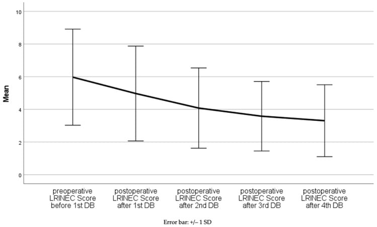
LRINEC. The Laboratory Risk Indicator for NECrotizing fasciitis scores six routine blood results:
- Range
- Less than 150
- Points
- 0
- Range
- ≥150
- Points
- +4
- Range
- Less than 15
- Points
- 0
- Range
- 15-25
- Points
- +1
- Range
- Greater than 25
- Points
- +2
- Range
- Greater than 135
- Points
- 0
- Range
- 110-135
- Points
- +1
- Range
- Less than 110
- Points
- +2
- Range
- ≥135
- Points
- 0
- Range
- Less than 135
- Points
- +2
- Range
- ≤141
- Points
- 0
- Range
- Greater than 141
- Points
- +2
- Range
- ≤10
- Points
- 0
- Range
- Greater than 10
- Points
- +1
The total is read in three bands:
- Below 6 - low risk (positive predictive value below 50%)
- 6-7 - intermediate risk; keep clinical vigilance high
- 8 or more - high risk (positive predictive value 75% or more); strongly consider surgery
The limitation. LRINEC was developed retrospectively and its sensitivity is only 60-80%, so a normal score does not exclude necrotising fasciitis. Clinical judgement remains paramount. [4]
Other blood tests. Each answers a different question:
- Blood cultures - positive in 20-50%, and identify the organism
- Lactate - raised lactate means tissue hypoperfusion and a worse prognosis
- Creatine kinase - raised if there is myonecrosis, which carries a worse prognosis
- Procalcitonin - may help separate bacterial infection from other causes
- Coagulation - PT/INR, APTT, fibrinogen and D-dimer, because DIC is common
- Arterial blood gas - a metabolic acidosis indicates severe sepsis
Imaging
Plain radiograph. A quick screen for gas. Gas tracking along the fascial planes is pathognomonic but present in only 25-50%: more often in Type I (polymicrobial) and Type III (clostridial), and it may be absent in Type II (GAS). Sensitivity is low, and the absence of gas does not exclude the diagnosis.
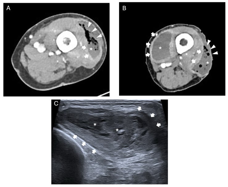
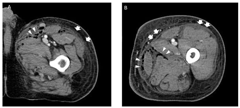
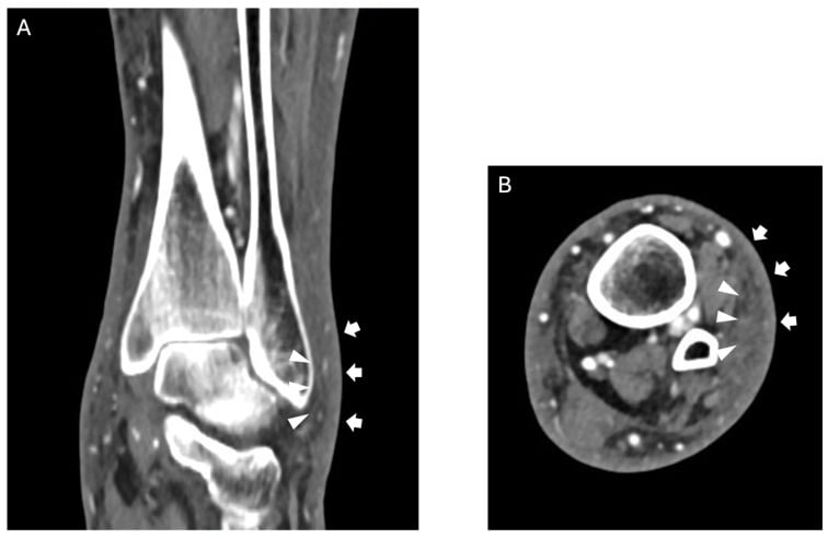
CT. The most useful modality if time permits. It is quick to acquire, shows the extent of disease for surgical planning, and picks up gas that a radiograph misses. The findings:
- Gas tracking along the fascial planes (specific, but not always present)
- Fascial thickening and enhancement
- Fluid collections along the fascia
- Non-enhancement of the fascia, which indicates necrosis
- The extent of soft tissue involvement
A normal CT does not exclude necrotising fasciitis.
MRI. The most sensitive modality, and rarely indicated acutely. It takes 30-60 minutes, may not be available after hours, and is reserved for the equivocal case where there is time. The findings:
- T2 hyperintensity along the fascia (oedema)
- Fascial thickening greater than 3mm
- Non-enhancement with gadolinium (necrosis)
- Fluid tracking along the fascial planes
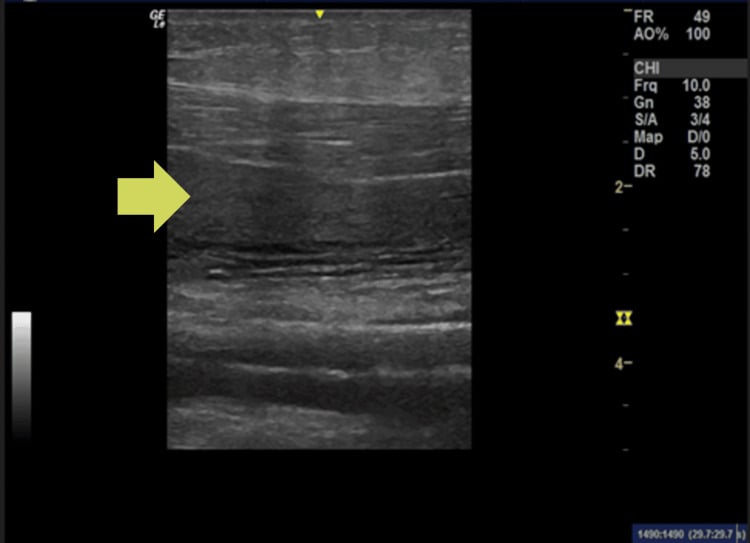
Ultrasound. A bedside tool of limited use: available at the bedside, no radiation, and it can guide aspiration, but it is operator dependent, of limited sensitivity, and cannot assess the deep fascial planes. The findings:
- Subcutaneous thickening
- Fascial thickening greater than 4mm
- Fluid along the fascial planes
- Hyperechoic foci with dirty shadowing (gas)
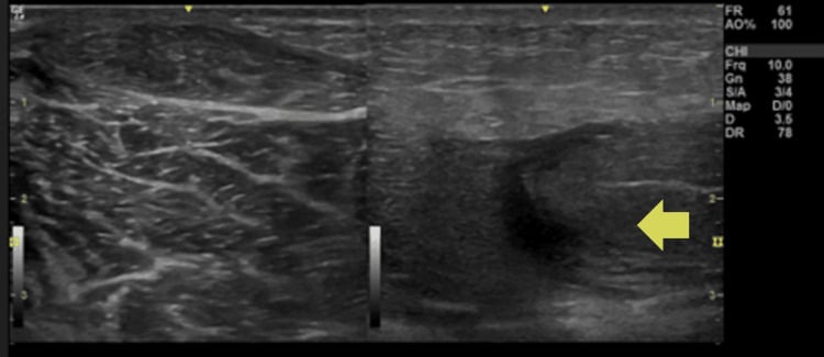
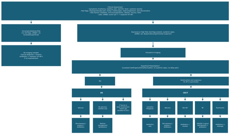
The Finger Test (Bedside Diagnosis)
When. Clinical suspicion is high but the diagnosis is uncertain. It is done under local anaesthesia at the bedside or in theatre.
How. Make a 2cm incision through skin and subcutaneous tissue down to the fascia, and look for:
- Lack of bleeding, because the vessels are thrombosed
- "Dishwater" grey pus, which is pathognomonic
- Necrotic fascia - grey and stringy, separating easily on finger dissection
- A positive finger test - the finger passes along the fascial plane with no resistance
If positive, proceed immediately to extensive debridement.
Management Principles
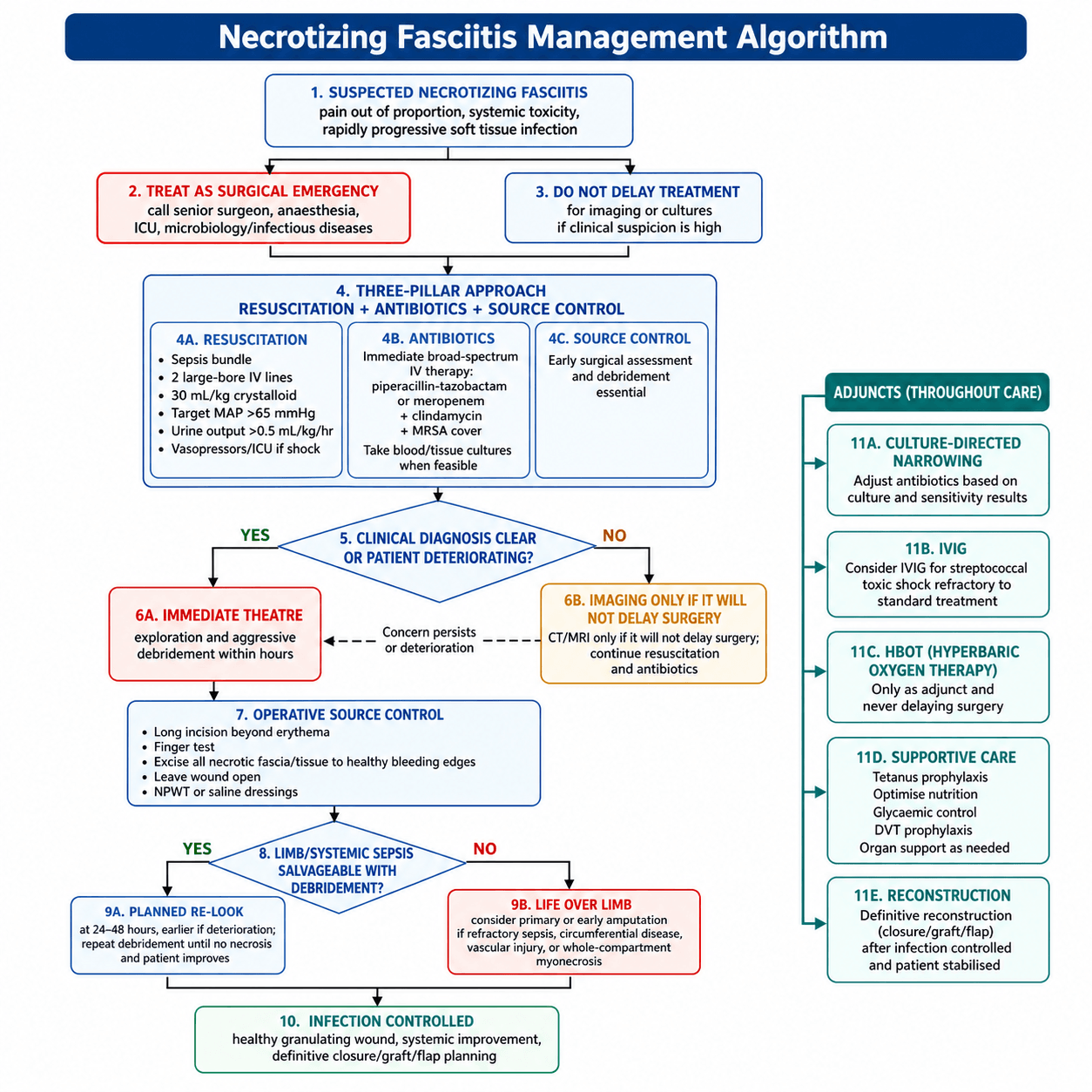
Three pillars, run at once. Resuscitation (fluids, vasopressors, intensive care), debridement (early, extensive and repeated) and antibiotics (empirical and broad-spectrum, with clindamycin for toxin inhibition). None waits for the others.
Recognise the sepsis. Apply the qSOFA/SOFA criteria. Most patients meet the criteria for sepsis, and many have septic shock requiring vasopressors.
Fluids. Large-bore intravenous access, ideally two cannulae, then a crystalloid bolus of 30 mL/kg in the first 3 hours, titrated to a mean arterial pressure above 65 mmHg and a urine output above 0.5 mL/kg/hour. Central venous access is placed for monitoring and for vasopressors.
Vasopressors. Noradrenaline is first-line if the patient remains hypotensive after fluids, to the same MAP target of 65 mmHg. More than one agent may be needed.
Intensive care. Every patient needs ICU or HDU admission for ongoing resuscitation, monitoring and the repeated trips to theatre, and may need renal replacement therapy or mechanical ventilation.
Coagulopathy. DIC is common with GAS (Type II). Transfuse blood products as needed, with FFP, platelets and cryoprecipitate for active bleeding.
Surgical Debridement
Surgery is both diagnostic and therapeutic, and it is the definitive treatment: the infection cannot be controlled with antibiotics alone, and no amount of them substitutes for adequate debridement of necrotic tissue.
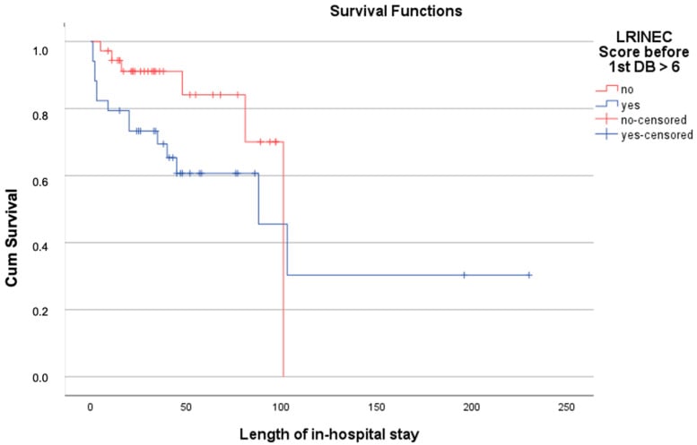
Timing. Operate as soon as the patient is resuscitated enough for anaesthesia, within hours rather than days, and do not wait for imaging when the clinical diagnosis is clear. Delay to debridement is the strongest modifiable predictor of death: in McHenry's series the mean time from admission to surgery was 90 hours in non-survivors against 25 hours in survivors, and Kobayashi found that delay beyond 12 hours markedly increased the number of debridements, septic shock and acute kidney injury.
Principles. The debridement is aggressive and extensive: all necrotic tissue is removed, and since healthy tissue bleeds, you debride until you see bleeding edges. Add fasciotomies if compartment syndrome is suspected.
Step by step.
- Incise longitudinally over the area of maximal involvement, extending beyond the visible erythema into healthy tissue
- Assess the fascia with the finger test: necrotic fascia is grey and stringy and separates from the underlying muscle with finger dissection, while healthy fascia is glistening white and adherent
- Debride the fascia, excising everything that offers no resistance until it bleeds and is adherent, extending the incisions to follow the necrosis
- Assess the muscle: healthy muscle is pink, contracts to stimulation and bleeds when cut; necrotic muscle is dull, non-contractile and does not bleed, and is debrided (myonecrosis means a worse prognosis)
- Assess the skin: skin that is viable now may necrose over the next 24-48 hours, so preserve what is viable (it can be excised at the re-look) and excise only what is overtly dead
- Leave the wound open, under saline-soaked dressings or NPWT, and plan the return to theatre
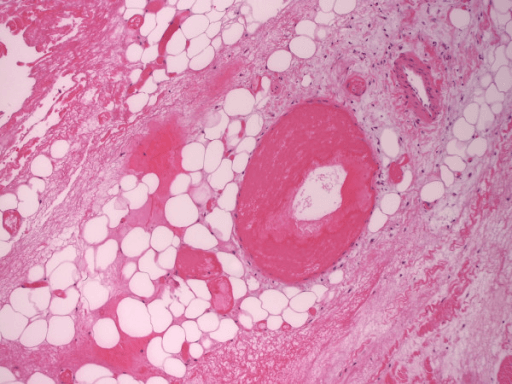
Complications and Prognosis
Complications
Early. The complications of the acute illness:
- Septic shock - multi-organ failure, DIC, death
- ARDS
- Acute kidney injury - from sepsis and rhabdomyolysis
- Limb loss - amputation is required in 15-20%
- Exsanguinating haemorrhage - from eroded vessels
- Toxic shock syndrome - especially in Type II (GAS)
Late. The survivor is left with:
- Chronic wounds - prolonged healing, skin grafts
- Scarring and contractures - which may limit function
- Chronic pain - phantom limb pain after amputation
- Psychological morbidity - PTSD, depression, body image
- Functional impairment - particularly after amputation
Prognostic Factors
- Relative Risk
- Strongest modifiable risk
- Comments
- McHenry; Kobayashi - the figures are under Surgical Debridement, Timing
- Relative Risk
- 2x mortality
- Comments
- Recognition and surgical referral delays
- Relative Risk
- 2-3x mortality
- Comments
- Reduced physiological reserve
- Relative Risk
- 1.5-2x mortality
- Comments
- Immunocompromise, vascular disease
- Relative Risk
- 30-70% mortality
- Comments
- Cytokine storm, DIC
- Relative Risk
- Higher mortality
- Comments
- Harder to debride, vital structures
- Relative Risk
- Poor prognosis
- Comments
- Extremes of immune response
- Relative Risk
- 2x mortality
- Comments
- Marker of severe sepsis
- Relative Risk
- 2x mortality
- Comments
- Must debride to healthy tissue
Survival Outcomes
Mortality. In the pre-antibiotic era it was 70-80%. With modern treatment it is 20-40% overall, falling to 10-25% with early recognition and aggressive surgery, and 50-70% when treatment is delayed.
What improves survival.
- Recognition within 24 hours of the onset of symptoms
- Surgery within 6-8 hours of presentation
- Adequate debridement, with multiple re-looks
- Intensive care with multi-organ support
- Clindamycin in the antibiotic regimen
- IVIG in streptococcal toxic shock
Guidelines, Registries & Global Practice
Global Epidemiology
Necrotising fasciitis is uncommon but its incidence appears to be rising worldwide. Reported population incidence is approximately 0.4-1.0 per 100,000 per year in high-income countries, with overall mortality of 20-40% even in modern series. The McHenry single-centre series reported 29% mortality and identified delay to debridement as the dominant determinant of death PMID 7748037, while the Stevens & Bryant NEJM review summarises the global microbiology and management of necrotising soft-tissue infections PMID 29211672.
- Predominant pattern
- Type I polymicrobial in diabetics/elderly; Type II GAS in healthy adults
- Notable organisms / risks
- Rising invasive GAS; MRSA in some regions
- Predominant pattern
- Marine-acquired NSTI
- Notable organisms / risks
- Vibrio vulnificus (warm seawater, liver disease); Aeromonas (freshwater)
- Predominant pattern
- Type I polymicrobial; high diabetes prevalence
- Notable organisms / risks
- Klebsiella, Vibrio, marine exposure
- Predominant pattern
- Late presentation, higher mortality
- Notable organisms / risks
- Delayed access to theatre/ICU is the key driver
Guideline Comparison
There is broad international consensus on the core principles - early surgical debridement, broad-spectrum antibiotics with clindamycin for toxin suppression, and aggressive resuscitation - although no high-quality randomised data exist for the surgical strategy itself.
- Position on diagnosis & timing
- Surgery is diagnostic and therapeutic; do not delay debridement for imaging; LRINEC supportive not exclusionary
- Antibiotic & adjunct guidance
- Broad-spectrum empirical cover + clindamycin; IVIG may be considered in STSS
- Position on diagnosis & timing
- Urgent surgical exploration when NSTI suspected; tissue Gram stain/culture
- Antibiotic & adjunct guidance
- Empirical broad-spectrum + clindamycin for GAS; narrow on culture
- Position on diagnosis & timing
- Recognise sepsis early, immediate senior surgical referral, no delay for imaging
- Antibiotic & adjunct guidance
- Local microbiology-guided broad-spectrum + clindamycin; sepsis-six resuscitation
- Position on diagnosis & timing
- High clinical suspicion mandates exploration; LRINEC adjunctive only
- Antibiotic & adjunct guidance
- Piperacillin-tazobactam or meropenem + clindamycin ± vancomycin; benzylpenicillin + clindamycin for confirmed GAS
Almost all guideline recommendations on the surgical management of NSTI are based on observational data and expert consensus (low-grade evidence) - there are no RCTs of debridement timing or strategy. The strongest comparative evidence in this field is the Fernando 2019 meta-analysis on diagnostic accuracy PMID 29672405 and the Darenberg 2003 RCT of IVIG in STSS PMID 12884156. No dedicated NF registry exists; epidemiology is drawn from national infection surveillance (e.g. invasive GAS programmes) and single-centre cohorts.
Access, Equity & Wound-Healing Considerations
Globally, determinants of NF outcome extend beyond the pathogen and the operating theatre:
- Rural and remote access: In many countries patients first present to facilities without on-site ICU or surgical capability and require urgent transfer to a definitive centre; transfer time is itself a contributor to debridement delay and mortality.
- Disadvantaged and Indigenous populations: Worldwide, groups with higher rates of diabetes, chronic kidney disease and skin infection (including Indigenous populations) carry elevated NF risk; renal dosing of antibiotics, family involvement and culturally safe wound care should inform management.
- Smoking cessation: Smoking impairs graft take and wound healing, so supporting cessation (behavioural support plus nicotine replacement therapy) is important for reconstructive success in survivors.
Viva Practice Scenarios
Practise clinical reasoning and management decisions out loud
“A 55-year-old diabetic man presents with a painful, swollen right leg. He had a minor cut 3 days ago. He appears unwell with HR 120, BP 90/60, T 39.2°C. There is erythema from mid-calf to mid-thigh with woody induration. He has severe pain despite IV morphine. How would you manage this patient?”
“You are in theatre and have made an incision over the affected area. Describe what findings would confirm necrotizing fasciitis and your operative approach.”
“A previously healthy 35-year-old woman presents with rapidly progressive leg pain and redness 2 days after a minor skin abrasion. Her LRINEC score is 4. What are your concerns and how would you proceed?”
Classification
- Type I: Polymicrobial (aerobic + anaerobic), diabetics, post-surgical, Fournier's
- Type II: Monomicrobial (GAS, S. aureus), healthy adults, minor trauma
- Type III: Clostridial (gas gangrene), Vibrio vulnificus (seawater)
Clinical Features
- PAIN OUT OF PROPORTION - cardinal early sign
- Hard signs: Crepitus, necrosis, bullae, dishwater pus - immediate surgery
- Soft signs: Disproportionate pain, rapid spread, systemic toxicity, antibiotic failure
- Woody induration - tense swelling beyond erythema
LRINEC Score
- CRP greater than 150 (+4), WCC greater than 25 (+2), Hb less than 110 (+2)
- Na less than 135 (+2), Creatinine greater than 141 (+2), Glucose greater than 10 (+1)
- Score ≥6 intermediate risk, ≥8 high risk
- CRITICAL: Sensitivity only 60-80% - clinical judgement trumps score
Finger Test
- Bedside incision to fascia under local anaesthetic
- Lack of bleeding indicates vessel thrombosis
- Dishwater gray pus is pathognomonic
- Necrotic fascia separates easily with finger dissection
- If positive - proceed to extensive debridement
Antibiotics
- Empiric: Piperacillin-tazobactam + Clindamycin + Vancomycin
- GAS: Benzylpenicillin 2.4g IV q4h + Clindamycin 900mg IV q8h
- ALWAYS include clindamycin - inhibits toxin production (Eagle effect)
- Vibrio: Doxycycline + Ceftriaxone
Surgical Principles
- Time is tissue - delay to debridement drives mortality (90h vs 25h, McHenry)
- Debride until it bleeds - necrotic tissue does not bleed
- Healthy fascia is adherent - necrotic fascia separates easily
- Plan re-look at 24-48 hours - most need 2-4 debridements
- Life over limb - amputate if refractory sepsis
Prognosis
- Overall mortality 20-40%, up to 70% if delayed
- Delay to debridement is the strongest modifiable mortality driver
- GAS toxic shock - 30-70% mortality
- Amputation required in 15-20%
Key Exam Phrases
- Pain out of proportion = NF until proven otherwise
- When in doubt, cut it out
- The operating theatre is both diagnostic and therapeutic
- Surgery should not be delayed for imaging
- Clindamycin inhibits toxin production regardless of bacterial growth phase
Evidence Base
LRINEC Score Development
- Retrospective derivation/validation (145 NF vs 309 severe cellulitis/abscess)
- Six routine variables: CRP, WCC, Hb, sodium, creatinine, glucose
- Cut-off of 6 gave PPV 92.0% and NPV 96.0% in this cohort
- AUC 0.98 (developmental) and 0.976 (validation)
LRINEC, Exam & Imaging Accuracy (Meta-analysis)
- Systematic review/meta-analysis: 23 studies, n=5982
- LRINEC ≥6 pooled sensitivity only 68.2%, specificity 84.8%
- LRINEC ≥8 sensitivity 40.8%, specificity 94.9%
- CT sensitivity 88.5%/specificity 93.3%; plain film sensitivity 48.9%
A LRINEC of Zero Does Not Exclude NF
- Case report of a 37-year-old man with surgically confirmed NF
- LRINEC score was 0 despite established disease
- Illustrates the danger of using LRINEC to rule out NF
- Reinforces that NF remains a clinical diagnosis
Time to Surgery and Outcome
- 65 patients with necrotising soft-tissue infection
- Mean time admission-to-operation 90 h in non-survivors vs 25 h in survivors (p=0.0002)
- Average 3.3 debridements per patient; overall mortality 29%
- Early debridement was the key modifiable determinant of survival
Delay Increases Debridements & Morbidity
- Retrospective cohort of 47 NSTI patients (overall mortality 17%)
- Surgery delayed beyond 12 h required far more debridements (7.4 vs 2.3, p<0.001)
- Delay beyond 12 h associated with more septic shock and acute kidney injury
- Effect persisted after adjustment for confounders
Clindamycin Suppresses Streptococcal Toxins
- In vitro study of 14 fully susceptible S. pyogenes isolates
- Clindamycin superior to penicillin at reducing SPE-A and SPE-B production
- Effect independent of bacterial growth phase (rationale for the Eagle effect)
- Supports adding clindamycin even to penicillin-susceptible GAS
IVIG in Streptococcal Toxic Shock (RCT)
- European randomised, double-blind, placebo-controlled trial in STSS
- Terminated early for slow recruitment (21 patients enrolled)
- 28-day mortality 3.6-fold higher in placebo group (not statistically significant)
- Significant improvement in SOFA score and superantigen neutralisation with IVIG
For the exam, remember:
- LRINEC score has only 68% sensitivity - clinical judgement essential
- Delay to debridement is the key modifiable mortality driver (90h vs 25h, McHenry)
- Clindamycin inhibits toxin production - always include
- IVIG in STSS has one underpowered RCT (Darenberg) - supportive but not definitive
- Hyperbaric oxygen evidence is weak - should not delay surgery
References
-
Sartelli M, Guirao X, Hardcastle TC, et al. 2018 WSES/SIS-E consensus conference: recommendations for the management of skin and soft-tissue infections. World J Emerg Surg. 2018;13:58. doi:10.1186/s13017-018-0219-9
-
Stevens DL, Bryant AE. Necrotizing Soft-Tissue Infections. N Engl J Med. 2017;377(23):2253-2265. doi:10.1056/NEJMra1600673
-
Arif N, Yousfi S, Vinnard C. Deaths from necrotizing fasciitis in the United States, 2003-2013. Epidemiol Infect. 2016;144(6):1338-1344. doi:10.1017/S0950268815002745
-
Wong CH, Khin LW, Heng KS, Tan KC, Low CO. The LRINEC (Laboratory Risk Indicator for Necrotizing Fasciitis) score: a tool for distinguishing necrotizing fasciitis from other soft tissue infections. Crit Care Med. 2004;32(7):1535-1541. doi:10.1097/01.ccm.0000129486.35458.7d
-
Fernando SM, Tran A, Cheng W, et al. Necrotizing Soft Tissue Infection: Diagnostic Accuracy of Physical Examination, Imaging, and LRINEC Score: A Systematic Review and Meta-Analysis. Ann Surg. 2019;269(1):58-65. doi:10.1097/SLA.0000000000002774
-
Wilson MP, Schneir AB. A case of necrotizing fasciitis with a LRINEC score of zero: clinical suspicion should trump scoring systems. J Emerg Med. 2013;44(5):928-931. doi:10.1016/j.jemermed.2012.09.039
-
McHenry CR, Piotrowski JJ, Petrinic D, Malangoni MA. Determinants of mortality for necrotizing soft-tissue infections. Ann Surg. 1995;221(5):558-565. doi:10.1097/00000658-199505000-00013
-
Kobayashi L, Konstantinidis A, Shackelford S, et al. Necrotizing soft tissue infections: delayed surgical treatment is associated with increased number of surgical debridements and morbidity. J Trauma. 2011;71(5):1400-1405. doi:10.1097/TA.0b013e31820db8fd
-
Mascini EM, Jansze M, Schouls LM, Verhoef J, Van Dijk H. Penicillin and clindamycin differentially inhibit the production of pyrogenic exotoxins A and B by group A streptococci. Int J Antimicrob Agents. 2001;18(4):395-398. doi:10.1016/s0924-8579(01)00413-7
-
Darenberg J, Ihendyane N, Sjölin J, et al. Intravenous immunoglobulin G therapy in streptococcal toxic shock syndrome: a European randomized, double-blind, placebo-controlled trial. Clin Infect Dis. 2003;37(3):333-340. doi:10.1086/376630
-
Therapeutic Guidelines Limited. Therapeutic Guidelines: Antibiotic. Melbourne: Therapeutic Guidelines Limited.