Dense sclerosing osteitis with no pus, no sequestrum, no sinus — a diagnosis of exclusion balanced between low-grade infection and sterile autoinflammatory bone disease
- Garré's is defined by NEGATIVES: no abscess, no sequestrum, no sinus, no purulent discharge — only dense cortical thickening and medullary sclerosis.
- It is a descriptive/exclusion diagnosis; the modern working task is to separate genuinely infective sclerosing osteomyelitis from sterile chronic non-bacterial osteomyelitis (CNO/CRMO/SAPHO).
- Malignancy must be excluded before labelling a sclerotic diaphyseal lesion inflammatory — osteosarcoma and Ewing sarcoma both produce sclerosis and periosteal reaction.
- Osteoid osteoma is the single most commonly confused benign mimic: night pain dramatically relieved by NSAIDs, thin-slice CT shows the lucent nidus within the sclerosis.
- Biopsy is for histology PLUS multiple deep tissue samples for extended (14-day) culture including anaerobes, mycobacteria, fungi, and broad-range 16S rRNA PCR.
- When sterile, treat as inflammatory bone disease: NSAIDs first line, bisphosphonate infusion for refractory pain, rheumatology co-management — not repeated debridement.
- Course is relapsing-remitting over years and tends to burn out; the surgical temptation to keep operating causes iatrogenic morbidity.
- “Near-normal inflammatory markers with striking radiographic sclerosis is the classic mismatch that should raise CNO/SAPHO.
- “Smooth, solid, layered periosteal new bone favours a benign/inflammatory process; interrupted, spiculated or Codman triangle patterns demand oncology referral.
- “Whole-body MRI (STIR) is the investigation of choice when multifocal sterile bone disease is suspected — it finds clinically silent lesions and can avoid biopsy of the symptomatic site.
- “Culture-negative does not equal sterile — but repeatedly negative extended cultures plus non-specific chronic inflammatory histology supports CNO.
- “Mandibular disease: exclude odontogenic sepsis, medication-related osteonecrosis of the jaw, and osteosarcoma of the jaw before labelling diffuse sclerosing osteomyelitis.
Spiculated, sunburst, onion-skin interrupted periosteal bone, cortical destruction or a soft-tissue mass are not features of Garré's. Refer to a sarcoma unit for staging and image-guided biopsy through a planned resection corridor. Never biopsy a possible sarcoma in a non-specialist centre.
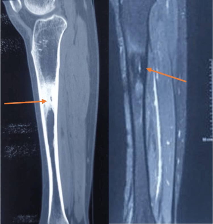
Thin-slice CT (1 mm) through the sclerotic segment: a rounded lucency less than 1.5 cm with central mineralisation is an osteoid osteoma, cured by CT-guided radiofrequency ablation or en-bloc excision. Missing it condemns the patient to years of antibiotics.
Empirical antibiotics before deep biopsy sterilise low-virulence organisms and forfeit the one chance at a culture-directed diagnosis. Take at least 5 separate deep samples with clean instruments for each, plus histology, before any antimicrobial.
Once CNO/CRMO/SAPHO is established, repeated decortication, sequestrectomy or drilling produces temporary relief followed by relapse and progressive bone loss. Escalate medically (NSAIDs, bisphosphonate infusion, DMARD/biologic) with rheumatology.
Definition, Nomenclature and Why the Exam Asks About It
- Setting / usage
- Historic eponym, general orthopaedics
- What it really denotes
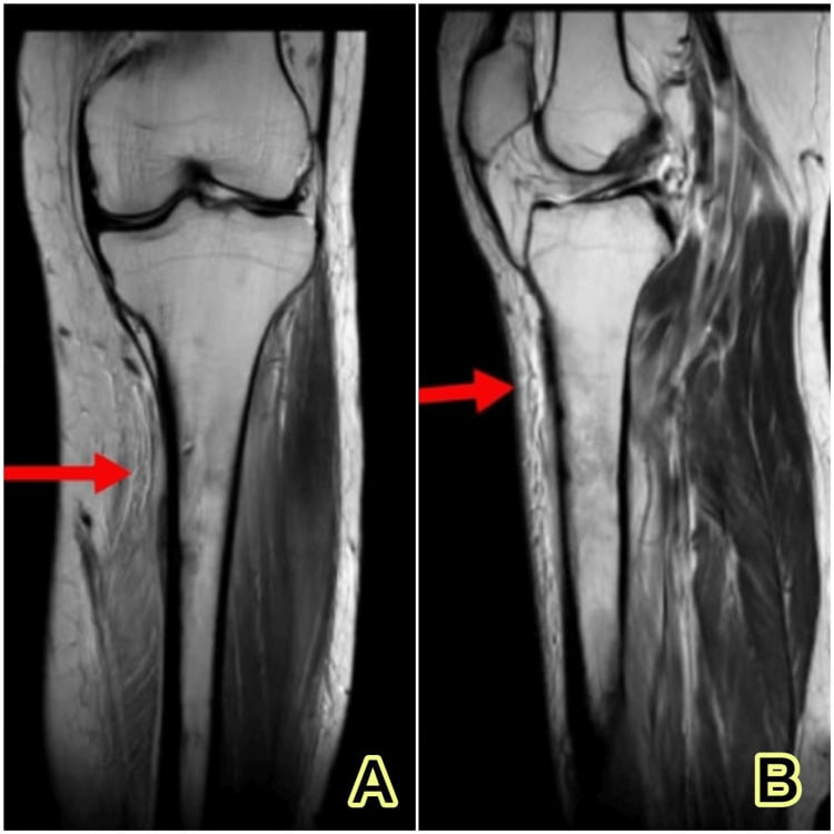
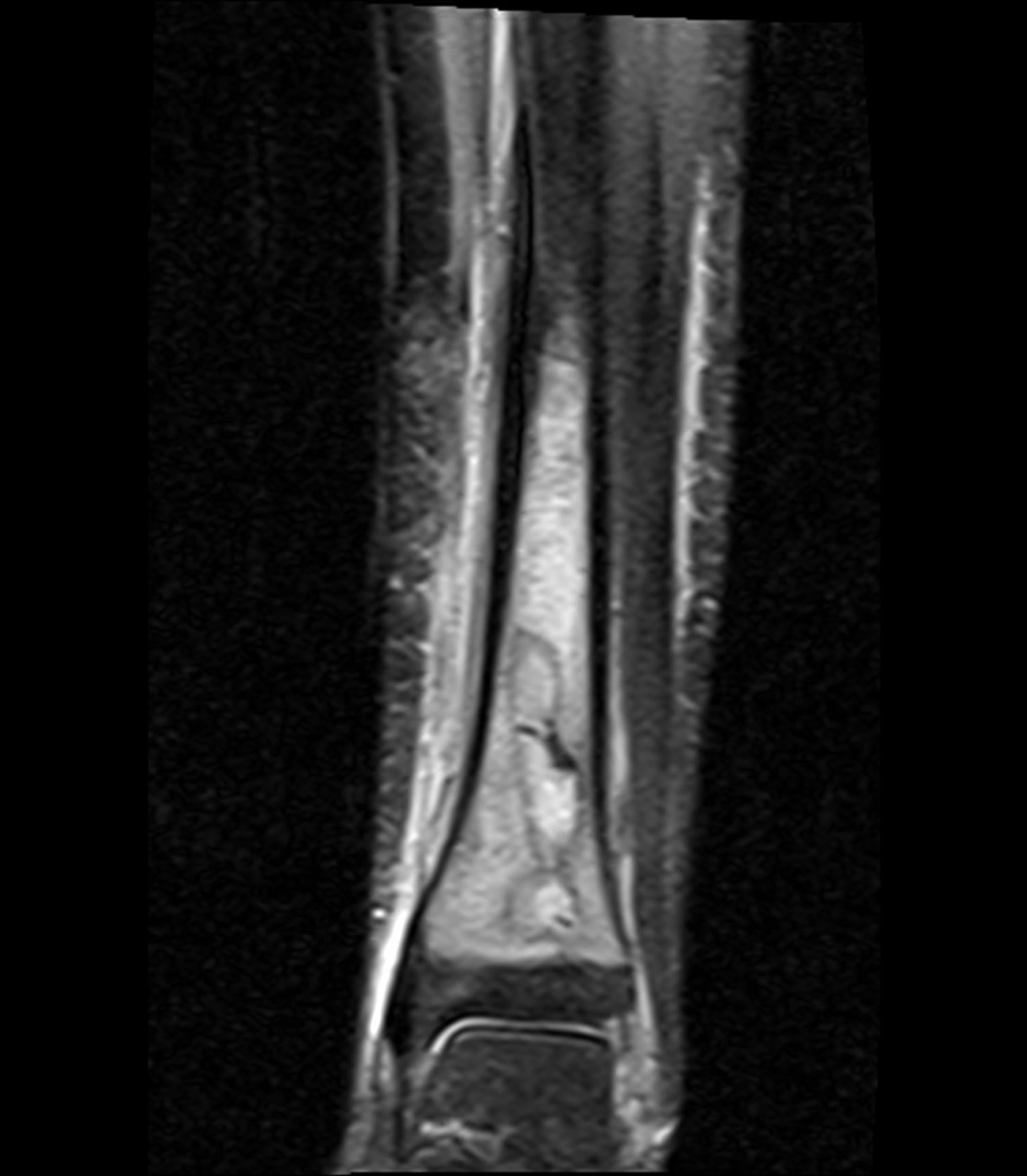
- Non-suppurative sclerosing osteitis: dense cortical thickening, medullary sclerosis with canal narrowing, smooth solid or laminated periosteal new bone, and no abscess, sequestrum, involucrum, cloaca or sinus
- Microbiology
- Undefined by the eponym itself — may be culture-positive or sterile
- Implication for management
- A radiological-clinical phenotype, not a microbiological diagnosis; a diagnosis of exclusion that must always be qualified by culture status
- Setting / usage
- Oral and maxillofacial surgery
- What it really denotes
- Recurrent painful mandibular hyperostosis, often linked to dental sepsis or masticatory overload
- Microbiology
- Frequently culture-negative
- Implication for management
- Dental sepsis control, NSAIDs, bisphosphonates
- Setting / usage
- Paediatric rheumatology
- What it really denotes
- Multifocal sterile autoinflammatory osteitis of childhood
- Microbiology
- Sterile by definition; extended culture negative
- Implication for management
- Whole-body MRI to map lesions; medical therapy — many historic 'Garré long bone' cases belong here
- Setting / usage
- Umbrella term, all ages
- What it really denotes
- Sterile osteitis whether unifocal or multifocal
- Microbiology
- Sterile
- Implication for management
- Preferred contemporary term; replaces loose use of the eponym
- Setting / usage
- Adult rheumatology
- What it really denotes
- Synovitis, acne, pustulosis, hyperostosis, osteitis — the adult equivalent of CRMO
- Microbiology
- Sterile (Cutibacterium acnes association debated)
- Implication for management
- Anterior chest wall hyperostosis is the clue; the skin history is diagnostic gold
- Setting / usage
- Craniofacial and orthopaedic infection literature
- What it really denotes
- Non-suppurative chronic osteitis with no preceding acute phase
- Microbiology
- Typically culture-negative on standard sampling
- Implication for management
- Combined medical-surgical protocol
- Setting / usage
- Assessment
- What it really denotes
- Chronic, low-grade, indolent inflammation with minimal systemic upset; near-normal white cell count and inflammatory markers
- Microbiology
- Requires biopsy with extended culture — imaging alone cannot separate infective from sterile
- Implication for management
- Place the patient in one of three boxes: tumour, infective (culture-positive) sclerosing osteomyelitis, or sterile inflammatory osteitis, because management diverges completely
Carl Garré (1893) described a non-suppurative sclerosing osteitis in which the bone reacted to chronic low-grade irritation with dense periosteal and endosteal new bone rather than with pus and necrosis. The eponym has since been applied loosely, and the fellowship examiner's real question is "what do you actually mean when you say Garré's?"
Defining features (all must be present):
- Chronic, low-grade, indolent bone inflammation
- Dense cortical thickening with medullary sclerosis and canal narrowing
- Smooth solid or laminated periosteal new bone
- Absence of abscess cavity, sequestrum, involucrum, cloaca or draining sinus
- Minimal systemic upset; near-normal white cell count and inflammatory markers
Two distinct modern usages the exam expects untangled:
- Setting / usage
- Historic, general orthopaedics
- What it really denotes
- Descriptive label for dense sclerosing osteitis without sequestrum
- Implication
- Diagnosis of exclusion — must be qualified by culture status
- Setting / usage
- Oral and maxillofacial surgery
- What it really denotes
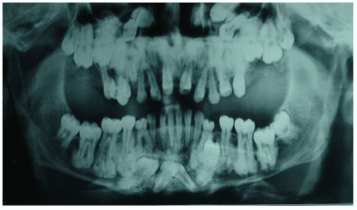
- Recurrent painful mandibular hyperostosis, often linked to dental sepsis or masticatory overload; frequently culture-negative
- Implication
- Managed with dental sepsis control, NSAIDs, bisphosphonates
- Setting / usage
- Paediatric rheumatology
- What it really denotes
- Multifocal sterile autoinflammatory osteitis of childhood
- Implication
- Whole-body MRI; medical therapy; many historic 'Garré long bone' cases belong here
- Setting / usage
- Umbrella term, all ages
- What it really denotes
- Sterile osteitis whether unifocal or multifocal
- Implication
- Preferred contemporary term
- Setting / usage
- Adult rheumatology
- What it really denotes
- Synovitis, acne, pustulosis, hyperostosis, osteitis — adult equivalent of CRMO
- Implication
- Anterior chest wall hyperostosis; skin history is diagnostic gold
- Setting / usage
- Craniofacial/orthopaedic infection literature
- What it really denotes
- Non-suppurative chronic osteitis without preceding acute phase
- Implication
- Combined medical-surgical protocol
The examiner's headline: Garré's is a radiological-clinical phenotype, not a microbiological diagnosis. Your job is to place the patient into one of three boxes — tumour, infective (culture-positive) sclerosing osteomyelitis, or sterile inflammatory osteitis — because management diverges completely.
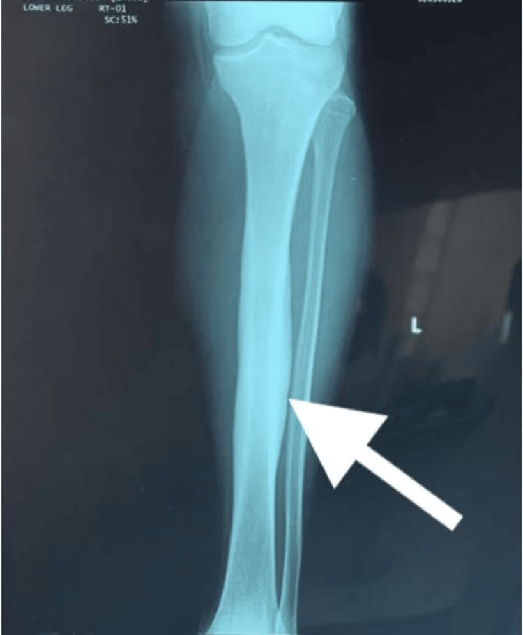
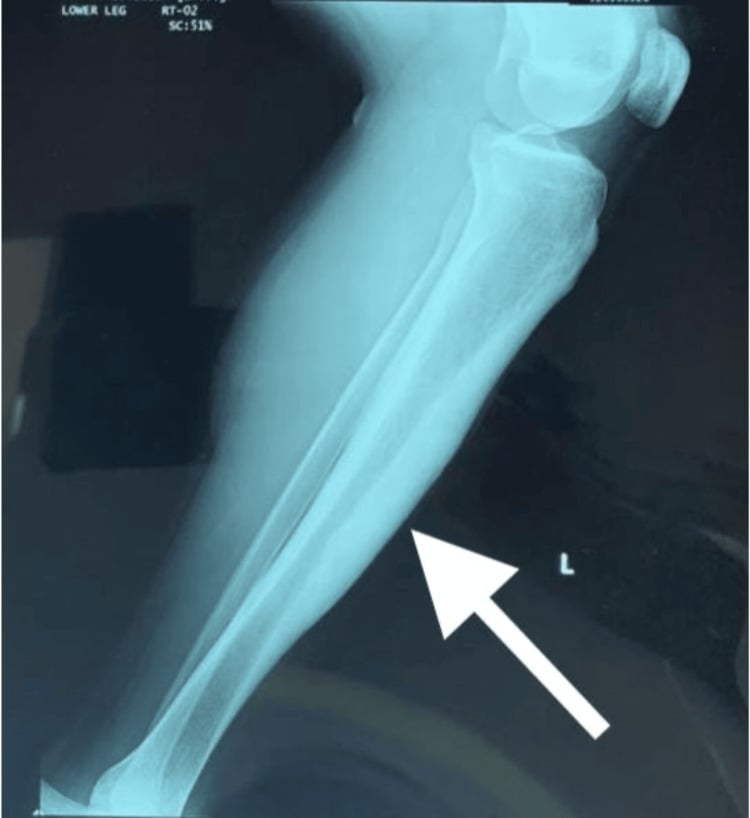
Aetiology and Pathophysiology
- Low-virulence organism in a bone with intact host defences produces a chronic reactive rather than destructive response.
- Organisms classically implicated: Actinomyces species (especially mandible, may follow dental extraction), Cutibacterium (Propionibacterium) acnes, coagulase-negative staphylococci, viridans streptococci, occasionally low-inoculum Staphylococcus aureus.
- Route: contiguous odontogenic spread in the jaw; haematogenous seeding in long bones; rarely post-traumatic.
- Chronic cytokine drive (IL-1, IL-6, TNF, RANKL/OPG shifts) stimulates periosteal and endosteal osteoblastic activity — the bone lays down more bone than it resorbs, giving sclerosis rather than lysis.
- Sclerotic, poorly vascularised bone is a pharmacological sanctuary: antibiotic penetration is poor, explaining the need for prolonged therapy and the tendency to relapse.
Biopsy — Technique and Microbiological Strategy
Biopsy is required in almost every case: to exclude malignancy, to obtain histology, and to attempt organism recovery.
If the lesion has ANY aggressive feature, the biopsy is performed in the sarcoma unit, along a tract that can be excised at definitive resection. An ill-placed tract can convert a limb-salvage case into an amputation.
No antimicrobials for at least 2 weeks before sampling in a clinically stable patient. Low-virulence organisms are easily suppressed below culture threshold.
Use STIR MRI oedema or CT to select the target. Sampling burnt-out sclerotic bone yields fibrosis and no organism.
Open or CT-guided core biopsy through a cortical window. Take at least five separate deep samples using a fresh sterile instrument and blade for each to interpret culture significance. Send tissue, not swabs — swabs are inadequate for bone infection.
Aerobic and anaerobic culture held for 14 days (essential for Cutibacterium and Actinomyces), mycobacterial and fungal culture, Gram stain, plus broad-range 16S rRNA PCR on tissue. Histology in formalin: request assessment for chronic inflammation, granulomas, malignancy; use special stains (Ziehl-Neelsen, PAS, Grocott).
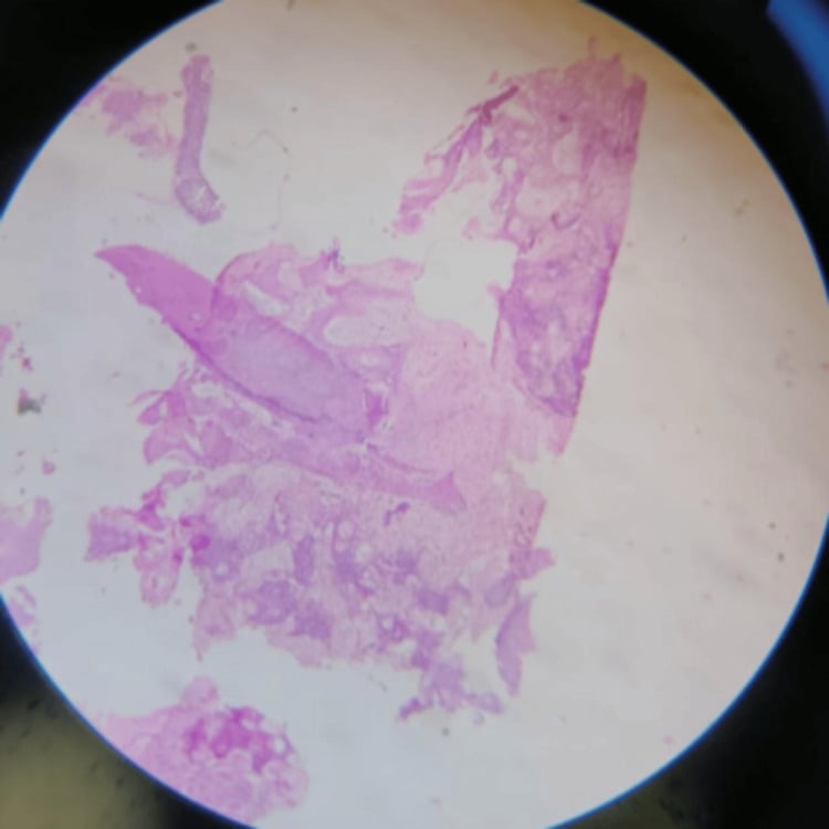
Growth of the same organism in 2 or more independent samples supports true infection. A single low-virulence isolate is usually contamination. Histology in sterile disease shows non-specific chronic inflammation with lymphocytes, plasma cells, fibrosis and prominent reactive new bone — with no organisms, no necrosis and no tumour.
A high proportion of sclerosing osteitis cases are culture-negative even with optimal technique. Repeatedly negative extended cultures and molecular testing, combined with compatible histology, imaging and a relapsing clinical course, constitute the practical case definition of chronic non-bacterial osteomyelitis.
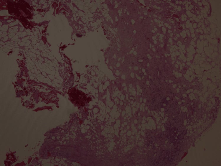
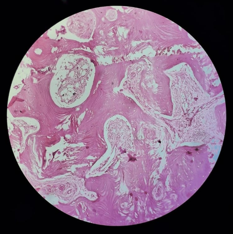
Natural History, Outcomes and Follow-up
- The course is relapsing-remitting over years, with flares of pain and swelling separated by quiescent intervals. Most patients burn out with residual bone thickening and deformity but resolution of pain.
- In children with CRMO, a majority achieve inactive disease over several years; a substantial minority have persistent or relapsing disease into adulthood.
- Long-term morbidity: vertebral compression and kyphosis in spinal CNO, limb-length discrepancy or angular deformity from physeal involvement, jaw deformity and malocclusion in mandibular disease, and chronic pain syndromes.
- Follow-up structure: clinical review with pain scores every 3 months while active; inflammatory markers if previously raised; STIR MRI of symptomatic sites at flares; whole-body MRI at diagnosis and to assess treatment response in multifocal disease; spinal imaging for any back pain; dental review in mandibular disease; bone health monitoring (calcium, vitamin D, DXA) in patients on bisphosphonates.
- Re-biopsy threshold: any change in pain character, rapid growth, new lytic destruction, a soft-tissue mass, or failure to respond as expected.
Clinical Presentation and Examination
The classic vignette: an adolescent or young adult with months of deep, aching, boring pain, notably worse at night and at rest, over the mandible or the diaphysis of the tibia or femur, with a firm, fusiform, non-fluctuant swelling fixed to bone. Symptoms flare and settle over years. There is little or no fever, no malaise, no weight loss, and no sinus.
- Why it matters
- Cyclical flares favour CNO/SAPHO; relentless progression favours tumour
- Why it matters
- Dramatic, reproducible relief within 30 minutes is near-pathognomonic of osteoid osteoma; partial relief is non-specific
- Why it matters
- Establishes SAPHO — often the diagnosis is made in the shoe department, not the operating theatre
- Why it matters
- Spondyloarthropathy/IBD association with CNO
- Why it matters
- Odontogenic source for mandibular disease and actinomycosis
- Why it matters
- Points to secondary chronic osteomyelitis, not Garré's
- Why it matters
- Medication-related or radiation osteonecrosis of the jaw mimics
- Why it matters
- Tuberculous and fungal osteomyelitis can be sclerotic and indolent
- Why it matters
- Red flags — malignancy or mycobacterial disease
- Why it matters
- Monogenic CNO variants
Marked radiographic sclerosis and hyperostosis with a well patient and near-normal CRP/ESR is the classic mismatch that should push you towards chronic non-bacterial osteomyelitis rather than pyogenic infection. Conversely, a very high CRP with sclerosis should make you re-examine for an occult abscess, sequestrum, or malignancy.
Investigation Strategy
- Expected in Garré's/CNO
- Normal or mild anaemia of chronic disease
- Interpretation
- Marked leucocytosis suggests pyogenic infection
- Expected in Garré's/CNO
- Normal to mildly raised
- Interpretation
- Very high values argue against pure sclerosing osteitis
- Expected in Garré's/CNO
- Normal or mildly raised
- Interpretation
- Markedly raised — consider osteosarcoma or Paget disease
- Expected in Garré's/CNO
- Normal
- Interpretation
- Raised in Ewing sarcoma / lymphoma
- Expected in Garré's/CNO
- Negative
- Interpretation
- Positive redirects to haematogenous pyogenic osteomyelitis
- Expected in Garré's/CNO
- Selected cases
- Interpretation
- Supports spondyloarthropathy-associated CNO
- Expected in Garré's/CNO
- If risk factors
- Interpretation
- Skeletal tuberculosis is a great sclerotic mimic
- Expected in Garré's/CNO
- Baseline
- Interpretation
- Required before bisphosphonate infusion
There is no blood test that makes the diagnosis. Normal markers never exclude low-grade infection, and raised markers never confirm it.
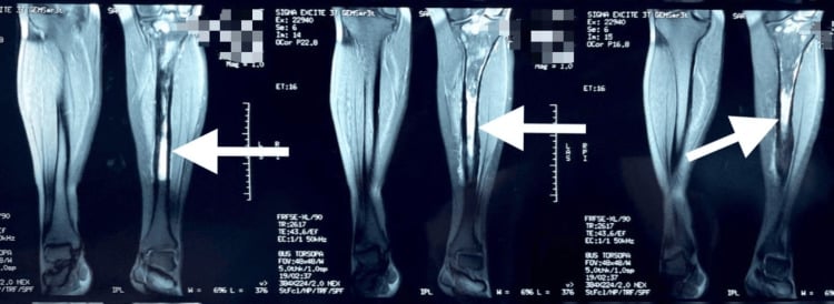
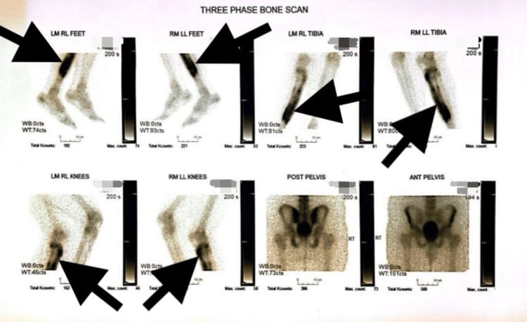
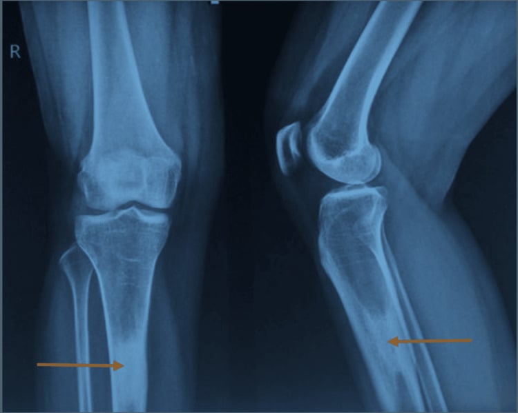
SCLEROSEDRadiographic criteria for Garré's sclerosing osteomyelitis
Hook:A SCLEROSED bone is dense everywhere and empty of dead bone — if you find a sequestrum, a nidus, or a mass, it is not Garré's.
Differential Diagnosis — The Heart of the Topic
- Feature that betrays it
- Progressive unremitting pain, palpable mass, raised ALP and LDH
- Test that settles it
- MRI plus radiographs: cortical destruction, sunburst spiculation, Codman triangle, soft-tissue mass
- Avoid the error by
- Treating any sclerotic lesion with a soft-tissue mass as malignant until proven otherwise; stage before biopsy
- If missed
- Antibiotics and delay while the tumour disseminates; a badly placed biopsy tract converts a limb-salvage case into amputation
- Feature that betrays it
- Fever, raised CRP/ESR, leucocytosis, systemic upset — mimics infection almost exactly
- Test that settles it
- MRI: permeative lysis, interrupted onion-skin periosteum, soft-tissue mass disproportionate to bone change
- Avoid the error by
- Never assuming infection in a febrile diaphyseal lesion; biopsy in the unit that will resect
- If missed
- Months of futile antibiotics in an adolescent with a chemo-sensitive but time-critical sarcoma
- Feature that betrays it
- Night pain relieved dramatically by NSAIDs within about 30 minutes
- Test that settles it
- Thin-slice CT: lucent nidus less than 1.5 cm with central mineralisation inside reactive sclerosis
- Avoid the error by
- Asking specifically about night pain and NSAID response before attributing sclerosis to infection
- If missed
- Prolonged antibiotics or open surgery for a lesion curable by CT-guided radiofrequency ablation
- Feature that betrays it
- Activity-related pain, athletic or military history, focal tenderness over thickened cortex
- Test that settles it
- CT or MRI: transverse or oblique lucent line in thickened cortex; 'dreaded black line' of anterior tibia
- Avoid the error by
- Taking a training-load history; correlating tenderness with the cortical line
- If missed
- Continued loading to complete fracture or anterior tibial non-union needing nailing or drilling
- Feature that betrays it
- Endemic exposure or immunosuppression, indolent course, cold abscess without heat
- Test that settles it
- Mycobacterial and fungal culture plus tissue PCR; imaging shows mixed lytic-sclerotic change with relatively preserved cortex
- Avoid the error by
- Sending tissue for AFB, fungal culture and PCR, not just routine bacteriology
- If missed
- Standard antibacterials fail; ongoing destruction, deformity and transmission risk
- Feature that betrays it
- Bisphosphonate or denosumab exposure with exposed necrotic mandibular bone after dental work
- Test that settles it
- Drug history plus imaging showing sclerosis, sequestra and periosteal new bone without a primary infective focus
- Avoid the error by
- Auditing antiresorptive and dental history in every sclerotic mandible
- If missed
- Radical debridement of a lesion needing conservative care; progression to pathological jaw fracture
- Discriminating clinical feature
- Night pain with dramatic NSAID relief within 30 minutes
- Discriminating imaging feature
- Thin-slice CT: lucent nidus less than 1.5 cm with central mineralisation
- Action
- CT-guided radiofrequency ablation — curative
- Discriminating clinical feature
- Progressive pain, mass, raised ALP/LDH
- Discriminating imaging feature
- Cortical destruction, sunburst spiculation, Codman triangle, soft-tissue mass
- Action
- Urgent sarcoma unit referral, staging then biopsy
- Discriminating clinical feature
- Fever, raised inflammatory markers, systemic upset — mimics infection closely
- Discriminating imaging feature
- Permeative lysis, interrupted onion-skin periosteum, large soft-tissue mass out of proportion to bone change
- Action
- Sarcoma unit; never assume infection in a diaphyseal lesion with fever
- Discriminating clinical feature
- Previous open fracture, implant, or sinus; discharge
- Discriminating imaging feature
- Sequestrum within a lytic cavity, involucrum, cloaca
- Action
- Culture-directed antibiotics plus radical debridement / Ilizarov reconstruction
- Discriminating clinical feature
- Localised pain, may be night predominant
- Discriminating imaging feature
- Well-defined lucency with sclerotic rim, penumbra sign on T1 MRI
- Action
- Curettage and culture-directed antibiotics
- Discriminating clinical feature
- Activity-related pain, athletic or military history, focal tenderness
- Discriminating imaging feature
- Transverse or oblique lucent line in thickened cortex; 'dreaded black line' in anterior tibia
- Action
- Load modification; intramedullary nailing/drilling for non-union anterior tibial cortex
- Discriminating clinical feature
- Anterolateral tibial bowing, painless, first decade
- Discriminating imaging feature
- Anterior tibial cortex intracortical lucencies with sclerotic rim; ground-glass
- Action
- Observation; biopsy if atypical to exclude adamantinoma
- Discriminating clinical feature
- Second to third decade, anterior tibia, slow growth
- Discriminating imaging feature
- Multilobulated 'soap bubble' anterior tibial cortical lucencies, may extend to medulla
- Action
- Biopsy and wide resection — it is a low-grade malignancy
- Discriminating clinical feature
- Relapsing course, multifocal, skin/bowel/joint features, sterile cultures
- Discriminating imaging feature
- Metaphyseal and clavicular lesions on whole-body STIR, symmetrical distribution
- Action
- Medical management with rheumatology
- Discriminating clinical feature
- Endemic exposure, immunosuppression, indolent course
- Discriminating imaging feature
- Mixed lytic-sclerotic with relatively preserved cortex, cold abscess
- Action
- Mycobacterial and fungal culture plus tissue PCR
- Discriminating clinical feature
- Symmetrical or 'dripping candle wax' pattern; family history
- Discriminating imaging feature
- Characteristic pattern: flowing hyperostosis or symmetrical diaphyseal cortical thickening
- Action
- Genetic/metabolic assessment, no antibiotics
- Discriminating clinical feature
- Older patient, bone pain, deformity, deafness
- Discriminating imaging feature
- Cortical thickening with trabecular coarsening, bone expansion, raised ALP
- Action
- Bisphosphonate therapy
A febrile adolescent with diaphyseal pain, raised CRP/ESR, leucocytosis and periosteal reaction is the classic Ewing-mimicking-osteomyelitis scenario. The discriminator is the soft-tissue mass disproportionate to the bone destruction on MRI and the permeative rather than reactive pattern. If in doubt, biopsy — and biopsy in the unit that will resect.
Management — Decision Thresholds
- Aggressive imaging features or diagnostic uncertainty → sarcoma unit staging (local MRI, chest CT, bone scan/PET) and planned biopsy. Everything else waits.
- CT nidus present → osteoid osteoma → radiofrequency ablation or en-bloc excision. Cure rate is high; recurrence managed by repeat ablation.
- Sequestrum, abscess or sinus present → this is secondary chronic osteomyelitis, not Garré's → radical debridement, dead space management (antibiotic-loaded carrier), stabilisation and culture-directed antibiotics.
- Organism recovered from 2 or more deep samples → prolonged culture-directed antibiotics, typically 3 to 6 months, with agents of good bone penetration; consider adjunctive fenestration if pain persists.
- No organism, compatible histology and course → treat as CNO/SAPHO: NSAIDs first line, escalate medically, rheumatology co-management, whole-body MRI to confirm multifocality.
- Refractory pain despite medical escalation → consider medullary fenestration/decortication understanding relief is variable and often temporary; document informed consent accordingly.
Surgical Technique — Medullary Fenestration / Decortication
refractory disabling pain in a confirmed sclerosing osteitis of a long bone after failure of appropriately escalated medical therapy, or the need for a diagnostic tissue sample where core biopsy has been non-diagnostic.
suspected malignancy (biopsy in the sarcoma unit instead), active multifocal sterile disease responding to medical therapy, unrealistic patient expectations of cure.
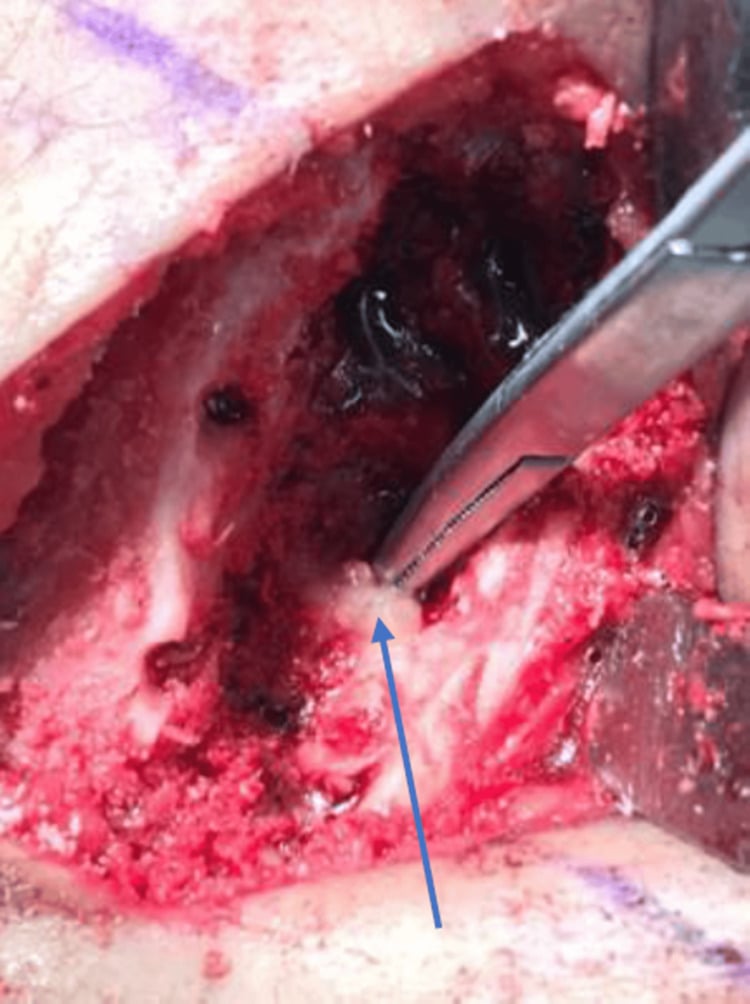
it decompresses raised intraosseous pressure, removes a segment of avascular sclerotic bone, permits definitive tissue sampling, and where infection exists improves antibiotic delivery. Reamed intramedullary nailing is an alternative in the tibia/femur when canal reaming is desired with immediate stabilisation, but it seeds any organism along the whole canal and is generally reserved for infective disease with mechanical need.
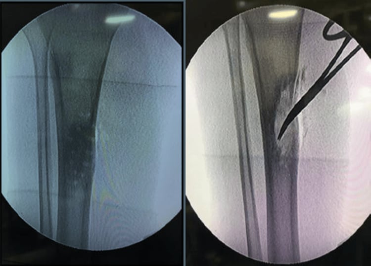
Position. Supine on a radiolucent table; sandbag under the ipsilateral buttock for the tibia; tourniquet high on the thigh (used but consider deflating before closure to secure haemostasis). Image intensifier from the contralateral side.
Imaging/equipment. Fluoroscopy; preoperative CT reformats on screen defining the sclerotic segment and canal; oscillating saw, fine osteotomes, high-speed burr, flexible reamers, bone biopsy trephine, multiple sterile specimen pots, antibiotic-loaded carrier (calcium sulphate beads) if infection suspected.
Preparation. Confirm no antibiotics given preoperatively unless septic; brief scrub team on the "clean instrument per sample" protocol; consent for possible sarcoma pathway conversion; group and save.
Approach. Tibia — direct anteromedial incision over the subcutaneous border, 1 cm medial to the anterior crest to avoid a wound over bone. Femur — lateral approach splitting the iliotibial band, vastus lateralis elevated anteriorly off the lateral intermuscular septum.
Dissection. Preserve periosteum where possible; incise longitudinally and elevate only the width of the intended window. Excessive stripping devascularises already compromised cortex.
Reconstruction/decompression step. Mark a rectangular window with rounded corners (stress risers at sharp corners cause fracture), typically 1 cm wide and no more than 30 to 50 percent of the bone diameter, sited away from the tension side. Use a drill-hole outline and fine osteotome or saw with continuous irrigation to avoid thermal necrosis. Harvest the cortical block as a specimen. Open the sclerotic medullary canal with a burr and gentle reaming until healthy bleeding bone is seen. Take the five deep samples. Irrigate with saline. Consider local antibiotic carrier if infection confirmed.
At-risk structures. Tibia — saphenous nerve and vein medially, anterior tibial artery deep to the interosseous membrane if over-penetrating laterally. Femur — perforating branches of the profunda femoris along the linea aspera (control with diathermy or ligation before they retract into the septum), sciatic nerve if dissection strays posteriorly.
Fixation. Usually none. If more than 50 percent of the diameter or more than 30 percent of the circumference is removed, or the bone is a weight-bearing femur, prophylactic intramedullary nailing or plating should be considered.
Closure. Layered closure over a drain if bleeding; avoid tight closure over the tibial subcutaneous border.
Aftercare. Protected weight bearing with crutches for 6 to 8 weeks with progressive loading; avoid impact and torsion for 3 months given the cortical window; radiographs at 6 weeks and 3 months to confirm window healing; continue medical therapy — surgery does not replace it.
Pitfalls. Sharp-cornered window causing fracture; over-resection; sampling burnt-out sclerosis giving false-negative histology; performing surgery for sterile inflammatory disease without medical escalation; failing to send tissue for mycobacterial culture.
Salvage of failure. Recurrent pain — repeat imaging to reassess for a missed nidus or malignancy before assuming relapse; escalate medical therapy; postoperative fracture treated with intramedullary nailing; established segmental infection may require resection with bone transport or vascularised fibular reconstruction.
STOPWhen NOT to operate on a sclerotic bone lesion
Hook:STOP before you cut a dense bone — most sclerotic pain is treated with a prescription, not an osteotome.
Guidelines, Registries & Global Practice
Global epidemiology. Sclerosing osteitis is uncommon everywhere but the balance of underlying causes varies markedly by setting. In high-income systems with early antibiotic access, sterile chronic non-bacterial osteomyelitis accounts for the majority of chronic sclerosing long-bone lesions in children and adolescents; incidence estimates for CNO are broadly comparable to those for other rare paediatric rheumatological diseases and it is likely under-recognised. In regions with high tuberculosis burden, restricted early antibiotic access, or a high incidence of untreated odontogenic disease, genuinely infective sclerosing osteomyelitis — including actinomycosis and tuberculous osteitis — remains proportionally far more common, and empirical antituberculous therapy is a legitimate consideration where testing is unavailable.
Society and consensus guidance that genuinely addresses this topic:
- Relevance
- Directly addresses sterile chronic non-bacterial osteomyelitis
- Key message
- NSAIDs first line for a defined trial; escalation to bisphosphonate, methotrexate or TNF inhibition; whole-body MRI for staging
- Relevance
- Address open fracture and implant-related bone infection
- Key message
- Provide the sampling discipline (multiple deep tissue samples, no swabs, antibiotics withheld until sampled) that should be applied here, but do not cover sterile osteitis
- Relevance
- Diagnostic definitions for bone and joint infection
- Key message
- Multiple concordant deep samples required to attribute causation; supports extended culture and molecular testing
- Relevance
- Biopsy of any suspicious bone lesion
- Key message
- Biopsy only in the unit that will perform definitive resection, through a resectable tract
- Relevance
- Mandibular form
- Key message
- Combined anti-inflammatory, dental sepsis control and selective decortication; bisphosphonate infusion for refractory pain
Registries. There are no implant registries relevant to this condition. Evidence comes instead from national and multinational CNO/CRMO patient registries and inception cohorts, which have driven the shift from empirical antibiotics and repeated surgery towards protocolised anti-inflammatory therapy, and from bone and joint infection registries which document culture yield and duration of therapy in low-virulence infection.
Practice variation by resource setting.
- Well-resourced settings: whole-body MRI, molecular microbiology, biologic therapy and multidisciplinary rheumatology-orthopaedics-radiology-microbiology meetings are available; the risk is over-investigation and delayed treatment.
- Intermediate settings: CT and conventional MRI available; extended culture may be, but broad-range PCR often is not; a pragmatic sequential trial of culture-directed or empirical antibiotics followed by NSAIDs is common.
- Limited-resource settings: radiographs and histology only. Here the priorities are (1) exclude malignancy by histology, (2) exclude tuberculosis, (3) trial NSAIDs before committing to prolonged empirical antibiotics with real toxicity and cost.
Controversies & Areas of Uncertainty
- Is Garré's osteomyelitis a real infective entity at all? Many authorities argue that most historic long-bone cases were sterile autoinflammatory osteitis, and that the eponym should be retired in favour of "chronic non-bacterial osteomyelitis" or "primary chronic osteomyelitis". Others maintain that a genuine low-virulence infective form exists, supported by culture-positive cases responding to antibiotics.
- Does culture-negative mean sterile? Proponents of an occult infective aetiology point to Cutibacterium recovered by molecular methods in SAPHO lesions and to occasional antibiotic responses; sceptics regard these as contaminants or as immune triggers rather than ongoing infection. The pragmatic position: a time-limited antibiotic trial may be justified where a plausible organism is isolated, but should not be repeated indefinitely.
- Duration of antibiotic therapy in culture-positive disease. No randomised evidence defines the endpoint; practice ranges from 3 to 12 months, guided by symptoms and markers rather than trial data.
- Role of surgery. Decortication and fenestration give unpredictable and often temporary relief. There is no randomised comparison against medical therapy, and publication bias favours positive surgical series.
- First-line agent in refractory CNO. Bisphosphonate versus TNF inhibitor as second line is not resolved; bisphosphonates are favoured for pain and vertebral disease, biologics where there is coexisting arthritis, skin or bowel disease.
- Antibiotic stewardship versus diagnostic uncertainty. Prolonged empirical antibiotics in a probably sterile disease is a common real-world harm; balancing this against the small risk of missing genuine infection remains a matter of judgement.
- Whether unifocal CNO and multifocal CRMO are the same disease at different points of expression, or biologically distinct — currently assumed to be a spectrum.
MCQ Practice Points
Q: Which feature is required to diagnose sclerosing osteomyelitis of Garré?
A: Absence of sequestrum, abscess and sinus. The condition is defined by dense cortical and medullary sclerosis with a purely reactive, non-suppurative response. A sequestrum within a lytic cavity indicates classic (secondary) chronic osteomyelitis and mandates surgical debridement. A draining sinus similarly excludes the diagnosis.
Q: A 14-year-old has a sclerotic tibial cortex and night pain relieved completely by ibuprofen within 30 minutes. What is the single most useful investigation?
A: Thin-slice CT of the affected segment. The history is characteristic of osteoid osteoma, and CT is the definitive test for the lucent nidus with central mineralisation. MRI may obscure the nidus in surrounding marrow oedema and can mislead towards infection or tumour. Treatment is CT-guided radiofrequency ablation.
Q: Which imaging investigation best supports a diagnosis of chronic non-bacterial osteomyelitis in a child?
A: Whole-body coronal STIR MRI. It detects clinically silent multifocal lesions in typical sites (clavicle, metaphyses, pelvis, spine, mandible), establishes the characteristic distribution pattern, guides biopsy site selection or avoids biopsy altogether, and involves no ionising radiation for repeated follow-up.
Q: In suspected low-grade sclerosing bone infection, which sampling strategy is correct?
A: At least five separate deep tissue samples using a fresh sterile instrument for each, sent for 14-day extended aerobic and anaerobic culture, mycobacterial and fungal culture, broad-range 16S rRNA PCR and histology, with antibiotics withheld beforehand. Swabs are inadequate; single samples cannot distinguish contamination from infection; short incubation misses Cutibacterium and Actinomyces.
Q: First-line therapy for culture-negative chronic non-bacterial osteomyelitis is what?
A: Full-dose non-steroidal anti-inflammatory drugs, which control a substantial proportion of cases. Refractory disease, and particularly spinal or hip involvement, is escalated to intravenous bisphosphonate infusion; methotrexate or TNF inhibition is used for further refractory disease or where arthritis, skin or bowel disease coexist. Prolonged antibiotics are of no benefit in sterile disease.
Q: Which finding most strongly suggests malignancy rather than sclerosing osteitis?
A: An interrupted, spiculated periosteal reaction with a Codman triangle and an associated soft-tissue mass. Garré's and CNO produce smooth, solid, continuous periosteal new bone with an intact cortex and no soft-tissue mass. Fever and raised inflammatory markers do NOT exclude malignancy — Ewing sarcoma classically mimics osteomyelitis systemically.
Exam Viva Scenarios
Practise clinical reasoning and management decisions out loud
“A 15-year-old presents with 8 months of deep aching pain over the mid-shaft of the right tibia, worse at night. There is a firm fusiform swelling over the subcutaneous border with no sinus, no erythema and no fever. Radiographs show fusiform cortical thickening, medullary sclerosis with canal narrowing, and smooth solid periosteal new bone. CRP is 6 and ESR is 18. How do you proceed?”
“A 22-year-old has had two decortication procedures on a sclerotic femoral diaphysis over three years, each followed by 6 months of intravenous antibiotics. All cultures at both operations were negative. Pain returns within months each time. She is now on long-term opioids and has been referred with a request for a third operation and an intramedullary reaming.”
“A 17-year-old has 6 months of recurrent right jaw pain and a firm fusiform swelling over the mandibular body with mild trismus. Orthopantomogram shows dense sclerosis of the mandibular body with periosteal new bone and no lytic cavity. There is a heavily restored lower molar. CRP is normal. Lower lip sensation is intact.”
“A 9-year-old girl has 12 months of intermittent left clavicular and right distal femoral pain with a firm swelling of the medial clavicle. Radiographs show clavicular sclerosis and hyperostosis and a metaphyseal sclerotic lesion at the distal femur. CRP is 12. A biopsy of the clavicle 6 months ago showed chronic inflammation with sterile cultures. She has palmoplantar pustulosis.”
Definition (all must hold)
- Chronic low-grade non-suppurative osteitis
- Fusiform cortical thickening plus medullary sclerosis with canal narrowing
- Smooth, solid, continuous periosteal new bone
- NO sequestrum, NO abscess, NO involucrum, NO cloaca, NO sinus
- Minimal systemic upset with near-normal inflammatory markers
Nomenclature to untangle
- Garré's — historic descriptive label; exclusion diagnosis
- Diffuse sclerosing osteomyelitis of the mandible — maxillofacial usage
- CRMO — sterile multifocal autoinflammatory osteitis of childhood
- CNO — umbrella term for sterile osteitis at any age
- SAPHO — adult equivalent: synovitis, acne, pustulosis, hyperostosis, osteitis
- Primary chronic osteomyelitis (Zurich) — non-suppurative from onset
Clinical picture
- Adolescent or young adult, months of deep aching pain worse at night
- Firm fusiform swelling fixed to bone — mandible, tibia or femur
- Relapsing and remitting over years, tends to burn out
- Examine skin (palmoplantar pustulosis, acne, psoriasis), clavicles and anterior chest wall, spine
- Lip numbness in mandibular disease is a red flag for malignancy
Investigation sequence
- Radiographs of whole bone, two planes, both joints
- Thin-slice CT — nidus? sequestrum? cortical breach?
- MRI with STIR and post-contrast — oedema, no rim-enhancing collection, no soft-tissue mass
- Whole-body STIR MRI if multifocal sterile disease suspected
- Bloods: FBC, CRP, ESR, ALP, LDH, blood cultures, IGRA if risk
- Biopsy: histology plus 5 deep samples, 14-day extended culture, mycobacterial/fungal, 16S PCR
Differential — must be able to list
- Osteoid osteoma — NSAID-responsive night pain, CT nidus
- Osteosarcoma and Ewing sarcoma — aggressive periosteum, soft-tissue mass, biopsy mandatory
- Classic chronic osteomyelitis with sequestrum / Brodie abscess
- Stress fracture — lucent line in thickened cortex
- Osteofibrous dysplasia and adamantinoma — anterior tibial cortex
- Tuberculous and fungal osteomyelitis
- Paget disease and sclerosing bone dysplasias
Management thresholds
- Aggressive features or uncertainty — sarcoma unit staging and biopsy first
- Nidus on CT — radiofrequency ablation
- Sequestrum or abscess — radical debridement plus culture-directed antibiotics
- Organism in 2 or more samples — prolonged culture-directed antibiotics with ID input
- Sterile — NSAIDs first line, then intravenous bisphosphonate, then methotrexate or TNF inhibitor
- Surgery (fenestration/decortication) only for refractory pain after medical escalation; relief often temporary
Fenestration technique essentials
- Rectangular window with ROUNDED corners to avoid stress risers
- Limit to less than 30 to 50 percent of bone diameter; consider prophylactic fixation if larger
- Irrigate while cutting to prevent thermal necrosis; preserve periosteum
- Take the cortical block plus 5 deep samples with fresh instruments
- Protected weight bearing 6 to 8 weeks; no impact for 3 months
- Femoral approach: control perforating vessels at the linea aspera before they retract
Highest-yield exam soundbites
- Garré's is a phenotype, not a microbiological diagnosis
- Marked sclerosis with normal markers equals think CNO/SAPHO
- Sclerotic medial clavicle in a child equals CRMO until proven otherwise
- Culture-negative is the norm; do not equate it with treatment failure
- Do not keep operating on sterile inflammatory bone disease
Evidence Base
Garre's Original Description of Non-Suppurative Sclerosing Osteitis
- NOT INDEXED IN PUBMED OR EUROPE PMC - this 1893 German paper predates indexing entirely and nothing attributed to it here can be checked against the record
- The eponym is attached to a description of acute infective osteomyelitis in which Garre reported no histological specimens and no radiographs, radiography not yet being available
- What is now called 'Garre's sclerosing osteomyelitis' - a chronic, non-suppurative, sclerosing process with no pus, no sequestrum and no sinus - is therefore NOT the entity Garre described
- The name has persisted through a century of secondary citation rather than through anything verifiable in the original
Subacute and Chronic 'Symmetrical' Osteomyelitis - the First Description of CRMO
- The paper that separated a sterile, symmetrical, relapsing multifocal bone inflammation of childhood from ordinary pyogenic osteomyelitis - the origin of what is now chronic recurrent multifocal osteomyelitis
- NO ABSTRACT IS INDEXED for this paper (PubMed records it as published in multiple languages), so the case numbers and radiographic criteria cannot be read from the record; the MeSH indexing confirms only the population (child, preschool child), the chronicity, and that radiography and immune-system disease were central
- The MeSH record also lists Streptococcus pneumoniae isolation, which is a caution against reading the original series as uniformly culture-negative in the modern sense
- Its lasting contribution is conceptual: symmetry, multifocality and relapse define the entity, not the organism
Acne-Pustulosis-Hyperostosis-Osteitis: National Survey of 85 Cases - the Paper That Coined SAPHO
- A national survey organised by the French Society of Rheumatology collecting 85 cases: 13 with severe acne, 44 with palmoplantar pustulosis, and 28 with hyperostosis and NO skin disease at all
- That last group of 28 is the point most often lost: a third of the original cohort had the bone disease WITHOUT the dermatosis, so absent skin findings do not exclude the diagnosis
- The authors argued that conditions described under many different names shared common features and transition forms, justifying a single acronym - SAPHO
- The unifying lesion is the bone: anterior chest wall above all, but also vertebral and peripheral, described as an inflammatory osteitis analogous to synovitis in inflammatory arthropathy
- Connections to psoriasis were judged loose and to ankylosing spondylarthritis slightly firmer, on both clinical and immunogenetic grounds
Clinical Care of Children with Sterile Bone Inflammation - the Modern Treatment Algorithm
- Defines the group by what is present and what is absent: sterile bone inflammation with histological neutrophil infiltration, in the ABSENCE of autoantibodies and autoreactive T cells - some cases carrying a genetic mutation
- WHOLE-BODY MRI is helpful in detecting ASYMPTOMATIC lesions, which is what converts an apparently solitary sclerotic lesion into a multifocal diagnosis and away from biopsy
- First-line treatment is NSAIDs, and this is usually sufficient to control symptoms while the bone heals
- Escalate to BISPHOSPHONATE where lesions persist, fail first-line treatment, or involve the SPINE OR HIP - those sites are named because of the structural consequences of untreated inflammation there
- Anti-TNF agents are rarely required
Bristol Diagnostic Criteria for CRMO - How Often Biopsy Can Be Avoided
- Forty-one children diagnosed with CRMO over 8 years at Bristol Royal Hospital for Children; median age at symptom onset 9 years
- MEDIAN TIME TO DIAGNOSIS WAS 15 MONTHS, range 0 to 92 - the delay is this disease's defining clinical problem, not the imaging
- The harms of that delay are specified: prolonged antibiotic courses with inpatient care, unnecessary radiation from repeated radiographs and bone scans, and repeated surgery including bone biopsies
- Applying the resulting Bristol criteria retrospectively, 34 of 41 children (83 PER CENT) could have been diagnosed WITHOUT a biopsy
- Time to diagnosis fell significantly year on year after information about CRMO was circulated to regional orthopaedic surgeons - awareness measurably shortened the delay
Primary Chronic Osteomyelitis of the Jaw - the Zurich Classification
- Thirty cases treated over 30 years at Zurich, analysed to replace a confused and non-uniform terminology with a simple clinical classification
- Mean age at onset 35 years (range 5 to 76) with TWO PEAKS OF INCIDENCE - one in adolescence and one after age 50 - which is the observation the classification is built on
- Six of the 30 (five adults, one adolescent) had non-facial bone, joint and skin manifestations consistent with SAPHO or CRMO, so a fifth of apparently jaw-only disease was systemic
- Radiology showed sclerosis, osteolysis and periosteal reaction in variable stages in ALL cases, more extensive in younger patients; microabscesses were seen in 11 cases, six of them children or adolescents
- Outcomes after surgery, antibiotics and hyperbaric oxygen: 11 complete remission, 14 amelioration, 5 no significant improvement - so a third did not clearly improve