Sterile autoinflammatory osteitis with hyperostosis — the great mimic of osteomyelitis and bone tumour
- SAPHO is a sterile autoinflammatory osteitis — cultures are negative and antibiotics do not work; the orthopaedic error is repeated debridement for presumed osteomyelitis.
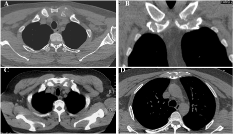
- The adult signature is anterior chest wall pain with hard hyperostotic swelling of the sternoclavicular region; the paediatric signature is multifocal metaphyseal and clavicular lesions (CRMO).
- It is a diagnosis of exclusion: infection, Paget disease, metastasis, lymphoma and small round cell tumours must be actively excluded, particularly when a lesion is solitary or atypical.
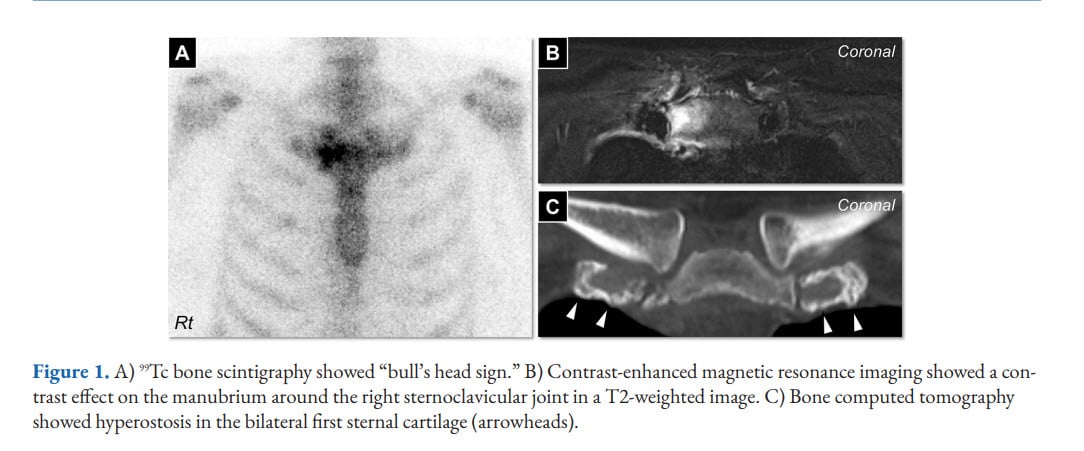
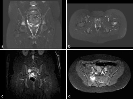
- Whole-body imaging (bone scintigraphy or whole-body MRI STIR) reveals clinically silent multifocality and supports the diagnosis — the bull's head sign is classic but not universal.
- Treatment is medical and led with rheumatology: NSAIDs, then bisphosphonate infusion, conventional immunomodulators, and TNF or IL-17/IL-1 directed biologics for refractory disease.
- Surgery is rarely indicated; hyperostosis reliably recurs after resection and wound problems are common.
- “Palmoplantar pustulosis is the skin lesion most tightly linked to anterior chest wall osteitis.
- “Sacroiliitis in SAPHO is frequently unilateral and adjacent to hyperostotic bone, unlike the symmetrical sacroiliitis of ankylosing spondylitis.
- “Inflammatory markers rise modestly; a markedly septic picture with high CRP and rigors should push you back towards infection.
- “HLA-B27 is positive in only a minority — a negative result does not exclude SAPHO and a positive one does not confirm it.
- “If you biopsy, warn the pathologist: early lesions show neutrophilic osteomyelitis-like change that can be misread as infection or malignancy.
Serial debridements, sequestrectomy and long-term antibiotics for a sterile disease. The bone gets more hyperostotic, the wound fails, and the diagnosis is still missed. Negative cultures from an adequate sample in a hyperostotic clavicle should trigger the SAPHO question, not another washout.
Sclerotic expanded clavicle or vertebral sclerosis is reported as osteosarcoma, lymphoma or metastasis. Conversely — never assume SAPHO for a solitary lesion. A solitary sclerotic lesion in a child is Ewing sarcoma or osteomyelitis until biopsy proves otherwise.
Skin manifestations are absent at presentation in a large proportion and may appear years later or never. Absence of pustulosis or acne does not exclude SAPHO. Ask about past acne fulminans, palmar pustules, and treated hidradenitis.
Sternoclavicular or clavicular resection for hyperostosis is followed by recurrence of bony overgrowth in a high proportion, plus scar pain and shoulder girdle instability. Escalate medical therapy instead. Surgery is reserved for genuine mechanical or neurovascular compromise.
Concept, Nosology and Pathogenesis
SAPHO is an acronym coined by Chamot and colleagues (1987) grouping Synovitis, Acne, Pustulosis, Hyperostosis and Osteitis into a single autoinflammatory entity. It is best understood as an innate-immune (autoinflammatory) sterile osteitis, not an autoimmune or infective disease.
Key nosological points
- SAPHO and chronic non-bacterial osteomyelitis (CNO) / chronic recurrent multifocal osteomyelitis (CRMO) sit on one disease spectrum. CRMO is the paediatric multifocal phenotype; SAPHO is the adult phenotype with the skin association and chest wall predilection. Many units now use "CNO/CRMO spectrum including SAPHO".
- It overlaps the spondyloarthritis family (sacroiliitis, enthesitis, psoriasiform skin disease, response to TNF blockade) but HLA-B27 positivity is only modestly increased and it is not a classical seronegative spondyloarthropathy.
- Monogenic phenocopies inform the biology: DIRA (IL-1 receptor antagonist deficiency), Majeed syndrome (LPIN2), and PAPA syndrome (PSTPIP1) all produce sterile osteitis, implicating IL-1 driven neutrophilic inflammation.
Proposed mechanism
- Genetic predisposition to dysregulated innate immunity (IL-1beta, IL-17, IL-23, TNF axes; neutrophil hyperactivity).
- A microbial trigger hypothesis — low-virulence Cutibacterium (Propionibacterium) acnes is cultured from a minority of bone biopsies. Whether this is causative antigenic drive or contamination remains unresolved; antibiotic trials show at best transient, non-sustained benefit.
- Result: sterile neutrophilic osteitis, followed by an exuberant osteoblastic reparative response producing the hallmark hyperostosis and enthesial new bone.
SAPHO is dominated by an osteoblastic response. Early lesions are oedematous and partly lytic; chronic lesions are sclerotic, thickened cortex with enthesial ossification and, at the chest wall, ossification of the costoclavicular ligament — this is why the clinical sign is a hard, enlarging, tender lump rather than a discharging sinus.
Clinical Presentation and Examination
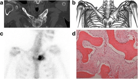
The adult classic — present in roughly two-thirds of adult SAPHO.
- History: insidious deep anterior chest ache, worse at night and in the early morning, better with movement and NSAIDs; weeks to years of relapsing episodes; often labelled "costochondritis" or "cardiac" and investigated as such.
- Distribution: sternoclavicular joints (often bilateral, may be asymmetric), manubriosternal joint, medial clavicle, first costochondral junctions.
Examination sequence
- Inspection — patient sitting, shoulders exposed to the nipple line, arms relaxed. Look for asymmetric fullness over the medial clavicle and the sternoclavicular region; overlying erythema and shininess in active flares; venous distension over the anterior chest wall or arm swelling suggests subclavian vein compression or thrombosis.
- Palpation — examiner stands in front, thumbs on the medial ends of both clavicles simultaneously to compare. Positive finding: a firm, bony, non-fluctuant enlargement that is warm and exquisitely tender. Bony hardness distinguishes hyperostosis from a fluctuant septic sternoclavicular abscess.
- Provocation — cross-body adduction and forced shoulder protraction load the sternoclavicular joint and reproduce pain.
- Vascular — assess for arm swelling, collateral veins, and compare radial pulses; document if thoracic outlet symptoms are present.
- False positives — sternoclavicular osteoarthritis in older women, condensing osteitis of the clavicle (usually unilateral, medial clavicle, in young women, no skin disease), post-traumatic and post-catheter septic arthritis.
SAPHORecalling the Acronym with Clinical Weight
Hook:Two skin letters, three bone letters — but you only need the bone letters to make the diagnosis.
Investigation Strategy
- Typical result in SAPHO
- Normal to modestly raised
- Interpretation
- A markedly septic profile favours infection
- Typical result in SAPHO
- Normal
- Interpretation
- Neutrophilia suggests pyogenic disease
- Typical result in SAPHO
- Negative
- Interpretation
- Take before any antibiotic if infection is plausible
- Typical result in SAPHO
- Negative
- Interpretation
- Excludes rheumatoid arthritis
- Typical result in SAPHO
- Positive in a minority
- Interpretation
- Neither confirms nor excludes
- Typical result in SAPHO
- Usually normal
- Interpretation
- Markedly elevated favours Paget disease
- Typical result in SAPHO
- Normal
- Interpretation
- Screening for sclerotic metastasis and myeloma in adults
SAPHO/CRMO is a diagnosis of exclusion. A single sclerotic or lytic bone lesion — especially in a child, adolescent, or an adult with a cancer history — must be investigated as a possible tumour or pyogenic infection, with staging imaging and a biopsy planned through a sarcoma unit before any intervention. Do not label a solitary lesion SAPHO on imaging alone.
Diagnostic Criteria
No universally validated criteria exist. Two working sets are quoted in vivas:
- Osteoarticular disease associated with acne conglobata, acne fulminans or hidradenitis suppurativa.
- Osteoarticular disease associated with palmoplantar pustulosis.
- Hyperostosis (axial or appendicular) with or without skin disease.
- Chronic recurrent multifocal osteomyelitis involving the axial or appendicular skeleton, with or without skin disease.
septic osteomyelitis, infectious chest wall arthritis, infectious palmoplantar pustulosis, palmoplantar keratoderma, diffuse idiopathic skeletal hyperostosis (DISH), retinoid-induced osteitis, and bone neoplasm.
For children, the Bristol and Jansson criteria for CNO emphasise typical radiological lesions plus either multifocality, a raised CRP, or a compatible biopsy, with malignancy and infection excluded.
"SAPHO is a clinico-radiological diagnosis of exclusion made on a characteristic multifocal sterile osteitis with hyperostosis, supported where present by palmoplantar pustulosis or severe acne, after infection and malignancy have been actively excluded."
Differential Diagnosis
- 1Step 1 — Presentation
Painful sclerotic hyperostotic lesion, classically anterior chest wall (sternoclavicular, manubriosternal), spine, mandible or metaphysis
Skin lesion plus typical site raises SAPHO high in the differential but does not confirm it
- 2Step 2 — Exclude sepsis first
Fever, rigors, high CRP and white cell count, abscess, sinus, intravenous drug use or indwelling line
Positive culture equals pyogenic or tuberculous osteomyelitis — treat as infection; missed sepsis risks mediastinitis and death
- 3Step 3 — Map the skeleton
Is disease multifocal or solitary?
Multifocal symmetrical anterior chest wall disease with typical skin lesions supports sterile osteitis
- 4Step 4 — Solitary lesion in a young patient
Solitary lesion, soft-tissue mass, permeative destruction, systemic symptoms or escalating pain
Excludes Ewing sarcoma and osteosarcoma; unplanned surgery costs limb and life
- 5Step 5 — Bloods and marrow screen
Night sweats, weight loss, cytopenias or blasts on film in a child; older adult with a known primary
Excludes lymphoma, leukaemia, Langerhans cell histiocytosis and sclerotic metastasis
- 6Step 6 — Exclude benign sclerotic mimics
Markedly raised alkaline phosphatase with cortical thickening, coarse trabeculae and bone expansion; or a young woman with isolated unilateral medial clavicle sclerosis and no skin disease
Avoids unnecessary immunosuppression and over-treatment of self-limiting disease
- 7Step 7 — Biopsy the bone if doubt persists
Solitary lesion, atypical site, no skin disease, or failure to settle
SAPHO shows sterile subacute or chronic inflammation with fibrosis and reactive new bone; cultures negative
- 8Step 8 — Accept sterile osteitis
Multifocal typical distribution, characteristic dermatosis, negative cultures, no malignancy on biopsy or imaging
Reassess if a new solitary aggressive lesion appears — the diagnosis remains one of exclusion
- Features that favour it
- Fever, rigors, high CRP and white cell count, abscess, sinus, intravenous drug use or indwelling line
- Discriminating test
- Aspiration and culture; MRI showing rim-enhancing abscess or sequestrum
- Consequence if missed
- Sepsis, mediastinitis, death
- Features that favour it
- Solitary lesion, soft-tissue mass, permeative destruction, systemic symptoms, rising pain
- Discriminating test
- MRI whole lesion plus staging and planned core biopsy through a sarcoma unit
- Consequence if missed
- Unplanned excision, limb and life loss
- Features that favour it
- Night sweats, weight loss, cytopenias, blasts on film, marrow signal change
- Discriminating test
- Blood film, LDH, marrow biopsy
- Consequence if missed
- Delayed oncological treatment
- Features that favour it
- Older adult, known primary, multiple axial lesions, raised alkaline phosphatase or PSA
- Discriminating test
- Staging CT, PSA, tumour markers, biopsy
- Consequence if missed
- Missed systemic malignancy
- Features that favour it
- Older patient, cortical thickening with trabecular coarsening and bone expansion, markedly raised alkaline phosphatase, usually painless bone enlargement
- Discriminating test
- Alkaline phosphatase and characteristic radiographic architecture
- Consequence if missed
- Unnecessary immunosuppression
- Features that favour it
- Young woman, unilateral medial clavicle sclerosis, no skin disease, no other lesions
- Discriminating test
- Localised imaging with no multifocality
- Consequence if missed
- Over-treatment; benign self-limiting
- Features that favour it
- Symmetrical sacroiliitis, thin marginal syndesmophytes, HLA-B27, psoriasis plaques
- Discriminating test
- Pattern of syndesmophytes and sacroiliitis symmetry
- Consequence if missed
- Overlapping treatment — less critical error
- Features that favour it
- Endemic region, indolent course, cold abscess, vertebral body destruction with disc sparing
- Discriminating test
- Acid-fast stain, mycobacterial culture, PCR from biopsy
- Consequence if missed
- Progressive destruction and spread
STERILEWhat Must Be Excluded Before Labelling SAPHO
Hook:SAPHO is only STERILE once you have excluded everything on this list.
Management — A Medical Disease Run With Rheumatology
- 1Step 1 — NSAIDs and educationFull-dose NSAID with gastroprotection for at least 2 to 4 weeks before declaring failure; many achieve durable control. Explain the relapsing–remitting course, the sterile nature of the disease, and that antibiotics have no established role. Physiotherapy for spinal mobility and shoulder girdle function.
- 2Step 2 — Bisphosphonate infusion for refractory osteitisIntravenous pamidronate or zoledronic acid is the most reproducibly effective second-line agent for painful osteitis and vertebral CNO lesions, often giving rapid, prolonged pain relief and reduced marrow oedema. Warn of the acute phase reaction after the first infusion; check calcium and vitamin D for hypocalcaemia; dental review for osteonecrosis of the jaw risk.
- 3Step 3 — Conventional immunomodulatorsMethotrexate, sulfasalazine (better for peripheral synovitis) or ciclosporin. Corticosteroids relieve flares rapidly but rebound on withdrawal — avoid long courses. Steroid withdrawal and, paradoxically, isotretinoin can flare acne-associated disease.
- 4Step 4 — Biologics for refractory diseaseTNF inhibitors (infliximab, adalimumab, etanercept) carry the largest observational evidence base for refractory bone and axial disease, but may induce or worsen palmoplantar pustulosis. Alternatives: IL-17 inhibition (secukinumab), IL-1 blockade (anakinra, especially IL-1 driven paediatric disease), IL-23/IL-12 blockade (ustekinumab), and JAK inhibitors in emerging reports. Screen for latent tuberculosis and hepatitis before any biologic.
- 5Adjuncts — antibiotics and dermatologyLong-course antibiotics against Cutibacterium acnes (doxycycline, azithromycin) give inconsistent, non-sustained benefit and are not standard care. Dermatology co-management is essential for acne fulminans and hidradenitis; skin severity does not track bone severity.
- 6Top rung — surgery, narrowly indicatedReserved for mechanical or neurovascular compromise: diagnostic biopsy for atypical or solitary lesions, decompression of subclavian vein or thoracic outlet compromise from hyperostosis, decompression with or without fusion for spinal neurological compression or instability, debridement only with positive microbiology, and salvage sternoclavicular resection or arthrodesis after failed maximal medical therapy. Resect no more than the medial 1 to 2 cm and preserve or reconstruct the costoclavicular (rhomboid) ligament.
There are no large randomised trials; the ladder below reflects consensus practice and observational cohorts. The orthopaedic role is diagnosis, exclusion of mimics, safe biopsy where indicated, and referral — not resection.
Full-dose NSAID (with gastroprotection) for at least 2 to 4 weeks before declaring failure. Many patients achieve durable control. Explain the relapsing–remitting natural history, the sterile nature of the disease, and that antibiotics have no established role. Physiotherapy for spinal mobility and shoulder girdle function.
Intravenous pamidronate or zoledronic acid is the most reproducibly effective second-line agent for painful osteitis and for vertebral CNO lesions, often producing rapid and prolonged pain relief and reduction in marrow oedema. Counsel about the acute phase reaction after the first infusion, hypocalcaemia (check calcium and vitamin D beforehand), and dental review for osteonecrosis of the jaw risk.
Methotrexate, sulfasalazine (more useful for peripheral synovitis) or ciclosporin. Corticosteroids give rapid relief in flares but rebound on withdrawal; avoid long courses. Note that corticosteroid withdrawal and, paradoxically, isotretinoin can flare acne-associated disease.
TNF inhibitors (infliximab, adalimumab, etanercept) have the largest observational evidence base for refractory bone and axial disease. Caveat: TNF blockade may induce or worsen palmoplantar pustulosis (paradoxical psoriasiform reaction). Alternatives where TNF fails or skin flares: IL-17 inhibitors (secukinumab), IL-1 blockade (anakinra — especially in IL-1 driven paediatric disease), IL-23/IL-12 blockade (ustekinumab), and JAK inhibitors in emerging reports. Screen for latent tuberculosis and hepatitis before any biologic.
Long-course antibiotics targeting Cutibacterium acnes (doxycycline, azithromycin) produce inconsistent, generally non-sustained benefit and are not standard care. Dermatology co-management is essential for acne fulminans and hidradenitis; skin severity does not track bone severity.
Complications and Natural History
- Comment
- The dominant morbidity; flares over years to decades
- Prevention / management
- Stepwise medical ladder, pain team, self-management plan
- Comment
- From bulky sternoclavicular hyperostosis; arm swelling, collaterals
- Prevention / management
- CT venogram, anticoagulation, decompression only if refractory
- Comment
- Predominantly paediatric CNO with vertebral lesions
- Prevention / management
- Early bisphosphonate therapy for spinal lesions; bracing; rarely instrumentation
- Comment
- From bulky non-marginal bridging
- Prevention / management
- Physiotherapy, TNF inhibition for active axial disease
- Comment
- Repeated debridement, long antibiotics, unnecessary resection
- Prevention / management
- Recognise the diagnosis; involve rheumatology early
- Comment
- Bisphosphonate acute phase reaction and jaw osteonecrosis; biologic infection risk and paradoxical pustulosis
- Prevention / management
- Pre-treatment dental and tuberculosis screening; monitoring
- Comment
- Uncommon — SAPHO is generally non-destructive
- Prevention / management
- Reassure; reconsider the diagnosis if severe erosion develops
Prognosis — chronic and relapsing but generally non-destructive, with most patients maintaining function. Substantial spontaneous remission occurs, particularly in paediatric CNO, though a proportion of children have persistent or relapsing disease into adulthood. Poorer outcome is associated with spinal involvement, multifocal disease at onset and delayed diagnosis.
Guidelines, Registries & Global Practice
Global epidemiology
- Rare, with prevalence estimated well under 1 in 10,000 in white European populations; likely substantially under-diagnosed globally.
- Female predominance in most adult series; typical adult onset in the third to fifth decades. Paediatric CNO peaks around ages 9 to 12 with a female predominance.
- Regional phenotype differences are important for exams: large East Asian cohorts (notably from China and Japan) report a very high frequency of palmoplantar pustulosis and anterior chest wall disease, whereas European and North American series report proportionally more severe acne and axial disease. The same disease, different skin expression.
Society and consensus guidance
- Relevance to SAPHO/CNO
- Supports NSAIDs first line, bisphosphonates for refractory or vertebral disease, TNF inhibitors for refractory disease; endorses whole-body MRI for staging
- Relevance to SAPHO/CNO
- Standardised sequential plans for CNO: NSAID trial, then choice of methotrexate/sulfasalazine, TNF inhibitor, or bisphosphonate
- Relevance to SAPHO/CNO
- Any solitary or atypical bone lesion must be biopsied through the unit that would perform definitive resection — directly applicable to suspected SAPHO with a single lesion
- Relevance to SAPHO/CNO
- Emphasise adequate deep sampling before antibiotics and reappraisal of diagnosis when repeated cultures are sterile — the trigger to consider SAPHO
- Relevance to SAPHO/CNO
- Systemic therapy planning; awareness that isotretinoin may precipitate acne fulminans with osteitis
Note there is no arthroplasty or implant registry evidence base for SAPHO — joint replacement is not part of its management, and quoting registry data here would be inappropriate.
Practice variation by resource setting
- Where advanced imaging is limited, bone scintigraphy remains the practical whole-body screening tool; where MRI is available, whole-body STIR is preferred, particularly in children, to avoid radiation.
- In tuberculosis-endemic regions, mycobacterial and fungal disease must be excluded rigorously — empirical antituberculous therapy for a sterile osteitis is a recognised diagnostic pitfall.
- Access to biologic therapy varies widely; where unavailable, bisphosphonate infusion is the highest-value escalation because it is inexpensive, widely available and effective for osteitis pain.
Controversies & Areas of Uncertainty
- Is Cutibacterium acnes pathogenic or contaminant? Cultured from a minority of biopsies; antibiotic trials show inconsistent, non-durable benefit. Prevailing view: a possible antigenic trigger in a genetically primed innate immune system rather than a true infection.
- Is SAPHO a distinct disease or a spondyloarthropathy variant? Overlapping features and treatment response argue for spectrum membership; the modest HLA-B27 association and prominent hyperostosis argue for distinctness.
- Should SAPHO and CRMO be merged nomenclature? Many paediatric rheumatologists advocate a single "chronic non-bacterial osteomyelitis" umbrella; adult rheumatology retains SAPHO for the skin-associated chest wall phenotype.
- First-line escalation: bisphosphonate or TNF inhibitor? Bisphosphonates are cheaper, safer and excellent for osteitis pain and vertebral lesions; TNF inhibitors are better for axial inflammation and coexisting skin/joint disease. Practice varies by unit and access.
- Paradoxical pustulosis with TNF blockade — how often it forces a switch to IL-17 or IL-23 directed therapy is not well quantified.
- Role of whole-body MRI as a routine staging and monitoring tool — increasingly standard in children, less established in adults, with no validated activity score in wide use.
- Diagnostic criteria — Kahn's criteria are unvalidated and predate modern imaging; consensus criteria incorporating whole-body MRI are awaited.
MCQ Practice Points
Q: Which skin manifestation is most strongly associated with anterior chest wall osteitis in SAPHO?
A: Palmoplantar pustulosis. Sterile pustules on palms and soles show the tightest association with sternoclavicular and manubrial hyperostosis, and made up 44 of the 85 cases in Chamot's founding survey. Acne conglobata and acne fulminans are also recognised, hidradenitis suppurativa and pyoderma gangrenosum occur, and psoriasis vulgaris is a weaker association. Add the caveat that matters at the bedside: 28 of those 85 patients had hyperostosis with NO skin disease at all, so absent skin findings do not exclude the diagnosis.
Q: What does the 'bull's head' sign describe?
A: Increased technetium-99m uptake on bone scintigraphy in the manubrium (the skull) and both sternoclavicular joints with the medial clavicles (the horns). It is highly characteristic of SAPHO anterior chest wall disease but is neither universal nor pathognomonic — its absence does not exclude the diagnosis.
Q: A 12-year-old has a solitary lytic lesion of the distal femoral metaphysis with periosteal reaction and a soft-tissue mass. Which statement is correct?
A: This must be investigated as a possible Ewing sarcoma or osteosarcoma, with staging and a planned biopsy through a sarcoma unit. SAPHO and chronic non-bacterial osteitis are diagnoses of exclusion and characteristically multifocal; a solitary lesion with a soft-tissue mass is a tumour until proven otherwise.
Q: Which second-line agent is used for refractory SAPHO osteitis pain and vertebral lesions, and what is the quality of the evidence behind it?
A: Intravenous bisphosphonate — pamidronate or zoledronic acid. It reduces bone pain and MRI marrow oedema in reported series, is inexpensive and widely available, and is particularly valuable for vertebral lesions at risk of collapse; check calcium and vitamin D and arrange dental review before infusion. Now say the second half honestly, because it is examinable and frequently got wrong: there is no approved therapy for adult chronic nonbacterial osteitis, bisphosphonates are used off-label, and as of 2024 no adequately powered randomised controlled trial had been conducted — the first placebo-controlled trial of pamidronate was only then recruiting. The supporting literature is open-label series of a few dozen patients, some published as correspondence.
Q: Which finding argues AGAINST SAPHO and FOR pyogenic osteomyelitis?
A: A rim-enhancing intraosseous or soft-tissue abscess with sequestrum on MRI, high fever and marked leucocytosis. SAPHO shows marrow oedema, periosteal reaction and hyperostosis without a drainable collection or sequestrum, with only modest inflammatory marker elevation.
Q: What is the expected outcome of surgical resection of sternoclavicular hyperostosis in SAPHO?
A: Recurrence of hyperostosis in a substantial proportion, with additional risks of wound breakdown, shoulder girdle instability and major venous injury. Surgery does not address the inflammatory driver; it is reserved for neurovascular compromise, instability or diagnostic biopsy.
Q: How does SAPHO spinal disease differ radiographically from ankylosing spondylitis?
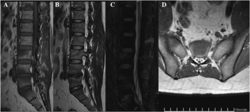
A: SAPHO produces bulky, non-marginal paravertebral bony bridges, vertebral corner erosions with dense reactive sclerosis and discitis-like endplate change, with frequently unilateral or asymmetric sacroiliitis. Ankylosing spondylitis produces thin marginal syndesmophytes with symmetrical sacroiliitis and squared vertebrae.
Exam Viva Scenarios
Practise clinical reasoning and management decisions out loud
“A 38-year-old woman presents with 8 months of anterior chest pain and a hard, tender swelling over the right medial clavicle and sternoclavicular joint. She has had two courses of oral antibiotics with no benefit. CRP is 22 mg/L, white cell count normal, temperature normal. Radiographs show sclerosis and expansion of the medial clavicle. How do you proceed?”
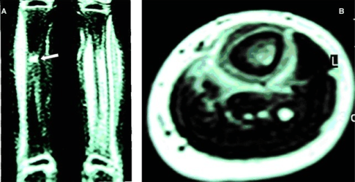
“A 10-year-old girl has 4 months of pain in the left clavicle and the right distal tibia, with a limp. She is systemically well. CRP is 18 mg/L. Radiographs show a sclerotic expanded medial clavicle and a metaphyseal lucency with surrounding sclerosis in the distal tibia. The referring team has started intravenous flucloxacillin. What is your assessment?”
“A 45-year-old man with established SAPHO and palmoplantar pustulosis has a large, tender, cosmetically distressing hyperostotic mass at the left sternoclavicular joint. He is on NSAIDs only and asks you to remove it. He also reports left arm swelling after exercise. What do you advise?”
“You debrided a 52-year-old woman's sclerotic sternoclavicular lesion 6 weeks ago for presumed chronic osteomyelitis. All cultures including extended anaerobic cultures are negative. Histology reports 'chronic sclerosing osteomyelitis'. She remains symptomatic and the wound is slow to heal. What now?”
Definition and spectrum
- Synovitis, Acne, Pustulosis, Hyperostosis, Osteitis — sterile autoinflammatory bone disease
- Adult phenotype of the CNO/CRMO spectrum; CRMO is the paediatric multifocal equivalent
- Innate-immune driven (IL-1, IL-17, IL-23, TNF); monogenic phenocopies DIRA, Majeed, PAPA
- Cutibacterium acnes cultured in a minority — trigger versus contaminant unresolved
Clinical signature
- Adult: anterior chest wall — sternoclavicular, manubriosternal, first costochondral; hard, warm, tender bony swelling
- Spine: thoracic predominance, corner erosions, sclerosis, non-marginal bulky bridges, discitis-like lesions
- Sacroiliitis often unilateral/asymmetric with iliac-side hyperostosis
- Child: metaphyseal and clavicular multifocal lesions, limp, systemically well
- Skin may precede by years, follow by years, or never appear
Investigations
- Bloods: CRP/ESR mildly raised, cultures negative, alkaline phosphatase normal, HLA-B27 positive in a minority only
- CT: hyperostosis, cortical thickening, erosion, costoclavicular ligament ossification
- MRI STIR/T2 fat-sat: marrow oedema (active); T1 low signal sclerosis (chronic); no abscess or sequestrum
- Bone scintigraphy: bull's head sign — sternum plus both sternoclavicular regions
- Whole-body STIR MRI preferred in children to map silent lesions without radiation
Biopsy rules
- Indicated for solitary or atypical lesions, systemic red flags, cancer history, or persistent diagnostic doubt
- Plan through a sarcoma unit along a resectable tract; multiple cores
- Send microbiology (including extended anaerobic, mycobacterial, fungal) AND histology
- Warn the pathologist SAPHO is in the differential
- Histology: neutrophilic early, lymphoplasmacytic mid, fibrosis and sclerosis late; no organisms, no tumour
Differentials to exclude (STERILE)
- Septic osteomyelitis and septic sternoclavicular arthritis
- Tumour — Ewing sarcoma, osteosarcoma
- Eosinophilic granuloma / Langerhans cell histiocytosis
- Round cell malignancy — lymphoma, leukaemia
- Infective granulomatous — tuberculosis, fungal
- Lesions of metastasis and myeloma; Established Paget disease
- Also: condensing osteitis of the clavicle, DISH, retinoid-induced osteitis
Treatment ladder (with rheumatology)
- 1. Full-dose NSAID for 2 to 4 weeks plus education and physiotherapy
- 2. Intravenous bisphosphonate (pamidronate/zoledronic acid) for refractory osteitis and vertebral lesions
- 3. Methotrexate, sulfasalazine, ciclosporin; avoid long-term corticosteroids
- 4. Biologics: TNF inhibitors first line biologic; IL-17, IL-23 or IL-1 blockade if TNF fails or pustulosis flares
- Antibiotics are not standard therapy; benefit is inconsistent and non-sustained
Surgical role and pitfalls
- Indications: diagnostic biopsy, neurovascular compromise, spinal cord compression or instability, true superimposed infection
- NOT for pain or cosmesis — hyperostosis recurs at the resection margin
- Sternoclavicular resection: preserve or reconstruct the costoclavicular ligament; vascular surgeon on standby
- At-risk: subclavian/brachiocephalic veins, subclavian artery, brachial plexus, pleural apex
- Salvage of failure is medical escalation, not re-resection
Prognosis
- Chronic relapsing but generally non-destructive; most maintain function
- Main structural risks: vertebral collapse in children, subclavian vein compression at the chest wall
- Worse outcome with spinal involvement, multifocality at onset and delayed diagnosis
- Iatrogenic harm from repeated surgery and prolonged antibiotics is a major avoidable morbidity
Landmark Evidence
Acne-Pustulosis-Hyperostosis-Osteitis Syndrome: Results of a National Survey, 85 Cases
- A French Society of Rheumatology national survey collecting 85 cases of osteoarticular disease associated with severe acne, palmoplantar pustulosis and primary thoracic or peripheral hyperostosis
- The composition matters more than the total: 13 had severe acne, 44 had palmoplantar pustulosis, and 28 HAD HYPEROSTOSIS WITH NO SKIN DISEASE AT ALL
- The authors argued these previously separate entities share common features and transition forms, and proposed the acronym SAPHO
- Bone involvement - anterior thoracic above all, but also vertebral and peripheral - was identified as the common denominator, described as an inflammatory osteitis analogous to synovitis in inflammatory arthropathy
- Connections to psoriasis were described as loose and to ankylosing spondylitis as somewhat firmer, on clinical and immunogenetic grounds
Chronic Recurrent Multifocal Osteomyelitis: Clinical Outcomes After More Than Five Years of Follow-Up
- Inception cohorts from two tertiary paediatric centres; of 45 eligible children, 23 (51 PER CENT) were assessed, at a median of 13 years since diagnosis
- The authors' stated conclusion is that long-term outcomes appear GENERALLY GOOD, with most subjects showing no evidence of disease activity or sequelae
- 18 of 23 (78 per cent) had a Health Assessment Questionnaire score of 0 - no or minimal physical disability
- But 6 (26 per cent) still had ACTIVE disease and 6 still had pain attributable to CRMO
- Associated conditions accumulated over time: arthritis in 6, psoriasis in 5, sacroiliitis in 3, inflammatory bowel disease in 3, recurrent pustular rashes in 2
- Quality-of-life impairment appeared in all domains and was most marked in the NON-PHYSICAL ones, which the disability score does not capture
Pamidronate for Pain in Adult Chronic Nonbacterial Osteitis: Protocol of a Randomized, Double-Blind, Placebo-Controlled Trial
- This is a TRIAL PROTOCOL, not a result - and it is cited here for what its background section establishes about the state of the evidence
- It states plainly that THERE IS NO APPROVED THERAPY for adult chronic nonbacterial osteitis and that intravenous bisphosphonates are used OFF-LABEL
- Critically: 'NO ADEQUATELY POWERED RANDOMIZED CONTROLLED TRIALS HAVE BEEN CONDUCTED' - as of 2024, for the drug that is standard second-line practice worldwide
- The trial randomises adults with persistent bone pain despite NSAIDs to two courses of intravenous pamidronate (30 mg daily for 3 consecutive days at 0 and 3 months) or placebo, with open-label pamidronate for all from 6 months
- Primary outcome is change in maximum pain score at 6 months; secondary outcomes include intralesional bone turnover on sodium-fluoride PET/CT, shoulder function, fatigue and work activity
- Registered EudraCT 2020-001068-27 and Dutch Trial Register NL68020.058.20
Short-Term Efficacy of Zoledronic Acid in the Treatment of 30 Cases of SAPHO Syndrome
- Indexed by PubMed as a LETTER, occupying two journal pages, with NO structured abstract available in the indexed record
- Reports short-term outcomes of zoledronic acid in 30 patients with SAPHO syndrome from a single Chinese centre
- Included here as a representative example of the evidence base that actually underpins bisphosphonate use in this disease: small, uncontrolled, short-term and often published as correspondence rather than as full papers
Whole-Body MRI at Initial Presentation of CRMO, Juvenile Idiopathic Arthritis, Their Overlapping Syndrome and Non-Specific Arthropathy
- 173 children imaged by whole-body MRI at a single centre, with final diagnoses of CRMO (15.0 per cent), juvenile idiopathic arthritis (29.5 per cent), an overlapping syndrome (4.6 per cent) and non-specific arthropathy (50.9 per cent)
- BONE MARROW OEDEMA was present in 100 PER CENT of CRMO, 88 per cent of overlap syndrome, 55 per cent of juvenile idiopathic arthritis and 11 per cent of non-specific arthropathy
- The bones of the LOWER EXTREMITY were the most affected group in every diagnosis
- Effusion separated the arthritides: 62.5 per cent in overlap syndrome and 52.9 per cent in juvenile idiopathic arthritis, but only sporadic in CRMO and non-specific arthropathy
- Enthesitis (7.8 per cent of JIA, 3.8 per cent of CRMO) and myositis (12.5 per cent of overlap, 3.9 per cent of JIA) were uncommon and absent in non-specific arthropathy