An area of increased bone density — the question is always: incidental, blastic metastasis, or aggressive primary?
- Sclerosis is not a diagnosis — it is either reactive (host bone responding), neoplastic osteoid (tumour making bone), or infarcted/dead bone.
- Age splits the list brutally: under 30 think osteoid osteoma, osteoblastoma, chronic osteomyelitis, osteosarcoma; over 40 think metastasis first.
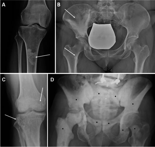
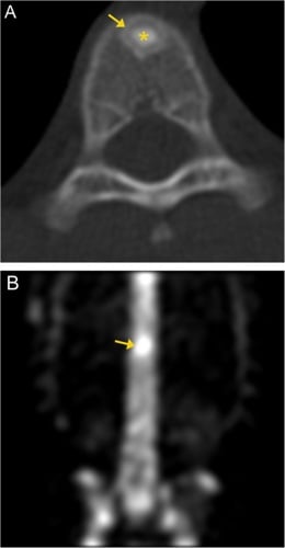
- A bone island (enostosis) has spiculated brush borders blending into trabeculae and shows no or minimal uptake on bone scan; a blastic metastasis has an amorphous margin and is hot.
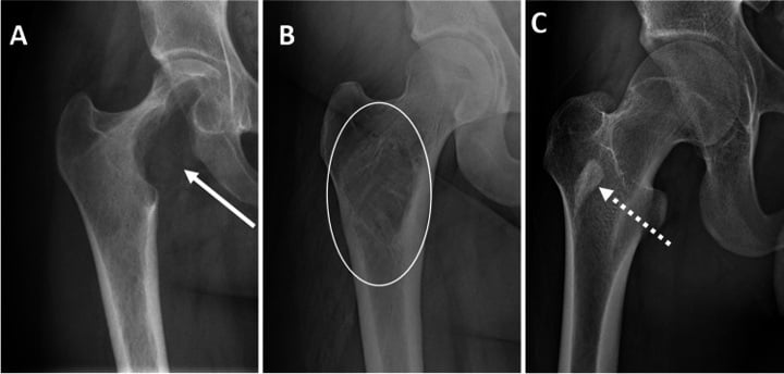
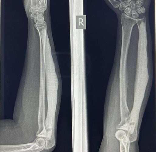
- Osteoid osteoma is a lucent nidus less than 1.5 cm within reactive sclerosis — the sclerosis is the host response, not the lesion.
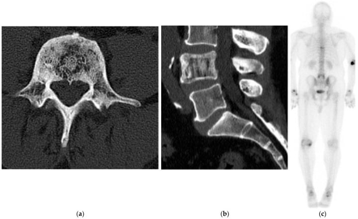
- Diffuse sclerosis in an older man is prostate carcinoma until PSA proves otherwise; check for pedicle involvement (the ivory vertebra).
- Cortical destruction, periosteal reaction and a soft tissue mass alongside sclerosis equals osteosarcoma — refer, do not biopsy locally.
- “Say the four descriptors aloud: site in bone, zone of transition, matrix, host response. That sequence alone earns the mark.
- “Ivory vertebra triad: metastasis (prostate, breast), lymphoma, Paget disease — Paget expands the vertebra, the other two do not.
- “Growing bone island (greater than 25 percent size change in six months, or 1 cm per year) is no longer safely benign — image further.
- “Osteopoikilosis is multiple bone islands clustered periarticularly, symmetrical, and cold on bone scan — never biopsy it.
Bone islands are cold or barely warm on bone scan and have brush borders. An amorphous, hot, painful sclerotic focus in a patient over 50 with a known carcinoma is a metastasis. Never reassure without checking margins and symptoms.
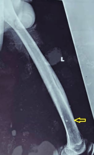
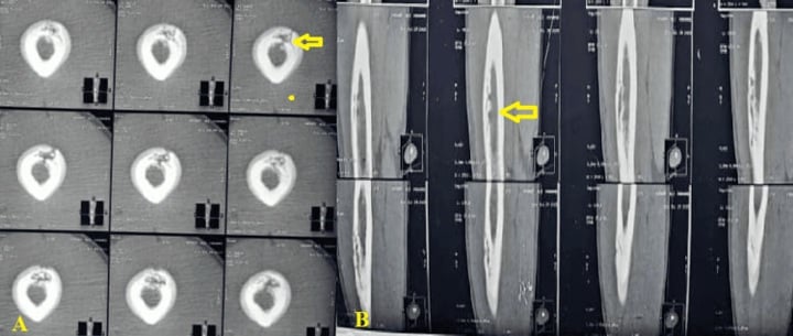
A dense cortical thickening in a teenager with night pain relieved by NSAIDs is osteoid osteoma. Plain films often hide the nidus inside the reactive bone — thin-slice CT is the diagnostic test, not MRI, which overcalls the oedema.
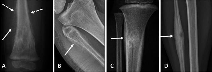
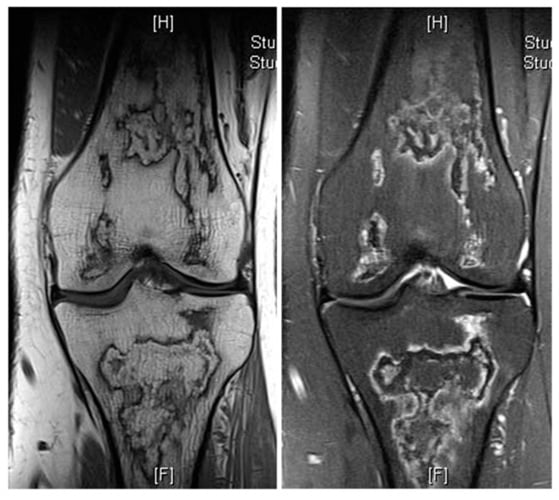
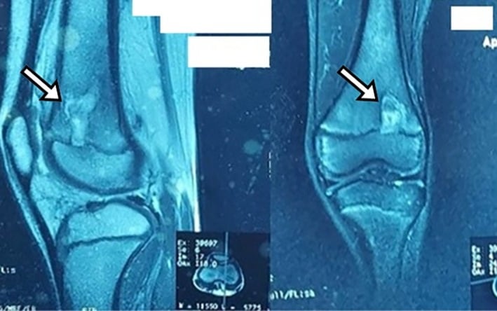
Mixed sclerotic lesion, aggressive periosteal reaction, soft tissue mass in a 15-year-old metaphyseal knee. Any unplanned biopsy tract contaminates the limb salvage plan. Refer to the sarcoma unit for staging and biopsy.
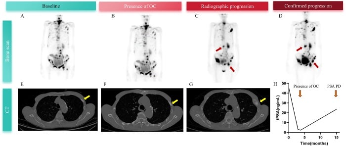
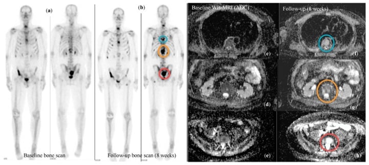
Lytic metastases that have responded to chemotherapy or hormonal therapy sclerose. New sclerosis after treatment may be healing, not progression — correlate with the pre-treatment films before declaring failure.
Recognising the Pattern
Definition. A sclerotic bone lesion is a focal or diffuse area of increased radiodensity relative to adjacent normal bone. Density arises from one of three mechanisms:
- Reactive/host bone — the bone responds to an irritant (osteoid osteoma nidus, chronic infection, stress).
- Tumour osteoid or matrix — the lesion itself makes bone or dense matrix (osteosarcoma, osteoblastic metastasis, enostosis).
- Dead or compressed bone — infarct, avascular necrosis, compressed trabeculae in a vertebral collapse.
Confirming it is genuinely present. Check two orthogonal views: overlapping structures, a bowel gas–free abdomen film, or a poorly penetrated projection all fake sclerosis. Confirm the density is intra-osseous, not surface calcification or an overlying soft tissue calcification. Compare with the contralateral side where possible.
Mimics / false positives.
- Overlapping cortex at a curved surface (femoral neck calcar, medial tibial plateau) — resolves on the lateral.
- Sesamoids, accessory ossicles and phleboliths projected over bone.
- Beam hardening and metallic artefact on CT.
- Healing fracture callus and post-surgical bone graft.
- Normal variants: dense metaphyseal growth lines, the sclerotic margin of a bone infarct on a knee film in an adult.
The viva script. "This is a plain radiograph of the [bone]. There is a focal sclerotic lesion in the [epiphysis/metaphysis/diaphysis], measuring approximately [x] cm, lying [centrally/eccentrically/cortically]. It has a narrow zone of transition with brush borders, there is no cortical destruction, no periosteal reaction and no soft tissue mass. The appearances are those of a latent, non-aggressive lesion, most consistent with a bone island. I would confirm with the clinical history — specifically pain, night pain, known primary malignancy — and, if there were any doubt, a bone scan."
Swap the second half if aggressive: "There is a wide zone of transition, cortical breach, aggressive periosteal reaction with Codman triangle and spiculation, and an associated soft tissue mass containing cloud-like osteoid matrix. This is an aggressive bone-forming lesion and I would treat it as osteosarcoma until proven otherwise."
Next Investigation
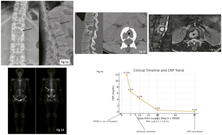
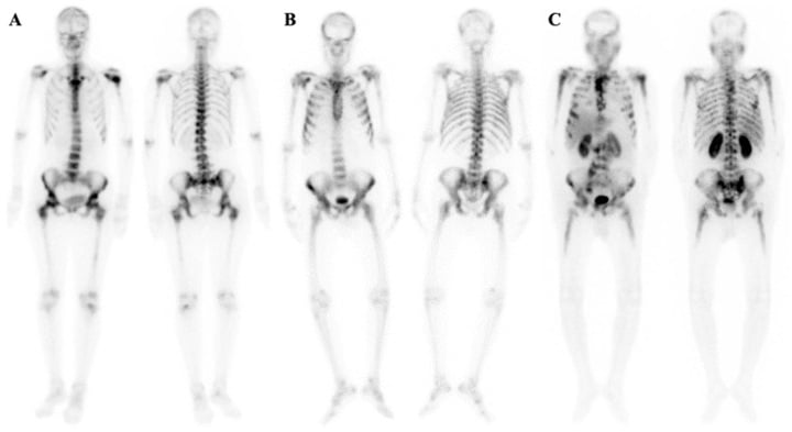
MLPIvory Vertebra — the classic triad
The Differential
- Typical age / setting
- Over 40; known prostate, breast, carcinoid, TCC bladder
- Discriminating feature
- Multiple, amorphous, axial skeleton and proximal long bones; ill-defined margins; hot on bone scan; often painful
- What confirms it
- Whole-body bone scan plus PSA, mammography, CT chest–abdomen–pelvis; biopsy if solitary and no known primary
- Typical age / setting
- 10–25 years; second peak over 60 in Paget bone or post-radiation
- Discriminating feature
- Metaphysis about the knee; mixed lysis and cloud-like osteoid, Codman triangle, sunburst spiculation, soft tissue mass
- What confirms it
- MRI whole bone for skip lesions, CT chest, biopsy at the sarcoma unit only
- Typical age / setting
- Any age, peak 40–60
- Discriminating feature
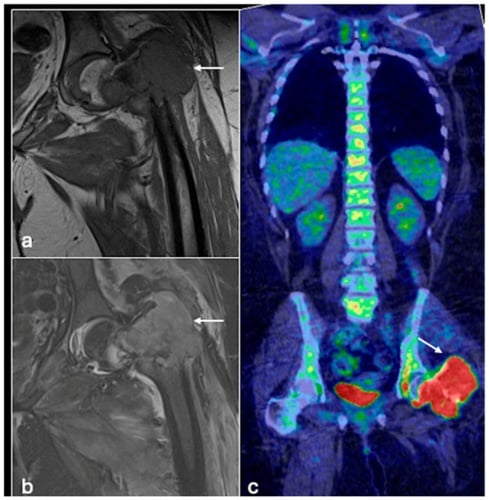
- Ivory vertebra or permeative diaphyseal sclerosis with a soft tissue mass grossly out of proportion to the bone change
- What confirms it
- MRI showing large extraosseous component; core biopsy with flow cytometry
- Typical age / setting
- Children and young adults; diabetics, IVDU, prior open fracture
- Discriminating feature
- Central lucency with thick surrounding sclerosis, sequestrum, involucrum, cloaca or sinus tract; serpiginous tract on MRI
- What confirms it
- MRI with contrast (rim enhancement), CRP/ESR, biopsy and culture before antibiotics
- Typical age / setting
- Any age; incidental, pelvis, femur, vertebral body
- Discriminating feature
- Usually less than 2 cm, spiculated brush borders blending with trabeculae, cold or barely warm on bone scan, stable over years
- What confirms it
- Comparison with old films; bone scan if any doubt — no further imaging if classic and asymptomatic
- Typical age / setting
- 10–25 years; male predominance; femoral neck, tibial diaphysis, posterior spinal elements
- Discriminating feature
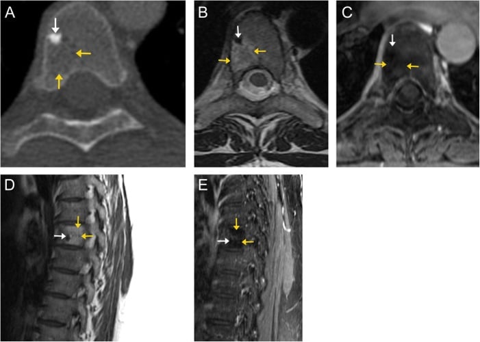
- Nidus less than 1.5 cm surrounded by dense reactive cortical sclerosis; night pain dramatically relieved by NSAIDs; painful scoliosis if spinal
- What confirms it
- Thin-slice CT to show the nidus; double-density sign on bone scan; CT-guided radiofrequency ablation is both proof and cure
- Typical age / setting
- 10–30 years; posterior elements of spine, sacrum, long bones
- Discriminating feature
- Greater than 2 cm, expansile, more lytic with sclerotic rim; pain not typically night-predominant nor NSAID-responsive
- What confirms it
- CT plus MRI to map the posterior element and canal; open biopsy and curettage
- Typical age / setting
- Over 55; pelvis, femur, tibia, skull, vertebrae
- Discriminating feature
- Bone is EXPANDED with cortical thickening and coarse trabeculae — expansion is the giveaway; picture-frame vertebra, blade of grass lytic front
- What confirms it
- Radiographs usually sufficient; raised ALP with normal calcium and phosphate; bone scan for extent
- Typical age / setting
- Adults; steroids, sickle cell, alcohol, caisson disease
- Discriminating feature
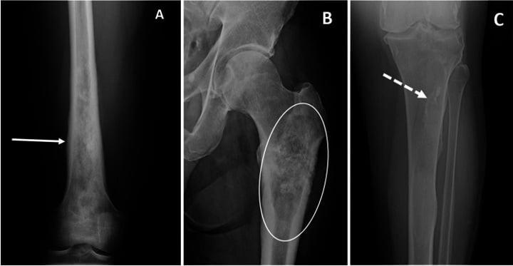
- Serpiginous, geographic sclerotic rim in metadiaphysis; medullary, not cortical; epiphyseal crescent sign if AVN
- What confirms it
- MRI double-line sign; no biopsy needed if classic
- Typical age / setting
- Athletes, recruits, elderly osteoporotic; tibia, metatarsal, femoral neck, sacrum
- Discriminating feature
- Linear sclerotic band PERPENDICULAR to trabeculae with localised periosteal reaction; activity-related pain
- What confirms it
- MRI STIR oedema with a low-signal line; clinical history of activity change
- Typical age / setting
- Any age; post-chemotherapy, healed non-ossifying fibroma, healed fibrous dysplasia
- Discriminating feature
- Sclerosis in the exact shape of a previously lytic lesion; NOF sclerosis fills in from the periphery in adolescence
- What confirms it
- Previous radiographs — the single most valuable investigation; no further imaging if the shape matches
- Typical age / setting
- Sclerosing bone dysplasias; osteopoikilosis often familial and asymptomatic
- Discriminating feature
- Osteopoikilosis: multiple symmetrical periarticular islands. Osteopathia striata: vertical striations. Melorheostosis: dripping candle wax along one sclerotome
- What confirms it
- Radiographs alone; cold bone scan in osteopoikilosis distinguishes it from blastic metastases
- Typical age / setting
- Children and young adults; clavicle, sternocostoclavicular region, metaphyses
- Discriminating feature
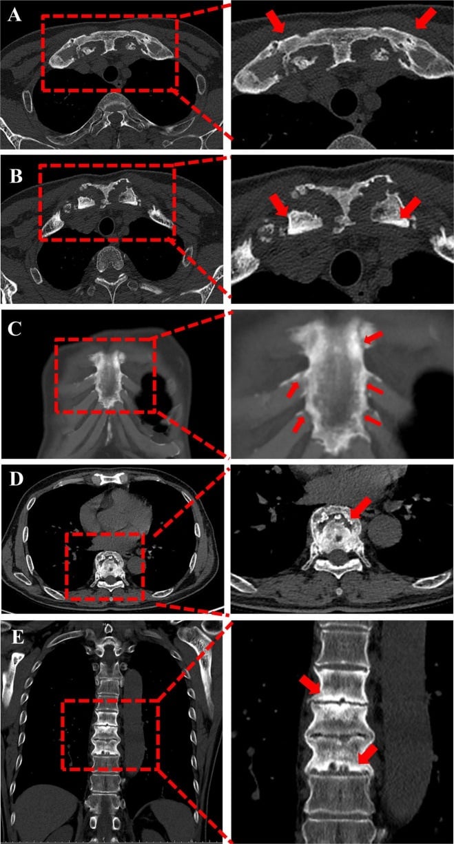
- Hyperostosis of the medial clavicle with palmoplantar pustulosis or acne; multifocal, culture-negative, relapsing
- What confirms it
- Whole-body MRI for multifocality; biopsy negative culture; response to NSAIDs or bisphosphonates
- Typical age / setting
- Adults; endemic fluoride areas, haematological disease, dialysis
- Discriminating feature
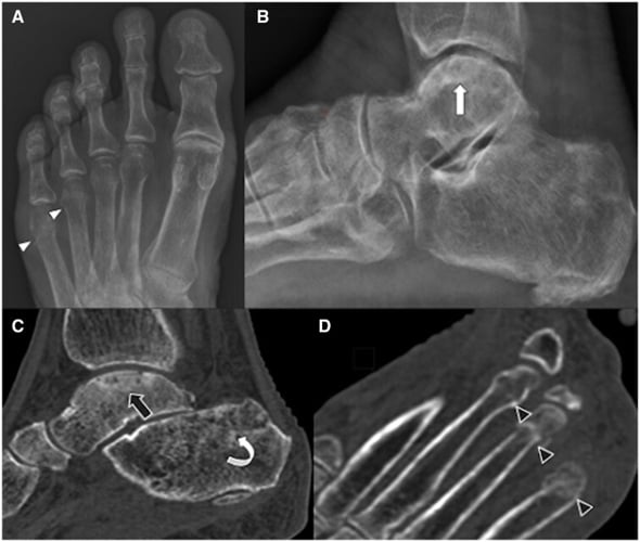
- DIFFUSE symmetrical sclerosis rather than focal; rugger-jersey spine in renal disease; ligamentous ossification in fluorosis
- What confirms it
- Biochemistry (calcium, phosphate, PTH, ALP), full blood count and film, marrow biopsy if myelofibrosis suspected
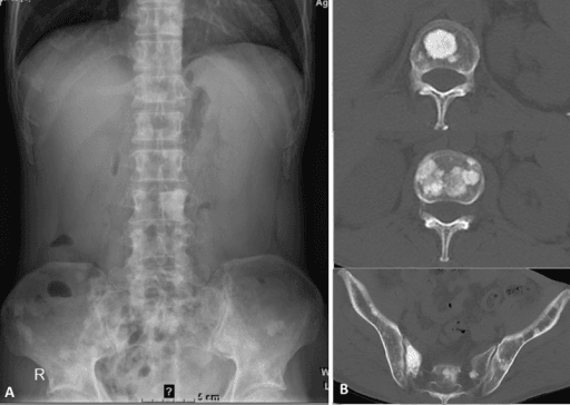
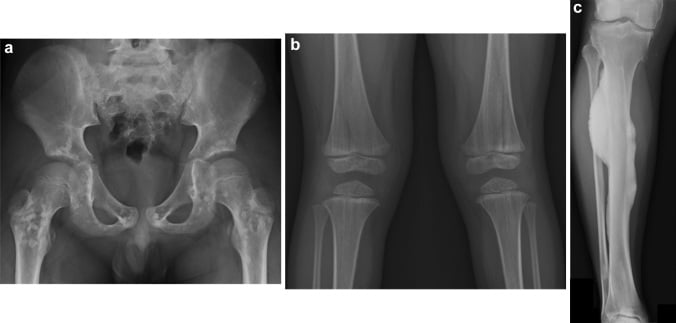
Narrowing It Down
- 1Step 1 — How old is the patient?
Establish the age before reading the film in any detail, and let it set the prior.
Under 30: osteoid osteoma, osteoblastoma, chronic osteomyelitis, osteosarcoma, CRMO, healed non-ossifying fibroma. Over 40: metastasis, lymphoma, Paget, myeloma variant, infarct. Age alone removes half the list and should be your opening sentence.
- 2Step 2 — Solitary or multiple?
Look for other lesions before characterising the one you were shown, and check the distribution.
Multiple, symmetrical and periarticular equals osteopoikilosis - cold on scan, reassure, never biopsy. Multiple, random, axial-predominant and hot equals metastases. Solitary keeps the whole primary tumour differential open. Note the base rate that makes this step matter: 89 per cent of healthy young adults have at least one bone island and 11 of 46 in Sala's series had more than three, so multiplicity ALONE does not imply disease.
- 3Step 3 — Where in the bone?
Name the segment and the compartment - cortical, medullary, epiphyseal, posterior elements - before naming a lesion.
Cortical with dense fusiform thickening equals osteoid osteoma or stress fracture. Medullary with a serpiginous rim equals infarct. Epiphyseal equals avascular necrosis or chondroblastoma. Posterior spinal elements equal osteoblastoma or osteoid osteoma. Metaphyseal about the knee in a teenager equals osteosarcoma.
- 4Step 4 — MEASURE the attenuation, then assess the margin
On CT, place the broadest possible region of interest at the centre of the lesion and record the mean Hounsfield value - then look at the border.
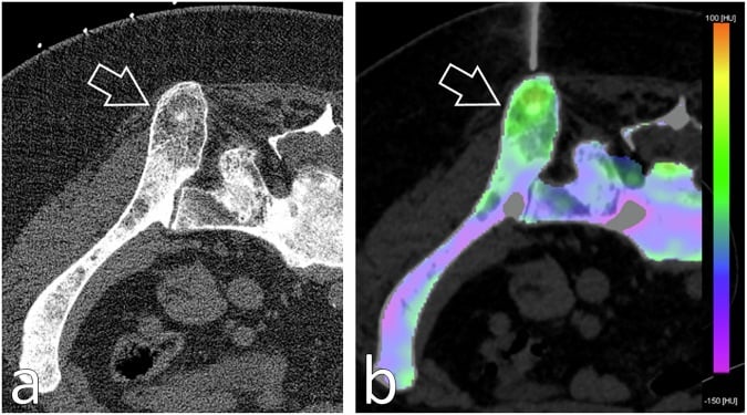
A mean above about 881 HU is an enostosis: Sala's bone islands averaged 1007 to 1052 HU against 712 to 728 for untreated osteoblastic metastases, giving 98 per cent sensitivity and 95 per cent specificity at that cut-off. Brush borders blending into trabeculae support it. A crisp sclerotic rim means a slow benign process; ill-defined, amorphous or permeative means aggressive - metastasis, lymphoma, osteosarcoma, infection. Check the threshold against your own scanner before trusting it, because attenuation depends on kVp and reconstruction kernel.
- 5Step 5 — Is the host bone reacting?
Look specifically for periosteal reaction and classify its pattern rather than simply noting its presence.
Solid, thick, single-layer reaction equals benign or chronic - osteoid osteoma, stress, osteomyelitis. Codman triangle, sunburst or lamellated onion-skin equals aggressive. Absent reaction with a cold, sharp lesion equals latent.
- 6Step 6 — Is the bone expanded?
Compare the overall calibre of the affected bone with the level above and below, and check the alkaline phosphatase.
Expansion with cortical thickening and coarse trabeculae in an older patient equals Paget disease. Metastases and lymphoma sclerose the vertebra WITHOUT expanding it. This single feature separates the three causes of an ivory vertebra.
- 7Step 7 — What are the systemic features, and if you must biopsy, where?
Take the history and bloods - night pain, fever, weight loss, known carcinoma, PSA, alkaline phosphatase, inflammatory markers - and if biopsy is needed, plan the target on the CT.
Night pain relieved by NSAIDs equals osteoid osteoma. Fever, raised CRP and a sinus equals osteomyelitis. Weight loss, known carcinoma and raised PSA or ALP equals metastasis. Pustulosis with clavicular hyperostosis equals SAPHO. When a biopsy is required, aim at the LEAST dense part of the lesion: Donners found 87 per cent of biopsies of mildly sclerotic bone (mean 244 HU) were tumour-positive against 56 per cent in dense sclerosis (622 HU), and needle gauge, core number and PET guidance made no difference.
The commonest safe endpoint in this algorithm is Step 4. If the borders brush, the lesion is under 2 cm, the patient is asymptomatic, and old films show stability — you are finished. Say so confidently rather than ordering an MRI.
MCQ Practice Points
Q: Which single objective CT feature best distinguishes a bone island from an untreated osteoblastic metastasis?
A: The mean attenuation of the lesion. Sala measured enostoses at 1007 to 1052 HU against 712 to 728 HU for untreated osteoblastic metastases, and a cut-off of 881 HU separated them with 98 per cent sensitivity and 95 per cent specificity. Place a broad region of interest at the centre of the lesion. Brush borders blending with trabeculae and a cold bone scan support the same conclusion, but they are subjective while the Hounsfield value is not. Caveat for the viva: the threshold was derived and tested on the same 66 patients, the two arms were different populations, and attenuation shifts with kVp and reconstruction kernel — so verify it locally rather than quoting it as a universal constant.
Q: What proportion of healthy young adults have at least one bone island on CT, and why does the number matter?
A: Eighty-nine per cent — 41 of 46 polytrauma patients aged 14 to 35 with no cancer in Sala's series, carrying 124 lesions between them, and 11 of the 46 had more than three. Commonest in the proximal femur (34 per cent), pelvis (22 per cent) and acetabulum (20 per cent). The finding is therefore close to normal anatomy, so an incidental dense focus in an asymptomatic patient needs no follow-up and no bone scan, and multiplicity alone does not imply metastatic disease.
Q: A sclerotic lesion in a cancer patient must be biopsied. What single planning decision most changes the chance of a diagnostic result?
A: Target the least dense part of the lesion, not the most conspicuous. Donners found 87 per cent of biopsies of mildly sclerotic bone (mean 244 HU) were tumour-positive against 56 per cent in dense sclerosis (622 HU), p equals 0.005; a 610 HU threshold gave an 89 per cent positive predictive value for tumour and 370 HU a 94 per cent predictive value for successful next-generation sequencing. Needle diameter, number of cores, tract length, lesion size, PET availability and tumour markers were all non-significant. Sixty-five biopsies, one operator, one centre — so treat the thresholds as a planning heuristic rather than a validated rule.
Q: Which of the ivory vertebra causes expands the vertebral body?
A: Paget disease. Metastasis and lymphoma sclerose the vertebra without expanding it. Paget also shows cortical thickening, coarsened trabeculae and an isolated raised alkaline phosphatase with normal calcium and phosphate. Lymphoma is the one to suspect when the soft tissue mass is out of proportion to the bone change.
Q: Multiple small symmetrical sclerotic foci clustered around the hips, shoulders and carpus in an asymptomatic 30-year-old. Diagnosis and action?
A: Osteopoikilosis. Autosomal dominant, LEMD3 mutation, benign. The distribution is the diagnosis: symmetrical, periarticular, sparing the skull and ribs. Confirm with a cold bone scan if there is diagnostic anxiety. No biopsy, no follow-up imaging; may be associated with Buschke–Ollendorff syndrome with dermatofibrosis lenticularis disseminata.
Q: What sclerotic lesion feature most strongly predicts aggression regardless of the underlying diagnosis?
A: A wide zone of transition. Combined with cortical destruction, aggressive periosteal reaction (Codman triangle, sunburst, onion-skin) and a soft tissue mass, it mandates staging and specialist referral before any tissue is taken. In a teenager with metaphyseal cloud-like osteoid, an unplanned local biopsy contaminates the limb salvage plan.
Q: A 17-year-old has night pain in the thigh relieved by ibuprofen and a plain film showing dense fusiform cortical thickening of the femoral diaphysis. What is the best investigation?
A: Thin-slice CT. It demonstrates the radiolucent nidus (less than 1.5 cm) hidden within the reactive sclerosis and guides radiofrequency ablation. MRI overestimates the surrounding oedema and may obscure the nidus altogether — a common cause of a missed diagnosis in a patient who has already had the "right" scan.
Exam Viva Scenarios
Practise clinical reasoning and management decisions out loud
“You are shown this pelvic radiograph of a 46-year-old woman taken for hip pain after a fall. There is a 1.2 cm rounded sclerotic focus in the intertrochanteric region. Describe it and give your differential.”
“You are shown this radiograph of the distal femur of a 15-year-old boy with six weeks of progressive thigh pain, now waking him at night. There is a mixed lytic and sclerotic metaphyseal lesion with cortical breach.”
“You are shown a lateral thoracolumbar radiograph of a 72-year-old man with back pain. L2 is uniformly dense compared with adjacent vertebrae. How do you approach this?”
Describe in this order
- Bone and site within it: epiphysis, metaphysis, diaphysis; central, eccentric, cortical, juxtacortical
- Size and number: solitary versus multiple
- Zone of transition: brush borders, sharp rim, ill-defined, permeative
- Matrix: homogeneous compact bone, cloud-like osteoid, ring-and-arc chondroid
- Host response: periosteal reaction type, cortical integrity, expansion, soft tissue mass
Age shortcuts
- Under 30: osteoid osteoma, osteoblastoma, osteosarcoma, chronic osteomyelitis, CRMO, healed NOF
- Over 40: osteoblastic metastasis, lymphoma, Paget disease, infarct, myelofibrosis
- Any age: bone island, osteopoikilosis, stress fracture, bone infarct
One-line discriminators
- Brush borders equals bone island
- Nidus less than 1.5 cm with night pain relieved by NSAIDs equals osteoid osteoma
- Greater than 2 cm, expansile, posterior spinal elements equals osteoblastoma
- Expanded vertebra with raised ALP equals Paget disease
- Soft tissue mass out of proportion to bone change equals lymphoma
- Cloud-like osteoid plus Codman triangle in a teenager equals osteosarcoma
- Serpiginous geographic rim in the medulla equals bone infarct
- Linear sclerosis perpendicular to trabeculae equals stress fracture
- Dripping candle wax along one sclerotome equals melorheostosis
Red flags demanding referral
- Wide zone of transition or permeative margin
- Cortical destruction or soft tissue mass
- Aggressive periosteal reaction
- Documented growth of a previously stable lesion
- Night pain not relieved by NSAIDs, weight loss, or new neurological signs
Investigation logic
- Old films first — stability is diagnostic reassurance
- CT for nidus, matrix, sequestrum and cortical detail
- Bone scan for solitary versus multiple and for lesion activity
- MRI whole bone before biopsy in any suspected primary bone tumour
- Staging CT plus PSA, mammography and myeloma screen when metastasis is suspected
- No further imaging for a classic asymptomatic stable bone island or osteopoikilosis
Evidence Base
Bone Islands Incidentally Detected on CT: Frequency of Enostosis and Differentiation from Untreated Osteoblastic Metastases by CT Attenuation
- 46 POLYTRAUMA patients aged 14-35 with no cancer and no previous fracture, imaged by thoracoabdominal CT, against 20 patients with untreated osteoblastic metastases
- 41 of the 46 (89 PER CENT) had at least one enostosis - 124 lesions in total, 2 to 15 mm; 11 patients had more than three
- Commonest sites: proximal femur 34 per cent, pelvis 22 per cent, acetabulum 20 per cent, proximal humerus 11 per cent, vertebrae 11 per cent, rib 2 per cent
- Mean attenuation of the 124 enostoses was 1007 plus or minus 122 HU (first observer) and 1052 plus or minus 107 (second); the 114 untreated osteoblastic metastases measured 728 plus or minus 163 and 712 plus or minus 178
- A CUT-OFF OF 881 HU for mean density gave 98 per cent sensitivity and 95 per cent specificity, with an area under the curve of 0.982
Spectral CT Assists Differentiation of Osteoblastic Bone Metastasis from Bone Island in Newly Diagnosed Cancer Patients
- 73 newly diagnosed cancer patients with 201 bone lesions - 92 osteoblastic metastases and 109 bone islands - all scanned with spectral (dual-energy) CT
- Measurements included CT values across 40-140 keV, spectral curve slope, effective atomic number, water and calcium density pairs, and iodine density
- EVERY feature differed significantly between the two groups (all p less than 0.05)
- A combined plain-plus-enhanced spectral model reached an area under the curve of 0.925 (95 per cent CI 0.879 to 0.957)
- That combined model significantly outperformed plain spectral CT alone (AUC 0.815, p less than 0.001) and enhanced spectral CT alone (AUC 0.901, p equals 0.024)
Optimising CT-Guided Biopsies of Sclerotic Bone Lesions in Cancer Patients
- 65 CT-guided sclerotic bone biopsies in cancer patients at one institution, reviewed against laboratory, imaging and procedural factors
- 74 per cent of biopsies were tumour-positive; next-generation sequencing was feasible in 22 of 30 prostate cancer patients (73 per cent)
- THE DENSITY OF THE TARGET DECIDED THE YIELD: 87 per cent of biopsies of MILDLY sclerotic bone (mean 244 HU) were positive against only 56 per cent in DENSE sclerosis (mean 622 HU), p equals 0.005
- A 610 HU threshold gave an 89 per cent positive predictive value for a tumour-positive biopsy; a 370 HU threshold gave 94 per cent for sequencing feasibility
- Blood parameters, tumour markers, PET availability, lesion size, lesion-to-cortex distance, needle diameter, tract length and number of samples were ALL non-significant
Sclerotic Bone Lesions Caused by Non-Infectious and Non-Neoplastic Diseases: A Review of the Imaging and Clinicopathologic Findings
- A pictorial review assembling the non-infectious, non-neoplastic causes of bone sclerosis - the half of the differential that is neither tumour nor infection
- Groups them as congenital and developmental disorders, depositional disorders, and metabolic diseases
- Named entities include fluorosis, Gaucher disease, pyknodysostosis, renal osteodystrophy and Ribbing disease
- The authors' framing is that the differential is narrowed by combining the imaging appearance with the clinical picture, not by the imaging alone
- Many of the entities covered exist in the literature only as case reports and small series