Intravertebral disc herniation through the cartilaginous endplate — an incidental finding that must be separated from infection, tumour and fracture
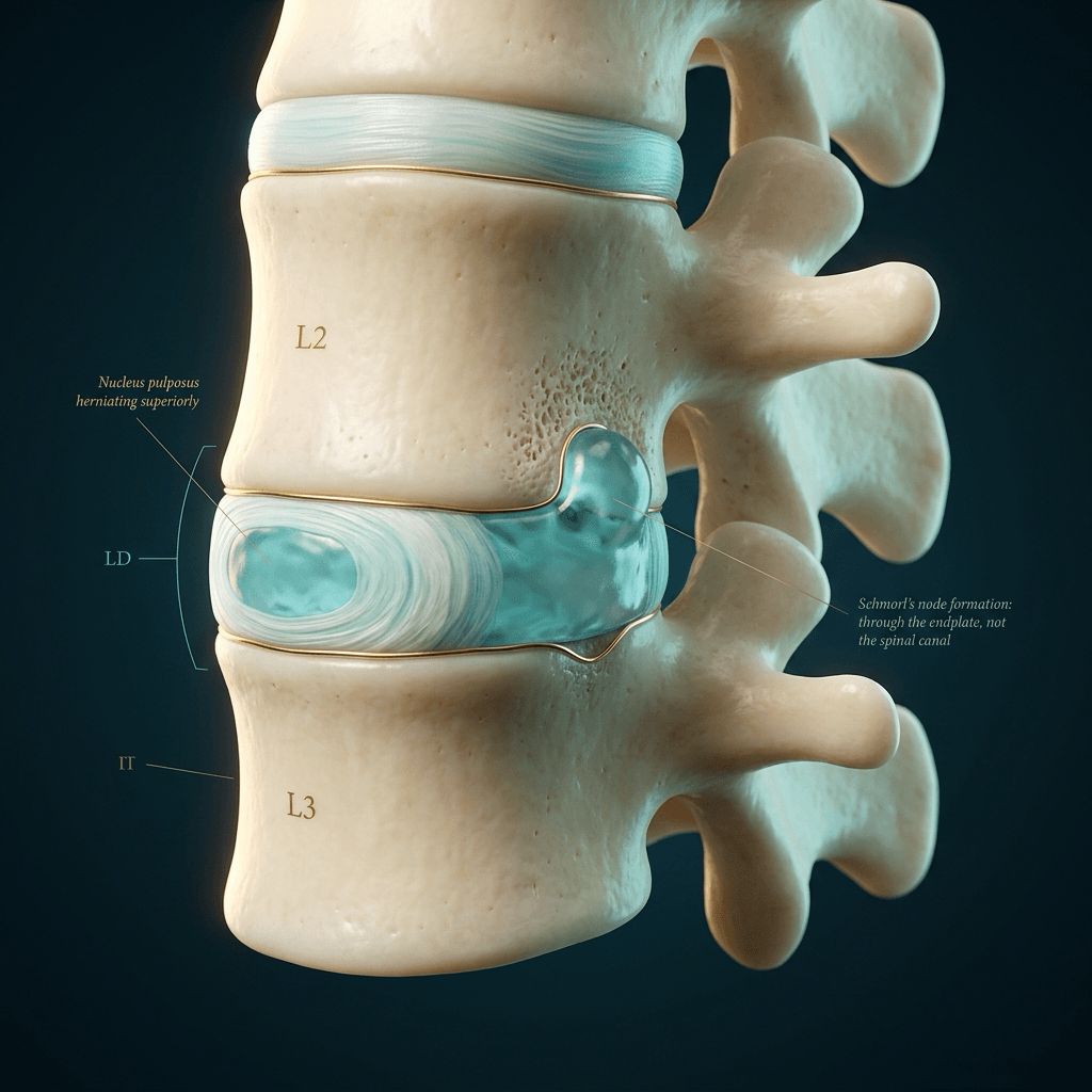
- A Schmorl node is herniation of nucleus pulposus vertically through the cartilaginous endplate into the vertebral body — the disc goes into the bone, not out of the back.
- Commonest in the lower thoracic and upper lumbar spine (T8–L2), where the sagittal curve transition concentrates axial load.
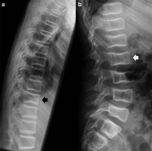
- The classic radiographic sign is a rounded or bracket-shaped indentation of the superior or inferior endplate with a corticated margin, disc height often preserved.
- A chronic Schmorl node is marrow-signal-neutral. An acute one has surrounding oedema on STIR and can be genuinely painful — that does not make it sinister.
- Giant Schmorl nodes can involve most of the vertebral body height and mimic a lytic lesion; the connection to the disc space on sagittal MRI is the giveaway.
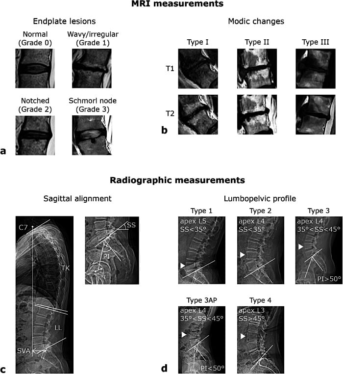
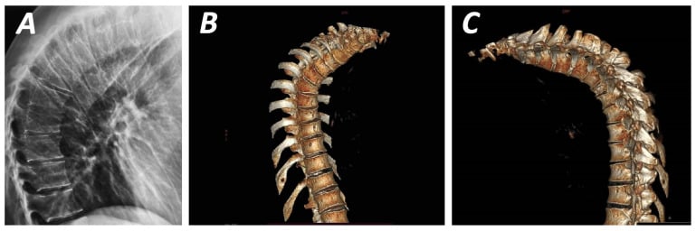
- Scheuermann disease is multiple Schmorl nodes plus anterior wedging of at least 5 degrees across three or more consecutive vertebrae with a thoracic kyphosis greater than 40 degrees.
- Discitis destroys two endplates either side of one disc and collapses the disc space; a Schmorl node spares the disc height and does not cross it.
- “Never call a solitary endplate lucency a Schmorl node unless you can trace a defect in the endplate cortex continuous with the disc.
- “A limbus vertebra is the anterior corner variant — a triangular ossicle separated by a corticated cleft, not a fracture.
- “Cupid's bow deformity is a normal parasagittal concavity of the lower lumbar inferior endplates and is bilateral and symmetric — do not report it as Schmorl nodes.
- “Multiple Schmorl nodes in a young athlete plus kyphosis equals Scheuermann; multiple in an older patient with fragility equals osteoporotic endplate failure.
- “If there is marrow oedema on both sides of a disc with loss of disc height and paraspinal soft tissue, that is infection until cultured otherwise.
Discitis begins in the subchondral endplate and looks focal and lucent early. The discriminators are: involvement of BOTH endplates facing the same disc, loss of disc height, T2 hyperintense disc, and paravertebral or epidural soft tissue. A Schmorl node has none of these.
A giant node can occupy most of the vertebral body with a sclerotic rim and surrounding oedema, and lights up on bone scan. Follow it to the endplate on sagittal MRI — a continuous track to the disc space with disc material inside proves the diagnosis. Metastasis has no such track.
An acute Schmorl node in an adolescent or after axial load causes real mechanical pain with focal STIR oedema. Reporting "degenerative, no acute finding" misses the source of pain. Say "acute intravertebral herniation with reactive oedema".
Cupid's bow (bilateral parasagittal concavity, L4–L5), notochordal remnant clefts, and the coronal cleft of infancy all indent the endplate. They are symmetric, bilateral and unaccompanied by marrow change.
Recognising the Pattern
Definition. A Schmorl node is a focal herniation of nucleus pulposus through a defect in the cartilaginous endplate and subchondral bone plate, into the cancellous bone of the vertebral body. It is an intravertebral disc herniation. The defect arises where the endplate is congenitally weak — the notochordal remnant, a vascular channel scar, or an area thinned by degeneration, osteoporosis, Paget disease or tumour.
Where it occurs. Lower thoracic and thoracolumbar spine predominantly (T8 to L2). Within the endplate it favours the posterior two-thirds, near the midline, and the inferior endplate slightly more than the superior. In Scheuermann disease the nodes cluster in the mid-to-lower thoracic spine with anterior wedging.
Confirming it is genuinely present.
- There must be a defect in the endplate cortex — a break in the thin white line, continuous with the disc.
- The lucency must be based on the endplate, not floating in the middle of the vertebral body.
- There should be a corticated or sclerotic rim around the intraosseous portion in chronic lesions.
- Disc height is preserved or only mildly reduced — the disc has moved up or down, not disappeared.
- On sagittal MRI, the herniated material follows disc signal (T2 bright if hydrated, dark if degenerate) and is contiguous with the parent disc.
Saying it out loud in a viva. "There is a rounded, corticated indentation of the inferior endplate of L1 in the posterior third, with an interruption of the endplate cortex and preserved disc height at T12–L1. There is no vertebral body collapse, no paravertebral soft tissue and no involvement of the opposing endplate. The appearances are those of a Schmorl node — an intravertebral disc herniation. I would look for further nodes and for anterior wedging to determine whether this is isolated or part of Scheuermann disease."
Mimics — the false positives.
- Cupid's bow deformity — bilateral, symmetric parasagittal concavities of the inferior endplates of L3–L5; on axial imaging two paramedian depressions, not a single midline defect. Normal variant.
- Coronal cleft vertebra — infantile, vertical lucency splitting the body, resolves with growth.
- Limbus vertebra — anterosuperior corner ossicle from herniation beneath the ring apophysis; corticated cleft, sharp triangular fragment. Not acute trauma.
- Endplate erosion of discitis — irregular, ill-defined, both sides of the disc, disc height lost.
- Osteoporotic endplate impaction — diffuse endplate concavity ("fish vertebra"), not a focal corticated defect.
- Focal fat deposition / Modic type 2 — signal follows fat on all sequences, no cortical break.
- Intraosseous haemangioma abutting the endplate — polka-dot on axial CT, T1 bright with fat, no endplate defect.
Next Investigation
Nuclear medicine bone scans and PET-CT are unhelpful discriminators here: acute and giant Schmorl nodes are avid and will be reported as suspicious. Anatomical imaging — the track to the disc — settles the question, not metabolic imaging.
The Differential
The differential is twofold: (a) causes of a genuine Schmorl node — that is, what weakened the endplate; and (b) lesions that mimic an endplate-based lucency. Both are listed because in a viva you must exclude the sinister before attributing to degeneration.
- Typical age / setting
- Any age; diabetic, IVDU, post-instrumentation, immunosuppressed
- Discriminating feature
- BOTH endplates around one disc destroyed, disc height lost, disc T2 hyperintense, paravertebral or epidural phlegmon
- What confirms it
- MRI with contrast; CT-guided biopsy and blood cultures
- Typical age / setting
- Older than 50, known primary, night pain, weight loss
- Discriminating feature
- Lesion centred on the vertebral body or pedicle, NOT the endplate; no track to the disc; pedicle destruction; disc spared
- What confirms it
- MRI whole spine T1 (marrow replacement, T1 darker than disc); biopsy
- Typical age / setting
- Adolescent or young adult; axial load, fall, weightlifting
- Discriminating feature
- Focal STIR oedema around a fresh endplate defect with sharp, non-corticated margins; matching mechanical pain
- What confirms it
- MRI STIR; CT shows the acute cortical break without sclerosis
- Typical age / setting
- Older adult; myeloma, lymphoma, renal cell
- Discriminating feature
- Endplate defect is irregular and lacks a cortical rim; posterior cortex bulges; soft tissue present
- What confirms it
- MRI plus staging CT; biopsy of the marrow lesion
- Typical age / setting
- Any adult; commonest cause overall
- Discriminating feature
- Solitary or few, corticated, T8–L2, no marrow oedema, disc degenerate but height preserved
- What confirms it
- Radiograph alone; no further imaging required if asymptomatic
- Typical age / setting
- Adolescent 12–17, thoracic kyphosis, rigid on extension
- Discriminating feature
- Multiple nodes PLUS anterior wedging at least 5 degrees over 3 or more consecutive vertebrae, kyphosis greater than 40 degrees, irregular endplates
- What confirms it
- Standing lateral whole-spine radiograph with Cobb measurement
- Typical age / setting
- Postmenopausal women, chronic steroids
- Discriminating feature
- Multiple nodes with generalised endplate concavity and reduced trabecular density; other fragility fractures
- What confirms it
- DXA; lateral spine radiograph or VFA showing wedge/biconcave fractures
- Typical age / setting
- Children and young adults, incidental
- Discriminating feature
- Strictly midline, posterior third, tiny, sharply corticated, no symptoms
- What confirms it
- No further imaging; recognise as normal variant
- Typical age / setting
- Middle-aged adults; often thoracolumbar
- Discriminating feature
- Occupies a large fraction of the body height but retains a continuous track to the disc and a sclerotic rim
- What confirms it
- Sagittal MRI showing disc-signal contiguity; CT shows the endplate defect
- Typical age / setting
- Older than 55
- Discriminating feature
- Cortical and trabecular thickening, picture-frame vertebra, enlarged body — endplate defect on abnormally coarse bone
- What confirms it
- Radiograph pattern plus raised alkaline phosphatase
- Typical age / setting
- Dialysis population
- Discriminating feature
- Rugger-jersey spine with subchondral sclerotic bands; endplate resorption elsewhere (phalanges, distal clavicle)
- What confirms it
- Serum calcium, phosphate, PTH; hand radiographs
- Typical age / setting
- Afro-Caribbean, childhood onset
- Discriminating feature
- H-shaped (Lincoln log) central endplate depression — square-shouldered, central, bilateral, not a focal node
- What confirms it
- Haemoglobin electrophoresis
- Typical age / setting
- Children and young adults, hepatosplenomegaly
- Discriminating feature
- Diffuse marrow T1 hypointensity with step-off endplate depressions and Erlenmeyer flask femora
- What confirms it
- Enzyme assay (beta-glucocerebrosidase)
- Typical age / setting
- Any age with a treatment field
- Discriminating feature
- Endplate change confined sharply to the radiation portal with fatty (T1 bright) marrow either side
- What confirms it
- Correlate treatment field; MRI showing geographic fatty marrow
Narrowing It Down
- 1Step 1 — Is it truly endplate-based and disc-contiguous?
Trace the endplate cortex on a sagittal image and look for continuity between the defect and the disc space.
A break continuous with the disc, with herniated material of disc signal inside, defines a Schmorl node. A lesion sitting in the body or pedicle with an INTACT endplate is a marrow lesion - metastasis, myeloma, infection or haemangioma - and the pathway changes entirely. This single question separates benign from sinister in most cases.
- 2Step 2 — One endplate or both sides of the same disc?
Look at the vertebra above AND below the disc, and measure the disc height.
A Schmorl node affects one endplate. Symmetrical destruction of the endplates on both sides of a single disc with loss of disc height is discitis-osteomyelitis until proven otherwise. Metastasis characteristically SPARES the disc and crosses to non-contiguous levels instead.
- 3Step 3 — Is the margin corticated?
Assess the rim of the defect specifically, on the plain film before the MRI.
A thin sclerotic rim means chronicity and stability - a healed, incidental node needing no action. An ill-defined, non-corticated, permeative margin means an active process: acute herniation, infection or tumour. Marginal cortication is the most reliable single reassuring feature on plain film.
- 4Step 4 — Solitary or multiple, and what is the pattern?
Count the nodes and look at the shape of the vertebral bodies around them, not just at the defect.
Solitary at T12-L1 in an adult equals degenerative. Multiple in a kyphotic adolescent with anterior wedging equals Scheuermann. Multiple with generalised osteopenia and biconcave bodies equals osteoporosis. Multiple with central H-shaped depression equals sickle cell. Multiple with dense subchondral bands equals renal osteodystrophy.
- 5Step 5 — Is there marrow oedema, and is it PROPORTIONATE?
Get STIR, and judge the oedema against the size of the defect rather than simply recording its presence.
A rim of STIR hyperintensity hugging the node is reactive and expected in an acute or symptomatic node. Oedema replacing the whole body, extending across the disc, or associated with epidural enhancement is NOT proportionate and mandates a search for infection or tumour.
- 6Step 6 — Are there systemic features?
Take the temperature, check CRP and ESR, and ask about weight loss, night pain and prior malignancy.
Fever and raised inflammatory markers point to infection. Weight loss, known malignancy, night pain and age over 50 point to metastasis - and a NEW endplate lesion in a patient with cancer is the one situation where this diagnosis genuinely matters, because the imaging features that distinguish it from a metastasis can avoid a biopsy. Neither red flag is required for a Schmorl node; their absence plus a corticated defect closes the case.
- 7Step 7 — Does it explain the patient's pain?
Ask whether the level matches, the pain is mechanical and axial, and there is corresponding oedema - all three, not one.
Set the answer against the base rate: at least one Schmorl node is present in 71 to 77 per cent of an unselected older population, so the prior probability that any given node is incidental is very high. If the node is signal-neutral and the level does not match, report it as incidental and keep looking for another cause. Calling a near-universal finding the source of the pain is the commonest error on this page.
MCQ Practice Points
Q: Which single feature most reliably distinguishes a giant Schmorl node from a vertebral metastasis on MRI? A: A continuous track of disc-signal material extending from the parent intervertebral disc through the endplate defect into the lesion. Metastasis has no communication with the disc and typically replaces marrow so that T1 signal becomes darker than the adjacent disc.
Q: A 14-year-old has a rigid thoracic kyphosis of 62 degrees. What radiographic criteria confirm Scheuermann disease? A: Anterior wedging of at least 5 degrees at three or more consecutive vertebrae, with endplate irregularity, Schmorl nodes and disc space narrowing, and a thoracic Cobb angle greater than 40 degrees. Postural kyphosis is flexible and has normal endplates and no wedging.
Q: Where within the vertebral endplate do Schmorl nodes most often occur, and why? A: In the posterior two-thirds near the midline, corresponding to the notochordal remnant and to vascular channel scars where the cartilaginous endplate and subchondral bone plate are congenitally thinnest. The thoracolumbar junction (T8 to L2) is the commonest region because axial load is concentrated at the kyphosis-to-lordosis transition.
Q: Which normal variant is most often misreported as bilateral Schmorl nodes in the lower lumbar spine? A: The Cupid's bow deformity — symmetric parasagittal concavities of the inferior endplates of L3 to L5, best appreciated on the frontal radiograph or axial CT as two paramedian depressions. It is bilateral, symmetric, without cortical breach and without marrow signal change.
Q: What single imaging feature most strongly favours discitis-osteomyelitis over a Schmorl node? A: Destruction of both endplates facing the same intervertebral disc with loss of disc height and abnormal T2 hyperintense signal within that disc, usually accompanied by paravertebral or epidural enhancing soft tissue. A Schmorl node affects one endplate and preserves the disc.
Q: An acute Schmorl node causes back pain in a 22-year-old weightlifter. What will the MRI show? A: A sharply marginated, non-corticated endplate defect with disc material within the body and a surrounding halo of STIR/T2 marrow oedema and thin marginal enhancement, without disc space narrowing or soft tissue mass. Management is analgesia, activity modification and time; the oedema resolves and the margin corticates.
Exam Viva Scenarios
Practise clinical reasoning and management decisions out loud
“You are shown a lateral lumbar radiograph of a 46-year-old office worker imaged after a minor road traffic collision. There is a rounded lucency indenting the inferior endplate of L1 with a thin sclerotic rim. Disc heights are preserved. Describe and interpret.”
“You are shown a standing lateral thoracolumbar radiograph of a 15-year-old rower with 6 months of mid-back pain and a rounded posture. There are multiple endplate irregularities with anterior wedging of T8, T9 and T10 and a thoracic kyphosis measured at 58 degrees.”
“You are shown a sagittal T2 and STIR MRI of a 63-year-old with 3 weeks of thoracolumbar pain. There is a large lesion at the superior endplate of L2 occupying half the vertebral body height with surrounding STIR hyperintensity. CRP is 14. How do you proceed?”
Definition and site
- Vertical herniation of nucleus pulposus through the cartilaginous endplate into cancellous bone
- Commonest at T8 to L2; posterior two-thirds of the endplate, near the midline
- Inferior endplate slightly more often than superior
- Arises at notochordal remnants, vascular channel scars, or endplates weakened by degeneration, osteoporosis, Paget or tumour
Benign features (report and stop)
- Thin sclerotic or corticated rim
- Preserved disc height
- One endplate only
- No marrow signal change, or oedema confined to a thin rim
- No paravertebral or epidural soft tissue
Red flag features (investigate)
- Both endplates around one disc involved plus disc height loss — discitis
- T1 marrow darker than adjacent disc — tumour
- Pedicle destruction or posterior cortical bulge
- Paravertebral or epidural enhancing soft tissue
- Non-corticated permeative margin with progressive collapse
Mimics to name in a viva
- Cupid's bow deformity — bilateral parasagittal, L3 to L5, normal variant
- Limbus vertebra — anterior corner ossicle with corticated cleft
- Coronal cleft vertebra of infancy
- Fish/biconcave vertebra of osteoporosis
- H-shaped Lincoln log vertebra of sickle cell disease
- Rugger-jersey spine of renal osteodystrophy
Scheuermann criteria
- Anterior wedging at least 5 degrees at 3 or more consecutive vertebrae
- Thoracic kyphosis greater than 40 degrees, rigid on extension
- Multiple Schmorl nodes, endplate irregularity, disc narrowing
- Atypical lumbar variant: nodes and pain without significant kyphosis
- Brace while growth remains for curves roughly 55 to 75 degrees; surgery for larger, painful, mature curves
Investigation ladder
- Asymptomatic corticated node — no further imaging
- Adolescent kyphosis — standing whole-spine lateral, Cobb angle, Risser grade
- Painful or atypical — MRI sagittal T1, T2, STIR
- Infection suspected — add gadolinium, CRP/ESR, blood cultures, CT-guided biopsy
- Avoid relying on bone scan or PET: giant and acute nodes are avid
Evidence Base
Prevalence and Distribution of Schmorl Node and Endplate Signal Change Across the Entire Spine: The Wakayama Spine Study
- 975 participants from a POPULATION-BASED cohort - 324 men (mean age 67.2) and 651 women (mean age 66.0) - imaged by MRI from C2/3 to L5/S1, the whole spine rather than one region
- At least one Schmorl node was present in 71 PER CENT OF MEN and 77 PER CENT OF WOMEN
- At least one endplate signal change was present in 57.9 per cent of men and 56.3 per cent of women
- The THORACIC region had the highest prevalence of both Schmorl nodes and endplate signal change in both sexes - which is why a lumbar-only study underestimates the finding
- Number of Schmorl nodes correlated with disc degeneration at r equals 0.41 (p less than 0.001) - a real but MODERATE association explaining under a fifth of the variance
Subtype Analysis of Schmorl's Nodes in the Lumbar Spine and the Association with Lumbar Degeneration: 2262 Abdominal CT Scans
- 2262 consecutive abdominal CT scans (mean age 45, 1534 men) reviewed by two independent readers, with 150 node-free controls for the degeneration comparison
- 440 patients (19 per cent) had 975 lumbar Schmorl nodes, classified as primarily DEVELOPMENTAL (SNd, 446 nodes) or primarily ACQUIRED (SNa, 529 nodes) on imaging features
- The two subtypes behave completely differently with age: SNa prevalence rose from 3 PER CENT TO 52 PER CENT across age groups (p less than 0.001), while SNd stayed flat at 7 to 10 per cent (p equals 0.230)
- They occupy different levels: SNa was commonest at L4 and L5, SNd at L2
- SNa was INDEPENDENTLY ASSOCIATED with higher lumbar degeneration scores (p less than 0.001); SNd WAS NOT (p equals 0.297)
- No sex difference for either subtype; inter-reader kappa for the classification was 0.67
Schmorl's Nodes: Demystification Road of Endplate Defects - A Critical Review
- 104 articles reviewed to April 2020, and the authors' first conclusion is about the literature rather than the disease
- THE LACK OF A CONSENSUAL DEFINITION of Schmorl's node among endplate defects is identified as the reason published results cannot be reliably compared
- More than half the available literature is epidemiological description or rare case reports with treatment options
- Schmorl's nodes are frequent at the thoracolumbar junction with ethnic and sex influence; LUMBAR nodes were frequently associated with disc degeneration and back pain
- THEIR PHYSIOPATHOLOGY REMAINS UNKNOWN - strain energy, vertebral morphology, genetic background and osteoimmunology are offered as possible mechanisms, none established
- A NEW Schmorl node can be mistaken for a bone metastasis in a patient with malignancy; imaging techniques to distinguish them are described, avoiding biopsy
- Treatment options for the rare painful presentation are few and of low evidence quality