Dense cortical thickening, with or without a lucent centre — the nidus question
- The cortex is the key: decide whether sclerosis is reactive (host bone reacting to something small) or productive (tumour matrix being laid down).
- Reactive sclerosis is dense, uniform and centred on a small lucency; tumour osteoid is cloud-like, ill-defined and extends into soft tissue.
- Night pain relieved by NSAIDs is close to pathognomonic for osteoid osteoma but is not universal — its absence does not exclude it.
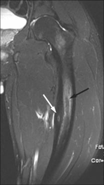
- MRI alone can mislead badly: oedema around a nidus, a stress fracture or a Brodie's abscess all produce a scary marrow and soft-tissue signal that mimics malignancy.
- Anterior tibial diaphyseal cortex is the classic site for osteofibrous dysplasia and adamantinoma — but 57 per cent of osteofibrous dysplasia needs no treatment at all, and Scholfield found NO progression from osteofibrous dysplasia to adamantinoma in 52 patients. The two are separated by age: mean 13.5 years versus 34.
- Adamantinoma's danger is late, not early: disease-specific survival is 93 per cent at ten years but 39 per cent at twenty. Resect with clear margins, never curette, and follow for decades.
- A solitary asymptomatic dense focus with no lucency, brush borders and no scintigraphic uptake is an enostosis — no follow-up required.
- “Say 'reactive sclerosis around a small lucent nidus' rather than 'sclerotic lesion' — it commits you to the correct pathway.
- “Multiple lesions push you towards osteopoikilosis, melorheostosis or metastases — check the whole film before speaking.
- “Age is the single most powerful filter: less than 5 years think infection and Langerhans cell histiocytosis; 5 to 25 think osteoid osteoma, Ewing sarcoma, adamantinoma; over 40 think metastasis, lymphoma, Paget disease.
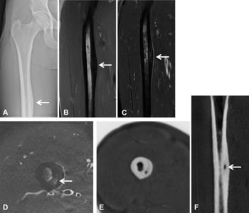
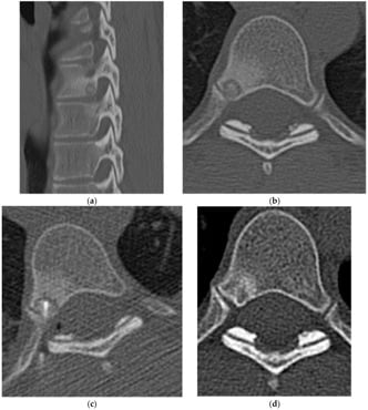
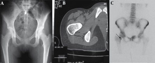
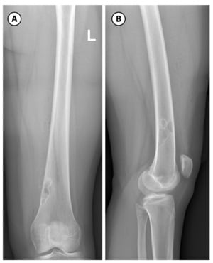
Fusiform cortical thickening with a tiny central lucency is the host bone reacting. The lesion is the nidus, not the sclerosis. Biopsying the sclerosis returns "reactive bone" and delays diagnosis.
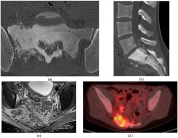
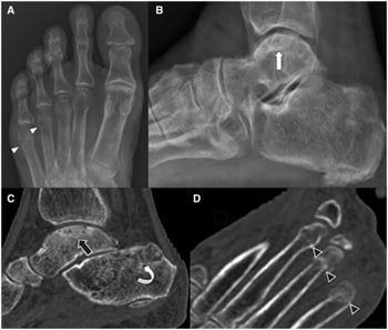
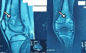
MRI in osteoid osteoma, stress fracture and Brodie's abscess shows florid marrow and periosteal oedema that reads as aggressive. Fat-suppressed sequences can obscure a small nidus entirely. Thin-slice CT is the better test — though Assoun found it beat MRI in only 63 per cent of proven cases, so read the radiograph, CT and MRI together rather than treating any one as decisive. Ask what the patient has been taking: anti-inflammatories significantly reduce the marrow and soft-tissue change, so a child already on ibuprofen presents a blander, harder study.
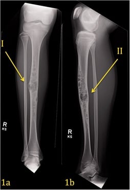
A lucent-and-sclerotic, multiloculated, eccentric anterior tibial diaphyseal cortical lesion in a CHILD is usually osteofibrous dysplasia — 57 per cent of Scholfield's 42 needed no treatment, and none of his 52 osteofibrous dysplasia or OFD-like patients progressed to adamantinoma. Adamantinoma presents two decades later (mean 34 years) and is the opposite error: curettage recurred in two of three, and disease-specific survival falls from 93 per cent at ten years to 39 per cent at twenty. Resect that one with clear margins and follow for decades.
In 407 published Brodie's abscesses, 84 per cent were afebrile and fewer than half had raised inflammatory markers — the median delay to diagnosis was 12 weeks. A normal temperature and CRP do not take subacute osteomyelitis off the list for a lucency with a sclerotic rim in a teenager.
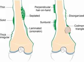
Look specifically for Codman triangle, sunburst spiculation and lamellated periosteum at the shoulders of the sclerosis. Their presence converts a benign-looking dense cortex into a staging problem requiring MRI of the whole bone, chest CT and referral before biopsy.
Recognising the Pattern
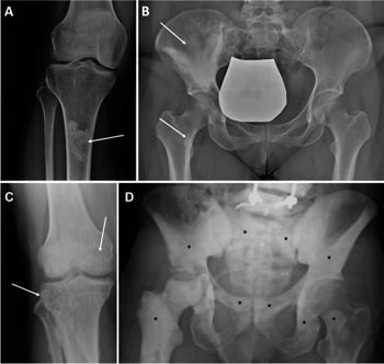
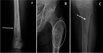
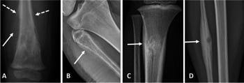
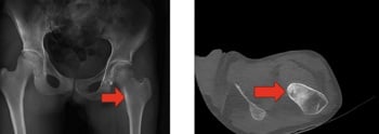
Definition. A sclerotic cortical lesion is an area of increased density arising within or immediately deep to the cortex, usually producing fusiform cortical thickening and narrowing of the medullary canal. It differs from a medullary sclerotic lesion (bone infarct, enostosis, blastic metastasis) in that the epicentre lies in the cortex itself.
Confirming it is genuine.
- Obtain two orthogonal views. Overlapping cortex on an oblique projection, and the normal thickened posteromedial femoral cortex, both mimic pathology on a single view.
- Trace the endosteal and periosteal margins independently. True cortical lesions expand or thicken one or both; projectional artefact does not.
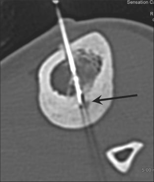
- Check for a lucency within the density on a bright light or by windowing. Reactive sclerosis without an identifiable nidus on radiograph still requires CT — the nidus is often below radiographic resolution.
- Confirm the lesion is not simply a healing fracture or a periosteal reaction to an adjacent soft-tissue process by asking about trauma, insertional tendinopathy and injection sites.
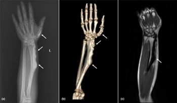
Saying it out loud. "This is an anteroposterior and lateral radiograph of the left tibia in a skeletally immature patient. There is dense fusiform thickening of the anteromedial diaphyseal cortex over approximately 4 cm, with narrowing of the medullary canal. Within the sclerosis there is a rounded lucency of about 8 mm containing a central density. The cortex is intact, there is no soft-tissue mass and the periosteal reaction is solid, not interrupted. This is a nidus with surrounding reactive sclerosis, and the first diagnosis is osteoid osteoma."
Mimics — the false positives.
- Normal anatomy: the linea aspera, the posteromedial proximal tibial cortex, the deltoid tuberosity, and the nutrient foramen (a lucency with a sclerotic rim in the wrong plane).
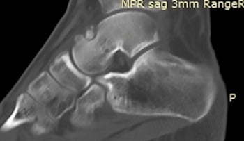
- Healing stress fracture: by 4 to 6 weeks the callus is dense and fusiform and closely resembles a nidus-bearing lesion; the "nidus" is the residual fracture line, which is linear and perpendicular to the cortex rather than round.
- Cortical desmoid (avulsive cortical irregularity): posteromedial distal femoral metaphysis at the adductor magnus insertion, in patients aged 10 to 15 — an irregular, saucerised cortex with a sclerotic base. Do not biopsy; it resolves.
- Chronic periostitis: venous stasis, hypertrophic osteoarthropathy and thyroid acropachy give solid, bilateral, symmetrical diaphyseal periosteal new bone that can look sclerotic.
- Post-injection or post-operative sclerosis: old screw tracks and drill holes sclerose and persist for decades.
Next Investigation
State explicitly when you would NOT image further. Recognising an enostosis and stopping demonstrates more judgement than ordering an MRI. The examiner is testing restraint as much as recall.
The Differential
- Typical age / setting
- 10 to 25 years; also over 60 with Paget disease or prior radiation
- Discriminating feature
- Cloud-like tumour osteoid extending beyond the cortex into soft tissue, with interrupted periosteum (Codman triangle or sunburst); pain is constant and unrelated to NSAIDs
- What confirms it
- MRI whole bone plus chest CT, then core biopsy through a planned resection tract at the tumour unit
- Typical age / setting
- 20 to 40 years; anterior tibial diaphyseal cortex (over 85 per cent tibia)
- Discriminating feature
- Multiloculated lucencies in a sclerotic anterior cortex with a 'soap bubble' appearance, often spanning several centimetres and bowing the tibia
- What confirms it
- Biopsy — epithelial nests in fibrous stroma, cytokeratin positive; requires wide resection not curettage
- Typical age / setting
- 5 to 20 years; femoral or tibial diaphysis, systemic upset and raised inflammatory markers
- Discriminating feature
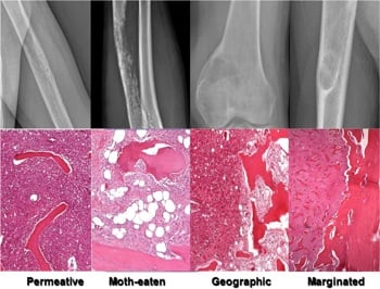
- Permeative cortical destruction with lamellated 'onion-skin' periosteum and a soft-tissue mass far larger than the bone change
- What confirms it
- MRI showing the soft-tissue component, then biopsy; small round blue cells, EWSR1 rearrangement
- Typical age / setting
- Over 50 years; known primary or systemic symptoms
- Discriminating feature
- Sclerosis that is ill-defined, non-fusiform and usually multifocal; cortical destruction disproportionate to the sclerosis in lymphoma
- What confirms it
- Whole-body bone scintigraphy or FDG-PET-CT, PSA, myeloma screen; biopsy if solitary
- Typical age / setting
- 10 to 25 years, male predominance; femoral neck, tibial diaphysis, posterior spinal elements
- Discriminating feature
- Nidus less than 1.5 cm, round, with central mineralisation and intense surrounding reactive sclerosis; night pain relieved within 20 to 30 minutes by NSAIDs
- What confirms it
- Thin-slice (1 mm) CT demonstrating the nidus; intense focal uptake with a 'double density' sign on bone scan
- Typical age / setting
- Athletes, military recruits, dancers; posteromedial tibia, second and third metatarsals, femoral neck
- Discriminating feature
- A linear lucency perpendicular to the cortex, becoming a transverse dense band with fusiform periosteal callus; pain worse with activity and better with rest — the opposite of osteoid osteoma
- What confirms it
- MRI showing a low-signal fracture line within marrow oedema, or CT showing a linear cortical breach
- Typical age / setting
- Children and young adults; metaphysis of tibia or femur, may be cortical
- Discriminating feature
- Serpiginous lucency with a thick sclerotic rim and a 'penumbra sign' on T1 MRI; may contain a sequestrum
- What confirms it
- MRI with contrast showing rim enhancement and a non-enhancing centre; aspiration and culture
- Typical age / setting
- Children and adolescents; mandible, tibia, femur
- Discriminating feature
- Dense diffuse sclerosis with cortical thickening and NO nidus, no sequestrum and no abscess cavity; low-grade pain and swelling over months
- What confirms it
- CT excluding a nidus; biopsy and culture, frequently negative; diagnosis of exclusion
- Typical age / setting
- Under 10 years; anterior tibial diaphyseal cortex, often bilateral
- Discriminating feature
- Intracortical lucencies with dense sclerotic margination and anterior tibial bowing, but no epithelial cells and no medullary involvement
- What confirms it
- Radiographic surveillance; biopsy only if progressive — histology shows osteoblastic rimming of woven bone
- Typical age / setting
- 10 to 30 years; posterior elements of the spine, also long bone diaphysis
- Discriminating feature
- Nidus-like lesion greater than 2 cm, less reactive sclerosis relative to size, pain not reliably relieved by NSAIDs and often expansile
- What confirms it
- CT for size and matrix; MRI for canal compromise; biopsy to exclude osteosarcoma
- Typical age / setting
- Any age; incidental, commonly pelvis, femur, spine
- Discriminating feature
- Homogeneous dense focus with radiating 'brush border' spicules blending into trabeculae; no lucency, no periosteal reaction, no pain
- What confirms it
- No further imaging needed if the appearance is classic; bone scan is cold or minimally warm if in doubt
- Typical age / setting
- Any age; incidental, familial, LEMD3 mutation
- Discriminating feature
- Multiple small round sclerotic foci clustered around joints in a periarticular distribution, symmetrical, asymptomatic
- What confirms it
- Radiographs alone; the symmetrical periarticular pattern excludes metastases
- Typical age / setting
- 20 to 40 years; monomelic, sclerotomal distribution
- Discriminating feature
- 'Dripping candle wax' undulating hyperostosis flowing along one cortical surface, crossing joints, with associated soft-tissue contracture
- What confirms it
- Radiographs are diagnostic; MRI only to assess soft-tissue involvement
- Typical age / setting
- Over 55 years; pelvis, femur, tibia, skull
- Discriminating feature
- Cortical thickening with coarsened trabeculae, bone enlargement (the bone is bigger) and a flame-shaped advancing lytic front in long bones
- What confirms it
- Radiographs plus raised alkaline phosphatase with normal calcium and phosphate
Bone enlargement is the single most useful sign in the elderly: metastasis and lymphoma sclerose the bone but do not enlarge it. Paget disease enlarges it. If the cortex is thickened AND the overall bone diameter has increased, think Paget first.
Narrowing It Down
- 1Step 1 — How old is the patient?
Fix the age before describing the film, and let it set the differential you are testing.
Under 5: infection and Langerhans cell histiocytosis dominate. 5 to 25: osteoid osteoma, osteoblastoma, Ewing sarcoma, osteosarcoma, osteofibrous dysplasia, stress fracture. 25 to 40: adamantinoma, chronic osteomyelitis. Over 40: metastasis, lymphoma, myeloma, Paget disease. Age alone removes half the list — and for the anterior tibia it does most of the work, separating osteofibrous dysplasia (mean 13.5 years) from adamantinoma (mean 34).
- 2Step 2 — Solitary or multiple?
Scan the whole film before characterising the lesion you were pointed at, and consider a bone scan or skeletal survey.
Multiple symmetrical periarticular foci equal osteopoikilosis. Multiple asymmetrical ill-defined foci over 40 equal metastases. A single flowing cortical hyperostosis down one limb equals melorheostosis. Solitary keeps the full tumour and infection differential live.
- 3Step 3 — Where in the bone, and where in the cross-section?
State the bone, the segment and the position within the cortex — anterior, posteromedial, eccentric, central — before naming anything.
Anterior tibial diaphyseal cortex: osteofibrous dysplasia in a child, adamantinoma in an adult. Femoral neck or proximal femoral cortex in a teenager: osteoid osteoma. Posterior spinal elements with painful scoliosis: osteoid osteoma or osteoblastoma. Posteromedial tibia or metatarsal shaft in a runner: stress fracture. Metaphysis with a lucent centre in a child: Brodie's abscess.
- 4Step 4 — Is there a lucency inside the sclerosis, what shape, and how big?
Measure the lucency rather than eyeballing it against the 15 mm convention.
Round with central calcification: nidus of osteoid osteoma. Round and over 2 cm: osteoblastoma. Treat 15 mm as a GRADIENT, not a border — Shields calls the cut-off arbitrary and found each extra millimetre multiplies post-ablation recurrence by 1.33, with 16.3 per cent recurring overall. Linear and perpendicular to the cortex: stress fracture. Serpiginous with a thick rim and possible sequestrum: Brodie's abscess. Multiloculated soap-bubble: adamantinoma or osteofibrous dysplasia. None at all: enostosis, Garré osteomyelitis, Paget disease or metastasis.
- 5Step 5 — Is the sclerosis reactive or is it tumour matrix?
Follow the cortical outline all the way round and decide whether the dense material stays inside it.
Reactive sclerosis is dense, uniform, sharply marginated and confined to bone — the lesion is the small lucency, not the dense bone around it. Tumour osteoid is fluffy, cloud-like, ill-defined and extends beyond the cortex into soft tissue. Cloud-like mineralisation outside the bone is osteosarcoma until proven otherwise.
- 6Step 6 — How aggressive is the margin and the periosteum?
Examine the shoulders of the sclerosis specifically, where periosteal reaction is most visible.
Solid, uninterrupted, thick periosteal reaction with a narrow zone of transition indicates a slow process — osteoid osteoma, stress fracture, chronic infection. Lamellated, spiculated or interrupted periosteum with a wide zone of transition and cortical destruction indicates Ewing sarcoma, osteosarcoma or aggressive infection.
- 7Step 7 — What are the systemic and symptom features, and what has the patient taken?
Take the pain history, check temperature and inflammatory markers, and ASK ABOUT ANTI-INFLAMMATORY USE before ordering or interpreting the MRI.
Night pain relieved by NSAIDs: osteoid osteoma. Activity-related pain relieved by rest: stress fracture. Weight loss with a known primary: metastasis. Asymptomatic incidental finding: enostosis or osteopoikilosis — stop here. Two cautions: normal bloods do NOT exclude infection (84 per cent of published Brodie's abscesses were afebrile and fewer than half had raised markers, with a 12-week median delay to diagnosis), and a patient already on ibuprofen has significantly less marrow and soft-tissue oedema on MRI, making a nidus easier to miss.
MCQ Practice Points
Q: Which single feature most reliably separates osteoid osteoma from osteoblastoma, and how firm is it?
A: Lesion size — but state its softness. A nidus under 15 mm with disproportionately florid reactive sclerosis is osteoid osteoma; a lesion over 2 cm with relatively less sclerosis, often expansile and frequently in the posterior spinal elements, is osteoblastoma. The histology is identical, and Shields' unit calls the 15 mm cut-off arbitrary: post-ablation recurrence rose 1.33-fold for every extra millimetre of lucent diameter, so the two entities behave as a continuum rather than a pair. The examiner wants the number; the better answer adds that it is a gradient.
Q: What recurrence rate should a patient be consented for before radiofrequency ablation of an osteoid osteoma?
A: Around one in six. Shields reported 16.3 per cent over a mean 72 months of follow-up — explicitly higher than the published literature, and the authors attribute part of that to the length of follow-up rather than technique. Risk scales with nidus size (hazard ratio 1.33 per millimetre), so a patient with a 13 mm lesion should be told a second procedure is a realistic possibility. Single centre, retrospective, no comparison arm.
Q: A 16-year-old has a metaphyseal lucency with a thick sclerotic rim, is afebrile and has a normal CRP. Does that exclude a Brodie's abscess?
A: No, and this is the commonest reason for delay. Across 407 published cases, 84 per cent were afebrile and fewer than half had raised serum inflammatory markers; the median time from symptom onset to diagnosis was 12 weeks. Pain was present in 98 per cent and swelling in half. Staphylococcus aureus grew in 67 per cent, treatment was surgical in 94 per cent, and 15.6 per cent recurred. Caveat: these are pooled case reports, and an afebrile presentation is exactly what makes a case publishable — so the true proportion is likely lower than 84 per cent.
Q: What is the penumbra sign and what does it indicate?
A: A rim of relatively high T1 signal at the margin of an intraosseous abscess cavity, representing granulation tissue lining the abscess. It is highly specific for subacute osteomyelitis (Brodie's abscess) and helps distinguish it from a tumour — useful precisely because the systemic markers so often will not.
Q: Why is MRI a poor first-line test for suspected osteoid osteoma, and how much better is CT?
A: The nidus is small and may be obscured by extensive marrow, periosteal and soft-tissue oedema on fluid-sensitive sequences, which is also readily misread as aggressive. Assoun's head-to-head in 19 histologically proven lesions found CT more accurate at detecting the nidus in 63 per cent of cases — better, not decisive — while MRI was superior for marrow and soft-tissue change in every case. Their conclusion was that MR should never be interpreted without the radiograph and CT beside it. A related and rarely taught point from the same paper: marrow and soft-tissue change correlated significantly with anti-inflammatory use, so a patient already on NSAIDs presents a blander study.
Q: A 9-year-old has a multiloculated lucent-and-sclerotic lesion of the anterior tibial diaphyseal cortex. Does it need resection to prevent adamantinoma?
A: No. Scholfield followed 42 osteofibrous dysplasia and 10 OFD-like adamantinoma patients and found NO progression to adamantinoma and no metastases; 57 per cent of the osteofibrous dysplasia group required no treatment at all. Curettage recurred in 3 of 13, resection in none. Adamantinoma is a different clinical animal presenting at a mean of 34 years, and there the error runs the other way: curettage recurred in 2 of 3, and disease-specific survival falls from 93 per cent at ten years to 39 per cent at twenty. Caveat: mean follow-up for the osteofibrous dysplasia group was 10.3 years from a mean age of 13.5, which does not reach the age at which adamantinoma typically appears — so non-progression is unobserved rather than disproved.
Q: A 55-year-old has generalised cortical thickening of the femur and coarse trabeculae. What single laboratory test is most useful?
A: Serum alkaline phosphatase. It is elevated in Paget disease with normal calcium and phosphate. Combined with bone enlargement and a flame-shaped lytic front, this confirms the diagnosis without biopsy. A sudden increase in pain warrants imaging for secondary osteosarcoma.
Q: Which cortical lesion mandates referral before biopsy, and why?
A: Any lesion with cortical destruction, a soft-tissue mass, interrupted periosteal reaction, or arising in the anterior tibial cortex. An unplanned biopsy tract contaminates compartments, may convert a limb-salvage resection into an amputation, and is one of the commonest avoidable errors in orthopaedic oncology. The tract must be planned by the surgeon who will resect it.
Exam Viva Scenarios
Practise clinical reasoning and management decisions out loud
“You are shown this radiograph of the tibia of a 15-year-old boy with a six-month history of night pain. There is dense fusiform thickening of the anteromedial diaphyseal cortex with a small central lucency. Describe and manage.”
“You are shown this radiograph of the tibia of a 28-year-old woman with a two-year history of aching and a palpable anterior swelling. There are multiloculated lucencies within a thickened, sclerotic anterior diaphyseal cortex, with mild anterior bowing. What is your differential and how do you proceed?”
“You are shown a pelvic radiograph performed for hip pain in a 62-year-old man. There is a well-defined 12 mm homogeneously dense focus in the ilium with fine radiating spicules at its margin. He has no other symptoms. What is it and what do you do?”
Describe in this order
- Patient age and skeletal maturity
- Bone, and region within it (epiphysis, metaphysis, diaphysis)
- Position in cross-section: intracortical, endosteal, periosteal, medullary
- Presence, size and shape of any internal lucency
- Matrix: reactive sclerosis versus cloud-like tumour osteoid
- Periosteal reaction: solid versus lamellated, spiculated or interrupted
- Soft-tissue mass, zone of transition, cortical integrity
Nidus shapes and their meaning
- Round, less than 1.5 cm, central density — osteoid osteoma
- Round, greater than 2 cm, expansile — osteoblastoma
- Linear, perpendicular to cortex — stress fracture
- Serpiginous with thick rim and sequestrum — Brodie's abscess
- Multiloculated soap bubble, anterior tibia — adamantinoma or osteofibrous dysplasia
- No lucency at all — enostosis, Garré osteomyelitis, Paget, metastasis
Site-specific reflexes
- Anterior tibial diaphyseal cortex — adamantinoma / osteofibrous dysplasia
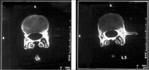
- Posterior spinal elements with painful scoliosis — osteoid osteoma or osteoblastoma
- Posteromedial distal femur, ages 10 to 15 — cortical desmoid, do not biopsy
- Posteromedial tibia in a runner — stress fracture
- Femoral neck in a teenager with night pain — osteoid osteoma
Red flags demanding sarcoma referral
- Cortical destruction or wide zone of transition
- Soft-tissue mass or mineralisation outside the cortical outline
- Codman triangle, sunburst or onion-skin periosteum
- Constant pain unrelieved by NSAIDs, especially with systemic upset
- Any anterior tibial cortical lesion in an adult
When to stop
- Classic enostosis: brush border, homogeneous, asymptomatic — no follow-up
- Osteopoikilosis: symmetrical periarticular foci — reassure
- Melorheostosis: dripping candle wax, monomelic — radiographs only
- Cortical desmoid at adductor magnus insertion — reassure, avoid biopsy
Evidence Base
Osteoid Osteoma: MR Imaging Versus CT
- 19 patients with HISTOLOGICALLY PROVED osteoid osteoma had both CT and MRI before excision
- CT was more accurate than MRI at detecting the nidus in 63 PER CENT of cases - not all of them
- MRI was better than CT at showing intramedullary and soft tissue change in ALL cases, which is precisely what produces the misleadingly aggressive appearance
- The presence or absence of marrow and soft tissue change correlated significantly with whether the patient was taking anti-inflammatory medication (p less than 0.05)
- The authors conclude MR images should not be interpreted without the plain radiograph and CT alongside them if serious diagnostic errors are to be avoided
Radiofrequency Ablation for Osteoid Osteoma: Recurrence Rates and Predictive Factors
- Consecutive patients with confirmed osteoid osteoma from a prospectively maintained database over ten years at one orthopaedic oncology unit
- Recurrence after radiofrequency ablation was 16.3 PER CENT at a minimum 21 months and mean 72 months of follow-up - HIGHER than the published literature
- Larger lucent diameter predicted recurrence on Cox regression (p equals 0.049, hazard ratio 1.33 per millimetre)
- The authors describe the 15 mm cut-off separating osteoid osteoma from osteoblastoma as ARBITRARY
- Their conclusion is that the distinction 'probably represents a progressive scale', with larger lesions responding less well to ablation
Does Osteofibrous Dysplasia Progress to Adamantinoma, and How Should They Be Treated?
- 73 patients from a single supraregional unit over 39 years - 42 osteofibrous dysplasia, 10 osteofibrous dysplasia-like adamantinoma, 21 adamantinoma - followed a mean 9 to 12 years
- NO PATIENT with osteofibrous dysplasia or the OFD-like variant developed metastases or progressed to adamantinoma
- 24 of the 42 osteofibrous dysplasia patients (57 per cent) required NO TREATMENT AT ALL and were simply observed; 3 of 13 curettages recurred (23 per cent), none of the resections did
- Mean age at diagnosis separates them: 13.5 years for osteofibrous dysplasia, 10.5 for OFD-like, but 34 years for adamantinoma
- Adamantinoma disease-specific survival was 93 per cent at ten years but had FALLEN TO 39 PER CENT BY TWENTY YEARS; 2 of 3 treated by curettage recurred
Brodie's Abscess: A Systematic Review of Reported Cases
- PRISMA systematic review of 70 articles reporting 407 patients, median age 17, male to female 2.1 to 1
- EIGHTY-FOUR PER CENT WERE AFEBRILE and FEWER THAN HALF had raised serum inflammatory markers
- Median duration of symptoms before diagnosis was 12 weeks; pain in 98 per cent, swelling in 53 per cent
- Diagnosis rested on plain radiography in 96 per cent, with MRI used in only 16 per cent and CT in 8 per cent
- Staphylococcus aureus in 67.3 per cent of cultures; surgery in 94 per cent, antibiotics alongside in 77 per cent; recurrence 15.6 per cent