One body, one endplate, or many — count before you name
- COUNT FIRST. One uniformly dense body, sclerosis confined to endplates, and a diffusely dense spine are three different questions with three different differentials — and the commonest error is answering the first when the film shows the second.
- For a SOLITARY uniformly dense body the full differential — metastasis, lymphoma, Paget disease, osteosarcoma, infection — is worked through on the Ivory Vertebra page. Everything here is about the patterns that are not that.
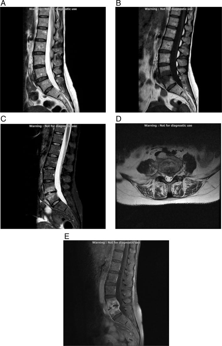
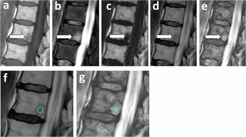
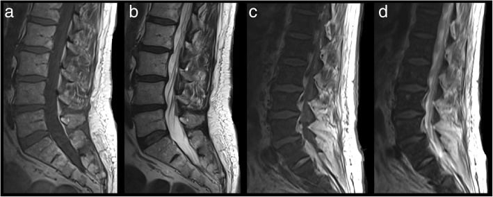
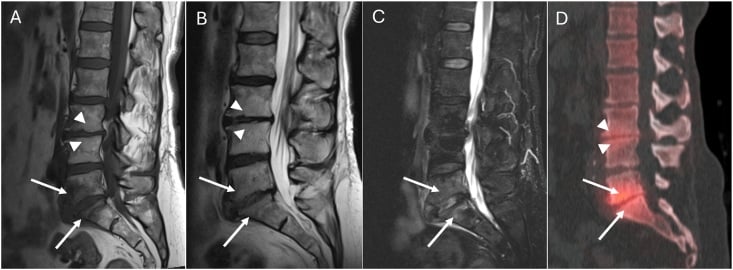
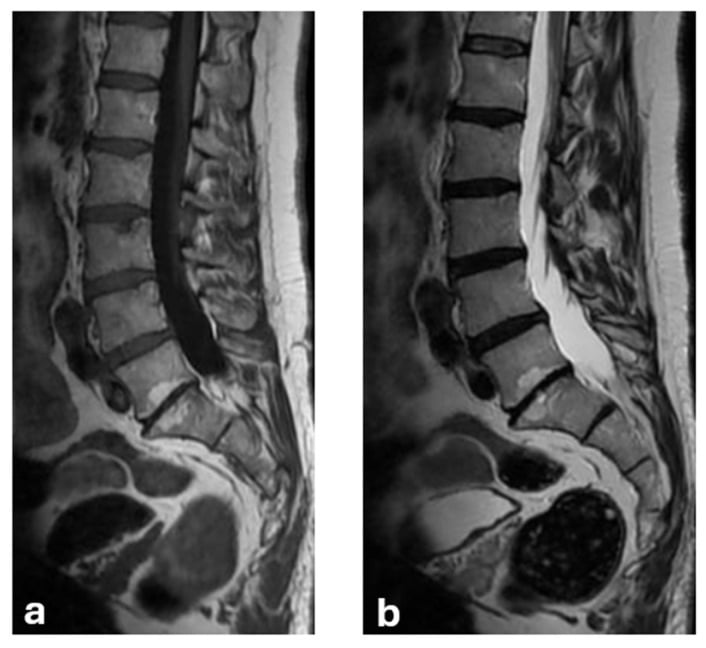
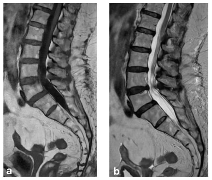
- Endplate sclerosis is the commonest vertebral sclerosis and the least diagnostic: 6 per cent of people without back pain have vertebral endplate signal change and 43 per cent of those with it do. It moves probability; it is not a cause.
- Modic change does NOT license an intervention at that level. Herlin's meta-analysis of 31 studies found associations with pain inconsistent — 15 positive, one negative — with only one study at low risk of bias.
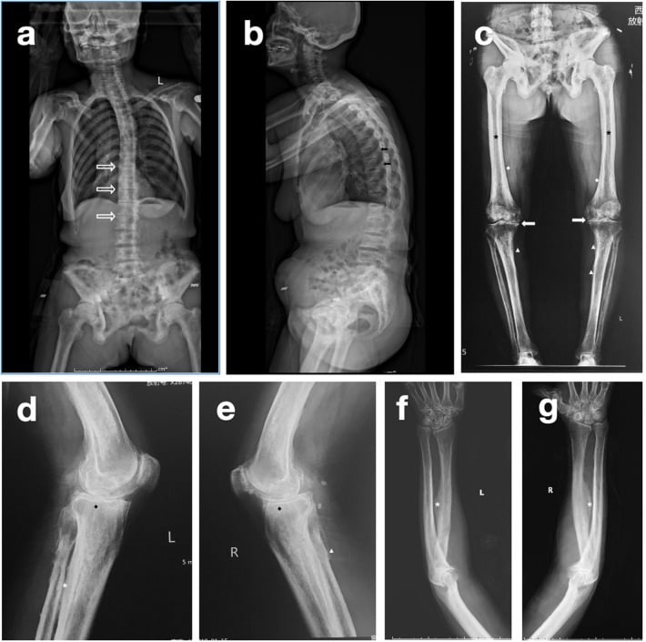
- Banded sclerosis sparing the mid-body across every level is rugger-jersey spine: renal osteodystrophy. The diagnosis is made on the biochemistry, not the film.
- Sandwich vertebra and endobone mean osteopetrosis, and its importance is surgical: intramedullary canal diameter under 5 mm predicted every postoperative periprosthetic femoral fracture in the Mayo series, and 58 per cent of those hips were reoperated within ten years.
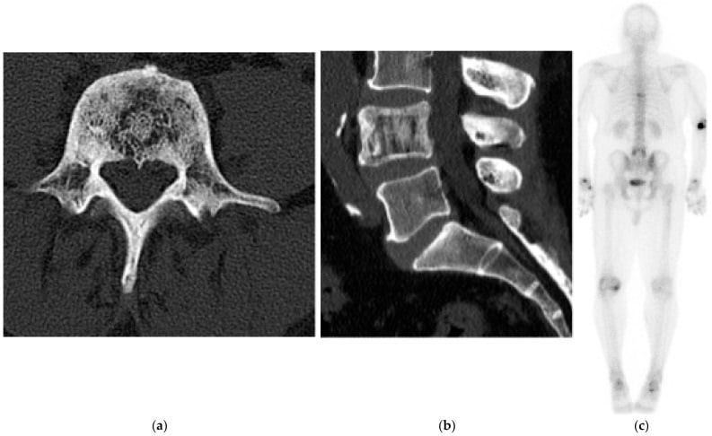
- An asymptomatic dense vertebra with normal size, intact pedicles and disc spaces and negative screening may need NOTHING. Carpineta followed two such patients for over a decade with no diagnosis ever reached and no consequence.
- “Always state whether the vertebral body is normal size, enlarged, or collapsed — this single observation splits the differential three ways.
- “Check the posterior elements: pedicle involvement favours metastasis; a diffusely dense pedicle with an expanded body favours Paget's.
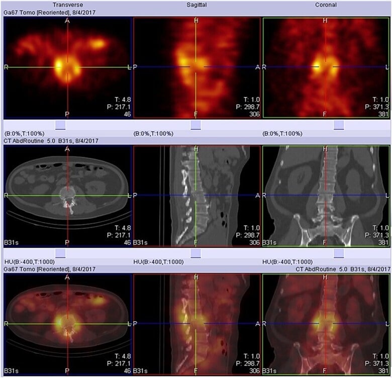
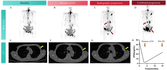
- “Sclerotic metastasis that becomes MORE sclerotic on treatment is a flare response, not progression — correlate with PSA or tumour markers.
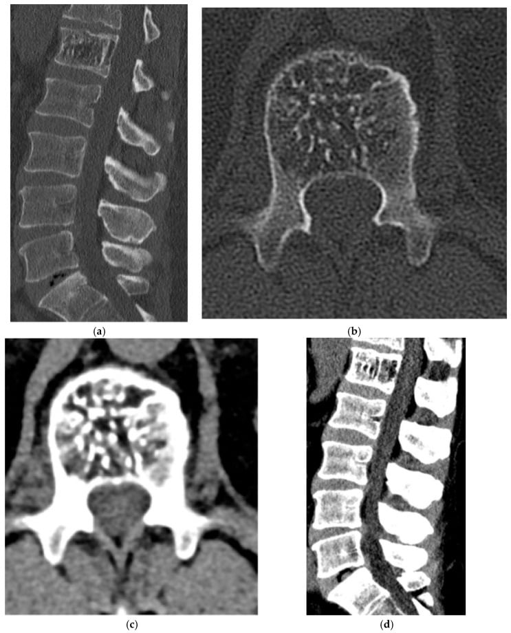
- “A dense vertebra with a coarse vertical striated pattern on CT and fat signal on MRI is a haemangioma — leave it alone.
In a patient under 40 with no known primary, lymphoma, chronic osteomyelitis and osteoblastic osteosarcoma matter more. Age-anchor before you commit.
Paget's disease is diagnosed by SIZE, not density. If you never comment on vertebral body dimensions you will miss the one diagnosis that needs no biopsy.
An overlying barium column, contrast in the aorta, an oblique projection or a sclerotic Schmorl node rim can simulate a dense body. Confirm on two orthogonal views or CT.
Committing to a solitary lesion on a coned lumbar film is a trap. Multiplicity changes the differential entirely — image the whole spine before you narrow.
Recognising the Pattern
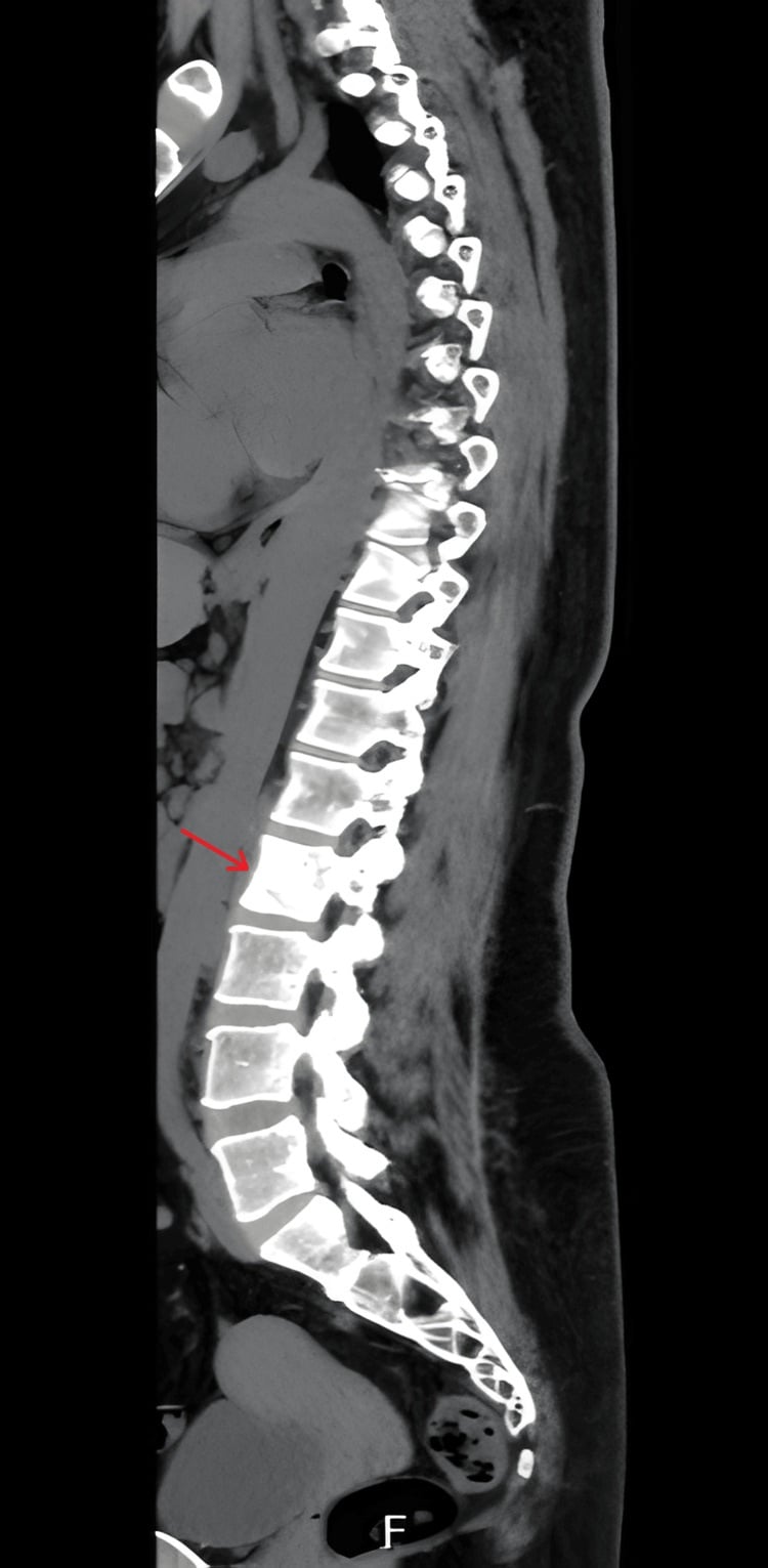
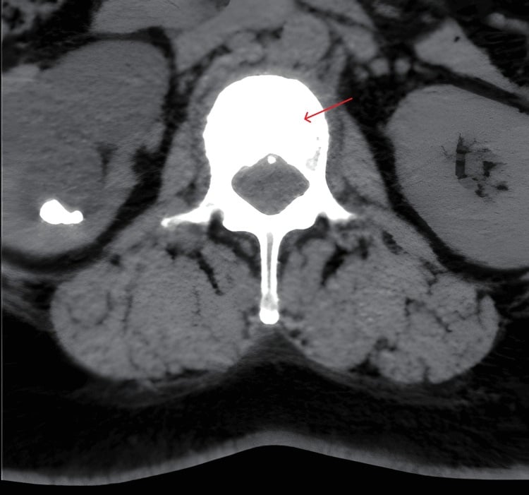
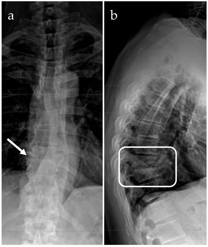
Definition. An ivory vertebra is a diffuse, homogeneous increase in radiodensity of a single vertebral body, sharply demarcated from the vertebrae above and below, with no significant loss of vertebral body height and no change in overall size. The term is descriptive, not diagnostic.
Confirming the sign is genuinely present.
- Compare density with at least two vertebrae above and below on the SAME film — window and exposure vary between studies.
- Confirm on the lateral as well as the AP. A dense body on AP only suggests overlying structure (bowel gas obscuring adjacent levels, contrast, a calcified aortic plaque, a pedicle screw).
- Assess vertebral body height on the lateral: preserved height supports true ivory vertebra; collapse points to fracture, infection or vertebra plana.
- Assess overall dimensions: an increased anteroposterior and craniocaudal dimension with cortical thickening equals Paget's disease.
- Look at the disc spaces: preserved disc spaces favour tumour; destroyed endplates with disc narrowing favour infection.
How to describe it out loud.
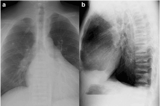
"This is a frontal and lateral radiograph of the thoracolumbar spine. There is a solitary vertebral body — the level appears to be T10 — which is diffusely and homogeneously sclerotic compared with the adjacent levels. The vertebral body height is preserved and the body is not enlarged. The pedicles appear intact, the adjacent disc spaces are preserved, and I cannot see a paraspinal soft tissue mass on this projection. This is the ivory vertebra sign. In a patient of this age my leading diagnoses are sclerotic metastasis, Paget's disease and lymphoma, and I would separate them by assessing vertebral size and by proceeding to MRI of the whole spine."
Mimics and false positives.
- Projectional/technical: underpenetrated adjacent levels, overlying barium, oblique rotation, hardware.
- Sclerotic Schmorl node or endplate reactive change: focal, adjacent to endplate, not whole-body.
- Vertebral haemangioma: appears dense on some projections but shows coarse vertical striations (corduroy) and fat attenuation on CT/MRI.
- Bone island (enostosis): focal, round, spiculated margin, cold on bone scan — not diffuse.
- Vertebra plana: collapsed and therefore apparently dense from superimposition; the height loss reveals the true nature.
Next Investigation
PLMIvory Vertebra — classic triad in the exam
The Differential
- Typical age / setting
- Over 50; known primary or new back pain
- Discriminating feature
- Vertebral body NOT enlarged; pedicle often involved with loss of pedicle cortex; frequently multiple non-contiguous levels
- What confirms it
- MRI marrow replacement low on T1; bone scan/PSMA PET; PSA or breast primary; biopsy if solitary
- Typical age / setting
- 15-40 and again over 55; night sweats, weight loss, nodal disease
- Discriminating feature
- Anterior vertebral body scalloping from adjacent nodal mass; paravertebral soft tissue with preserved disc spaces
- What confirms it
- CT chest/abdomen for nodal disease; core biopsy of node or bone; FDG PET
- Typical age / setting
- 10-25, or over 55 arising in Paget's/post-radiation
- Discriminating feature
- Cloud-like tumour bone extending beyond the cortex into a soft tissue mass; aggressive periosteal reaction
- What confirms it
- MRI extent, staging CT chest, biopsy through planned resection route
- Typical age / setting
- Any age; immunosuppression, IVDU, endemic TB
- Discriminating feature
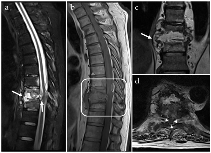
- Disc space destruction and endplate erosion — sclerosis is REACTIVE and follows destruction; large paravertebral abscess in TB
- What confirms it
- MRI with contrast; CT-guided biopsy and culture including AFB; inflammatory markers
- Typical age / setting
- Over 55; often incidental, raised ALP with normal calcium
- Discriminating feature
- Vertebral body ENLARGED with cortical thickening — picture-frame vertebra; coarse thickened trabeculae
- What confirms it
- Radiograph alone is often diagnostic; ALP; bone scan shows intense uniform uptake of the whole vertebra
- Typical age / setting
- Treated prostate or breast cancer
- Discriminating feature
- Increasing sclerosis with falling tumour markers — the flare phenomenon; margins become better defined
- What confirms it
- Serial imaging plus tumour markers; PET may show reduced metabolic activity
- Typical age / setting
- 30-60; usually incidental
- Discriminating feature
- Coarse vertical striations, polka-dot appearance on axial CT; fat signal high on T1
- What confirms it
- CT is diagnostic; MRI shows fat; no follow-up needed if classic
- Typical age / setting
- Over 65, osteoporosis, steroid use
- Discriminating feature
- Height loss with a linear band of condensation parallel to the endplate, not whole-body sclerosis
- What confirms it
- MRI shows fracture line with surrounding oedema; STIR high signal resolving over months
- Typical age / setting
- Dialysis-dependent chronic kidney disease
- Discriminating feature
- Band-like endplate sclerosis with a lucent central body — MULTIPLE levels, never solitary
- What confirms it
- Renal function, calcium, phosphate, PTH
- Typical age / setting
- Presents in childhood or found incidentally in adults
- Discriminating feature
- Sandwich vertebra: sharply defined dense endplate bands with a distinct margin; generalised skeletal sclerosis, bone-within-bone
- What confirms it
- Skeletal survey; family history; genetic testing
- Typical age / setting
- Over 60; splenomegaly, leucoerythroblastic film
- Discriminating feature
- Diffuse uniform sclerosis of ALL vertebrae with no focal lesion; sclerosis of ribs and pelvis
- What confirms it
- Full blood count and film; JAK2; bone marrow trephine (dry tap)
- Typical age / setting
- Afro-Caribbean, childhood onward
- Discriminating feature
- H-shaped or Lincoln-log vertebra from central endplate infarction; multiple levels
- What confirms it
- Haemoglobin electrophoresis
- Typical age / setting
- 30-60; flushing, urticaria pigmentosa, diarrhoea
- Discriminating feature
- Patchy mixed sclerotic and lucent marrow lesions throughout the axial skeleton
- What confirms it
- Serum tryptase; marrow biopsy with CD117 mast cells
- Typical age / setting
- Endemic areas, industrial exposure, chronic high fluoride intake
- Discriminating feature
- Dense vertebrae PLUS ligamentous and interosseous membrane calcification (forearm, pelvis)
- What confirms it
- Occupational/dietary history; urinary fluoride
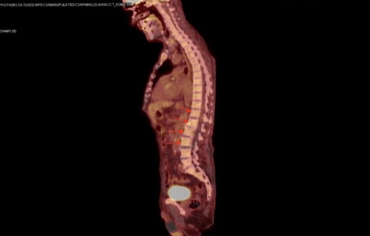
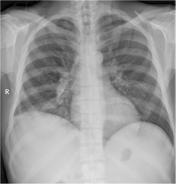
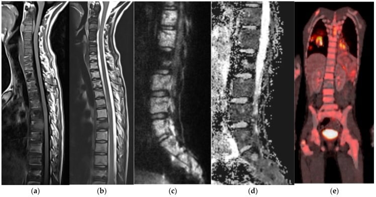
Narrowing It Down
- 1Step 1 — COUNT. How many, and where within the vertebra?
Image the whole spine before characterising anything, and state whether the sclerosis occupies whole bodies, endplates only, or everything.
A single uniformly dense body is the ivory vertebra — metastasis, lymphoma, Paget disease, osteosarcoma or infection, and that differential belongs on the Ivory Vertebra page. Sclerosis confined to endplates at one or two levels is degenerative Modic change. Banding at every level with a spared mid-body is rugger-jersey spine. Uniform density throughout points to a systemic marrow or metabolic disease. Answering the first question when the film shows the second is the commonest error here.
- 2Step 2 — If it is endplate change, resist explaining the pain with it
Note the level and Modic type, then go back to the history and examination for a cause of the symptoms.
Vertebral endplate signal change was present in 6 per cent of people WITHOUT back pain and 43 per cent of those with it, so the finding shifts probability without establishing cause. Herlin's meta-analysis of 31 studies found the association INCONSISTENT — 15 positive, one negative, only one study at low risk of bias — and the single poolable odds ratio of 4.01 came from a provocative discography subgroup. Report it; do not target it.
- 3Step 3 — How old is the patient?
Anchor the differential on age before proposing a diagnosis.
Over 50 with a solitary dense vertebra: metastasis first, Paget second. Under 40: lymphoma, chronic infection and osteoblastic osteosarcoma rise. Under 10: consider Langerhans cell histiocytosis, but that produces vertebra plana with height loss rather than a true ivory vertebra — a collapse pattern, not a sclerotic one.
- 4Step 4 — Is the vertebra ENLARGED, normal, or collapsed?
Compare the anteroposterior and vertical dimensions with the levels above and below rather than judging density alone.
Enlargement with cortical and trabecular thickening equals Paget disease and usually removes the need for biopsy. Normal size favours metastasis or lymphoma. Reduced height favours fracture, infection or histiocytosis. This single observation splits the differential three ways and is the one most often left unsaid.
- 5Step 5 — If the sclerosis is banded or generalised, look outside the spine
Request creatinine, calcium, phosphate and parathyroid hormone, a full blood count with film, and look at the hands, skull and pelvis on any available film.
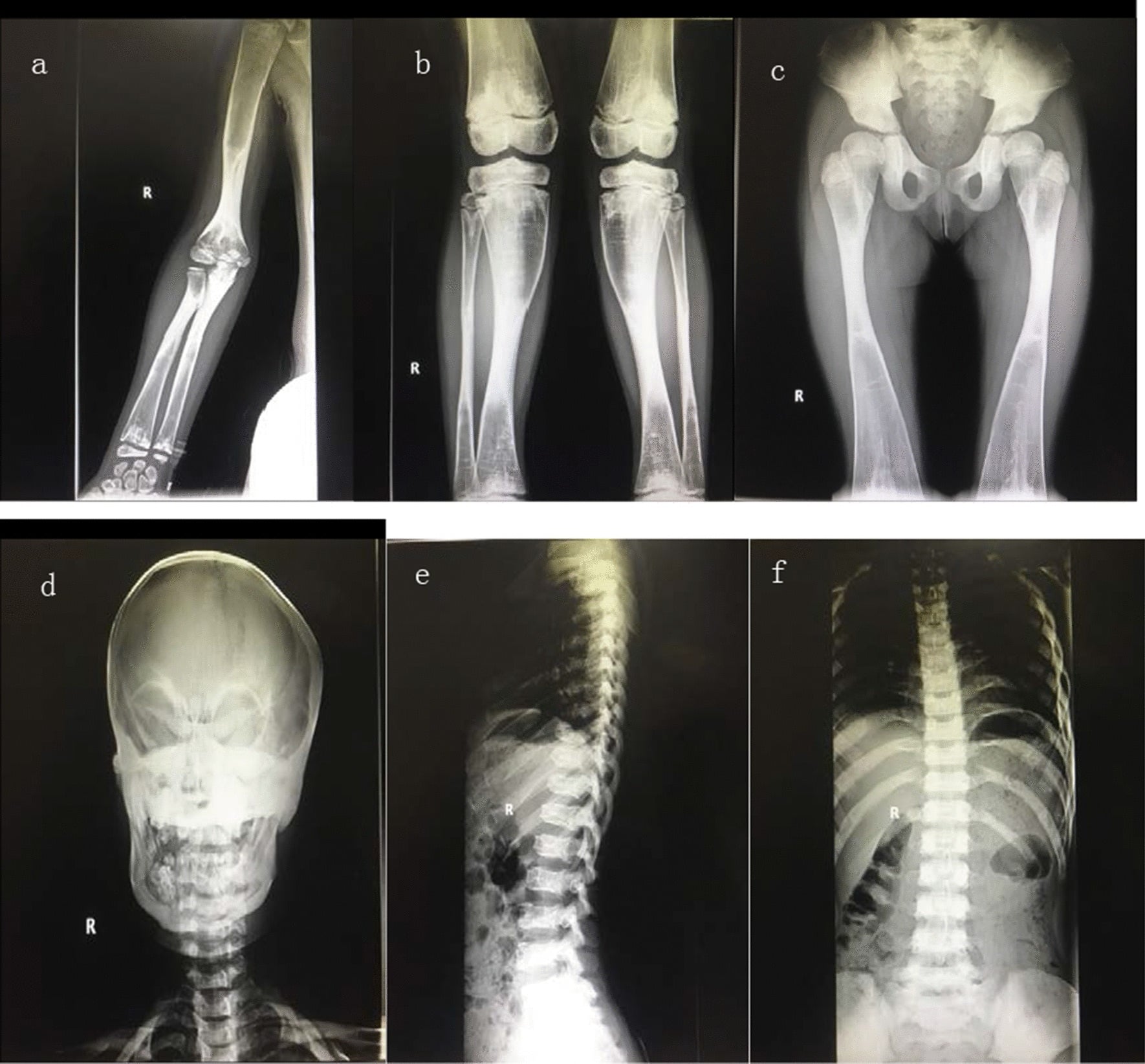
Rugger-jersey banding with a lucent mid-body is renal osteodystrophy with secondary hyperparathyroidism — subperiosteal resorption of the radial border of the middle phalanges and a pepper-pot skull confirm it. Sharper, denser sandwich vertebrae with endobone are osteopetrosis. A leucoerythroblastic film with splenomegaly is myelofibrosis. These are systemic diagnoses made off the spine film, not on it.
- 6Step 6 — What have the disc spaces, endplates and soft tissues done?
Look specifically at disc height, endplate integrity and the paravertebral outline on every study.
Preserved discs with intact endplates favour tumour. Endplate erosion with disc narrowing and a paravertebral collection favours pyogenic or tuberculous spondylodiscitis, and tuberculosis spares the disc longer while producing a large calcified paraspinal abscess. A circumferential mass with anterior body scalloping suggests lymphoma; cloud-like matrix within a mass suggests osteosarcoma; a rim-enhancing collection tracking under the anterior longitudinal ligament suggests infection.
- 7Step 7 — Commit, and be willing to commit to nothing
State one most likely diagnosis with two alternatives, then decide whether the next step is a test or a review appointment.
If it is not Paget disease or a classic haemangioma, proceed to whole-spine MRI, staging CT, and biopsy of the most accessible representative site — planned with the treating surgeon if a primary bone sarcoma is possible. But if the patient is ASYMPTOMATIC, the body is normal in size with intact pedicles and discs, and screening is negative, observation is a legitimate endpoint: Carpineta followed two such patients for 13 and 10 years, never reached a diagnosis despite biopsy in one, and neither came to harm.
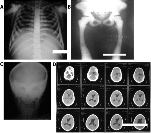
MCQ Practice Points
Q: An MRI shows Modic type 3 endplate sclerosis at L5/S1 in a patient with chronic low back pain. How much does it explain?
A: Less than it looks. Vertebral endplate signal change was present in a median 43 per cent of patients with non-specific low back pain but also in 6 per cent of non-clinical populations, so it is neither normal nor diagnostic — it shifts probability. Herlin's meta-analysis of 31 studies found the association with pain inconsistent: 15 significantly positive, one significantly negative, and only one study at low risk of bias. The only poolable odds ratio, 4.01 (1.52–10.61), came from studies using concordant pain on provocative discography, a test with its own false-positive problem. Report the finding; do not let it select a level for intervention.
Q: What is the first question to ask of a dense vertebra, before any differential?
A: How many, and where within the bone. One uniformly dense body of normal size is the ivory vertebra and carries the tumour-versus-Paget-versus-infection differential. Sclerosis confined to endplates at one or two levels is degenerative. Banded sclerosis at every level with a spared mid-body is rugger-jersey spine and a renal diagnosis. Uniform density throughout is a marrow or metabolic disease. These are four different questions, and reaching for the ivory vertebra list when the film shows banding is the commonest error on this topic.
Q: Multiple vertebrae show dense endplate bands with relatively lucent central bodies. What are the two leading diagnoses and how do you separate them?
A: Renal osteodystrophy (rugger-jersey spine — bands are indistinct and blurred, the patient is often dialysis dependent, PTH is raised, and subperiosteal resorption of the radial border of the middle phalanges confirms it) versus osteopetrosis (sandwich vertebra — bands are sharply demarcated, with generalised skeletal sclerosis and a bone-within-bone appearance). The separation is made on the biochemistry as much as the film.
Q: A patient with osteopetrosis and sandwich vertebrae needs a total hip replacement. What do you measure and what do you consent for?
A: Measure the intramedullary canal diameter 10 cm distal to the lesser trochanter. In the Mayo series of nine hips, EVERY postoperative periprosthetic femoral fracture occurred in a patient with a diameter under 5 mm. Quote the reoperation figure, not the survivorship one: 10-year survivorship free of revision was 89 per cent but free of ANY reoperation only 42 per cent, with a 44 per cent periprosthetic femoral fracture rate and intraoperative acetabular fracture in five of nine. Plan for a narrow stem, slow reaming with irrigation to avoid thermal necrosis, and prophylactic cabling. Caveat: nine hips over 47 years, so these are planning prompts, not validated rates.
Q: An asymptomatic patient has an incidental solitary dense vertebra of normal size with intact pedicles and disc spaces. Screening bloods, tumour markers and whole-spine MRI are all normal. What next?
A: Observation with periodic clinical review — not repeat imaging and not biopsy. Carpineta followed two such patients for 13 and 10 years with extensive repeated workup; no aetiology was ever found in either, Paget disease could not be confirmed even on bone biopsy in one, and both remained entirely asymptomatic. This is two patients, so it establishes that an idiopathic ivory vertebra can be benign, not how often it is — but it is the endpoint every algorithm omits, and saying it out loud shows you know when to stop.
Q: Which single radiographic feature best distinguishes Paget disease from osteoblastic metastasis in a sclerotic vertebra?
A: Enlargement of the vertebral body with cortical thickening — the picture-frame appearance. Metastasis increases density without increasing size. Trabecular coarsening and pedicle expansion support Paget, as does an isolated raised alkaline phosphatase with normal calcium and phosphate.
Q: A dense vertebra with anterior vertebral body scalloping in a 28-year-old with fever and weight loss — most likely diagnosis?
A: Hodgkin lymphoma. The scalloping results from pressure erosion by adjacent nodal mass. Disc spaces remain preserved, which separates it from spondylodiscitis.
Q: A treated prostate cancer patient develops increasingly sclerotic vertebral lesions with a falling PSA. Interpretation?
A: Osteoblastic flare — healing of previously lytic or occult metastases. Increasing sclerosis with falling markers indicates response, not progression. Correlate with tumour markers and metabolic imaging before changing therapy.
Q: Which finding most strongly favours infection over tumour in a sclerotic vertebra?
A: Endplate destruction with disc space narrowing and a paravertebral collection. Neoplastic disease characteristically spares the disc; pyogenic and tuberculous infection cross it. Tuberculosis spares the disc longer than pyogenic infection and produces a larger, often calcified, paraspinal abscess.
Exam Viva Scenarios
Practise clinical reasoning and management decisions out loud
“You are shown this lateral lumbar radiograph of a 68-year-old man with three months of mechanical back pain. L3 is uniformly dense. Describe what you see and take me through your reasoning.”
“You are shown this radiograph of a 24-year-old with three months of thoracic back pain, drenching night sweats and 8 kg weight loss. T8 is dense with subtle anterior vertebral body scalloping.”
“You are shown a lateral lumbar radiograph performed for renal colic. L4 is sclerotic, taller and deeper than adjacent bodies, with thickened cortical margins.”
Define the sign
- Homogeneously dense vertebral body, sharply demarcated from neighbours
- Height preserved, size normal — otherwise it is a different pattern
- Confirm on two views; exclude projectional and overlying causes
The big three
- Metastasis — normal size, pedicle involvement, over 50, prostate or breast
- Paget's — ENLARGED vertebra, picture frame, raised ALP, normal calcium
- Lymphoma — anterior scalloping, young adult, B symptoms, preserved discs
Aggressive alternatives
- Osteoblastic osteosarcoma — cloud-like matrix beyond the cortex
- Chronic osteomyelitis / TB — endplate destruction, disc narrowing, abscess
- Chordoma — midline sacrum or clivus, lytic with sequestered calcification
Multiple dense vertebrae
- Renal osteodystrophy — blurred rugger-jersey bands
- Osteopetrosis — sharp sandwich vertebra, bone-within-bone
- Myelofibrosis, mastocytosis, fluorosis, sickle cell (H-shaped)
- Widespread osteoblastic metastasis
Investigation ladder
- MRI whole spine with contrast — marrow, epidural disease, multiplicity
- CT for matrix and architecture; PET or bone scan for distribution
- PSA, ALP, calcium, phosphate, PTH, FBC and film, CRP, electrophoresis
- Biopsy any unexplained solitary lesion — plan the tract with the sarcoma unit
No further imaging required
- Classic enlarged Paget's vertebra with typical biochemistry
- Classic corduroy or polka-dot haemangioma
- Bone island with spiculated margins and no scan uptake
- Established rugger-jersey spine in known renal failure
Evidence Base
Vertebral Endplate Signal Changes (Modic Change): A Systematic Literature Review of Prevalence and Association with Non-Specific Low Back Pain
- 82 study samples from 77 original articles, MEDLINE, EMBASE and SveMED from 1984 to November 2007
- Median prevalence of any vertebral endplate signal change was 43 PER CENT in patients with non-specific low back pain or sciatica
- In NON-CLINICAL populations the median prevalence was 6 PER CENT - a sevenfold difference
- Prevalence rose with age and, importantly, was NEGATIVELY associated with the overall quality score of the study
- Seven of ten studies found a positive association with low back pain, odds ratios ranging enormously from 2.0 to 19.9
Modic Changes - Their Associations with Low Back Pain and Activity Limitation: A Systematic Review and Meta-Analysis
- PROSPERO-registered systematic review, 5210 citations screened, 31 studies included
- ONLY ONE of the 31 studies had a low risk of bias
- 15 studies (48 per cent) found a significant positive association with low back pain; one found a significant NEGATIVE association; the rest found none
- Meta-analysis was possible only for the subgroup using concordant pain on provocative discography, giving an odds ratio of 4.01 (95 per cent CI 1.52 to 10.61)
- Only one of seven studies found an association with activity limitation; the authors conclude the associations are INCONSISTENT and that low-bias studies will likely change their direction and strength
Total Hip Arthroplasty in Patients With Osteopetrosis
- 7 patients (9 hips) with osteopetrosis undergoing primary total hip arthroplasty from one joint registry between 1970 and 2017, mean age 48, mean follow-up 8 years
- 10-year survivorship free from revision or implant removal was 89 PER CENT - which sounds reassuring
- 10-year survivorship free from ANY REOPERATION was 42 PER CENT: a 58 per cent reoperation rate
- Periprosthetic femoral fracture rate 44 per cent; FIVE OF NINE hips sustained an intraoperative acetabular fracture and one an intraoperative femoral fracture
- EVERY postoperative femoral fracture occurred in a patient whose intramedullary diameter was less than 5 mm measured 10 cm distal to the lesser trochanter
The Ivory Vertebra: An Approach to Investigation and Management Based on Two Case Studies
- Two asymptomatic patients with an incidentally discovered solitary dense vertebra, followed for 13 and 10 years respectively
- Both underwent extensive repeated investigation; NO AETIOLOGY WAS EVER FOUND in either, and all tests remained normal for the whole period
- Paget disease was suspected in one but could not be confirmed even on BONE BIOPSY
- Both patients remained entirely asymptomatic with respect to the vertebra throughout follow-up
- The authors propose an algorithm: once baseline screening has excluded ominous pathology, observe with periodic clinical reassessment rather than investigate further