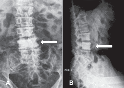
One vertebral body, uniformly dense, normal size, normal disc spaces — until proven otherwise this is tumour
- Definition: diffuse homogeneous increase in opacity of a single vertebral body, with preservation of vertebral size and shape and no loss of adjacent disc height.
- Preserved disc spaces are the key negative — disc involvement pushes you towards infection, not tumour.
- In an adult over 50 the differential is metastasis, lymphoma, Paget's disease. In a child it is lymphoma, osteoblastic metastasis (neuroblastoma), and osteosarcoma.
- Paget's disease is identified on the plain film alone: vertebral enlargement, cortical and endplate thickening giving the 'picture frame', and coarsened trabeculae.
- Plain radiographs need roughly 30 to 50 percent trabecular bone change before sclerosis is visible — a normal film never excludes marrow disease.
- A soft-tissue mass out of proportion to bone destruction, wrapping the vertebra circumferentially, is lymphoma until proven otherwise.
- Bone biopsy is required whenever no primary is known and the pattern is not diagnostic of Paget's disease.
- “Say the three features out loud before offering a differential: uniform density, normal size, normal discs.
- “An enlarged ivory vertebra is Paget's disease (or, rarely, a sclerosing metastasis with expansion). A normal-sized one is not.
- “Hodgkin lymphoma is the classic teaching answer for ivory vertebra in a patient under 40 with night sweats.
- “Osteopetrosis and other sclerosing dysplasias give sclerosis at every level — the sign requires a solitary vertebra, so multiplicity reframes the whole differential.
- “Check the pedicles: an absent pedicle alongside sclerosis at the same level means metastasis, not Paget's.
The sign requires diffuse, homogeneous sclerosis of the whole body. Patchy sclerosis, a sclerotic band, or endplate sclerosis are different problems with different differentials. Bone islands and healed benign lesions do not qualify.
An overlying structure — barium in bowel, contrast, a pacing lead, a calcified aortic plaque, or simply a lordotic vertebra viewed obliquely — can produce apparent density. Confirm on the orthogonal view and on CT before committing.
Preserved discs are what separates tumour from infection. Endplate erosion with disc-height loss and sclerosis is chronic pyogenic or tuberculous discitis, not an ivory vertebra.
No sclerotic solitary vertebra is adequately staged on plain film. Whole-spine MRI plus a search for the primary is the default. Reserving biopsy for 'if the scan is unclear' is the wrong answer in the viva.
Recognising the Pattern
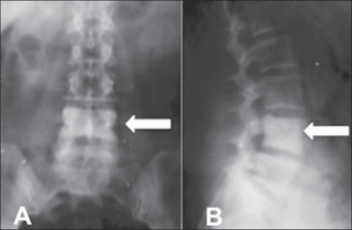
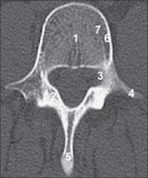
Definition. An ivory vertebra is a single vertebral body showing diffuse, homogeneous increase in radiographic opacity, in which the vertebra retains normal size and contour and the adjacent intervertebral disc spaces are preserved. It is a descriptive sign, not a diagnosis.
Confirming it is genuinely present
- The density must be uniform across the whole body, not focal or patchy, and must extend to the cortical margins.
- It must be one level — scan every visible vertebra above and below on the same film before you call it solitary.
- Disc heights above and below must be normal. Loss of disc height reframes the case as infective.
- Size and shape are normal. Vertebral enlargement, cortical thickening or endplate thickening moves you to Paget's disease.
- Confirm on the lateral as well as the AP. Projectional density disappears; true sclerosis does not.
- Check the posterior elements: pedicle, lamina and spinous process involvement, or an absent pedicle, changes the answer.
The words to use out loud
"This is a frontal and lateral radiograph of the thoracolumbar spine. There is diffuse homogeneous sclerosis of a single vertebral body — the ivory vertebra sign. The vertebral body maintains normal height and normal anteroposterior dimension, the cortex is not thickened, and the adjacent disc spaces are preserved. The remaining visible vertebrae have normal density. I would like to see the posterior elements and assess for a paravertebral soft-tissue mass. In an adult of this age my leading differential is osteoblastic metastasis, then lymphoma, then Paget's disease — and I would exclude Paget's on this film by the absence of vertebral enlargement."
What mimics the pattern (false positives)
- How to exclude it
- Repeat or orthogonal view; density crosses anatomical boundaries
- How to exclude it
- Density resolves on the true lateral
- How to exclude it
- Vertebral height is reduced — this is collapse, not ivory vertebra
- How to exclude it
- Vertical striations (corduroy) or polka-dot on axial CT, and fat signal on T1 MRI
- How to exclude it
- Focal, ovoid, brush-border margins, does not involve the whole body
- How to exclude it
- Density is markedly greater than bone, tracks along trabeculae, obvious history
- How to exclude it
- Sclerosis confined to a radiotherapy port that respects vertebral levels in a straight line
Next Investigation
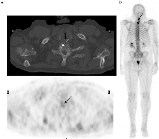
Do not send a patient with suspected primary bone sarcoma for biopsy locally. Any aggressive solitary vertebral lesion without an obvious primary should be discussed with a specialist bone tumour multidisciplinary team before tissue is taken — a poorly placed tract can convert a limb- or spine-sparing resection into a far larger operation.
MOPIvory Vertebra — the classic triad plus
The Differential
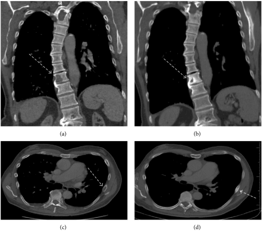
- Typical age / setting
- Men over 55, back pain, raised PSA
- Discriminating feature
- Often multifocal on further imaging; pedicle may be eroded or absent; body remains normal-sized
- What confirms it
- PSA, bone scan showing multiple hot spots, prostate MRI/biopsy
- Typical age / setting
- Women over 45, known or occult primary
- Discriminating feature
- Mixed lytic-sclerotic elsewhere in the skeleton; sclerosis may appear only after treatment response
- What confirms it
- Mammography, CT chest-abdomen-pelvis, bone biopsy
- Typical age / setting
- Adolescent to age 40, night sweats, weight loss, cervical nodes
- Discriminating feature
- Circumferential paravertebral soft tissue disproportionate to the bone change; anterior scalloping from nodal pressure
- What confirms it
- Nodal or bone core biopsy, PET-CT, mediastinal nodes on chest CT
- Typical age / setting
- Age 40 to 70, may be primary bone lymphoma
- Discriminating feature
- Extensive marrow replacement on MRI with a permeative pattern yet a nearly intact cortex on radiograph
- What confirms it
- Core biopsy with immunohistochemistry; PET-CT for staging
- Typical age / setting
- Over 55, often incidental, alkaline phosphatase raised with normal calcium
- Discriminating feature
- Vertebral ENLARGEMENT with cortical and endplate thickening — the 'picture frame' vertebra; coarse trabeculae
- What confirms it
- Plain film alone is usually diagnostic; alkaline phosphatase; bone scan shows intense uniform uptake
- Typical age / setting
- Teens/twenties, or over 60 arising in Paget's bone
- Discriminating feature
- Aggressive periosteal reaction, cortical breach and a soft-tissue mass with cloud-like tumour matrix
- What confirms it
- MRI plus biopsy at a bone tumour centre; new pain in known Paget's is the red flag
- Typical age / setting
- Any age, indolent back pain, may follow instrumentation
- Discriminating feature
- Endplate irregularity and disc-height loss — the discs are NOT preserved
- What confirms it
- MRI showing disc and endplate high T2 signal; CT-guided aspiration and culture
- Typical age / setting
- Endemic exposure, immunosuppression, insidious course
- Discriminating feature
- Anterior vertebral body involvement with subligamentous spread and a calcified paraspinal abscess
- What confirms it
- MRI, tissue for acid-fast bacilli culture and molecular testing
- Typical age / setting
- Children and young adults; palmoplantar pustulosis, acne
- Discriminating feature
- Sternoclavicular hyperostosis coexisting with the vertebral lesion; relapsing-remitting sterile course
- What confirms it
- Whole-body MRI showing multifocal sterile lesions; sterile biopsy
- Typical age / setting
- Any adult, usually incidental
- Discriminating feature
- Vertical coarse trabeculae (corduroy) on lateral; polka-dot pattern on axial CT
- What confirms it
- CT and MRI — fat and vascular signal; no marrow replacement
- Typical age / setting
- Age 10 to 25, night pain relieved by NSAIDs, painful scoliosis
- Discriminating feature
- Lesion is centred on the POSTERIOR elements with surrounding reactive sclerosis, not the body
- What confirms it
- Thin-slice CT to find the nidus; scoliosis concave to the lesion
- Typical age / setting
- Age 40 to 70, sacrum or clivus more than mobile spine
- Discriminating feature
- Midline lesion with amorphous calcification and a large soft-tissue mass; usually lytic with sclerotic rim
- What confirms it
- MRI (very high T2 signal), biopsy showing physaliphorous cells
- Typical age / setting
- Systemic disease, known haematological or renal history
- Discriminating feature
- Sclerosis is diffuse and MULTILEVEL — a solitary ivory vertebra is atypical; rugger-jersey banding in renal disease
- What confirms it
- Bloods (tryptase, full blood count and film, renal function); marrow biopsy
- Typical age / setting
- Congenital, presents in childhood or as an incidental finding
- Discriminating feature
- Every vertebra is dense with a bone-within-bone or sandwich appearance — never solitary
- What confirms it
- Skeletal survey; family history; no further work-up needed
Narrowing It Down
- 1Step 1 — Is it truly solitary?
Review every vertebra on the film, then image the whole spine.
Multiple dense vertebrae take you out of the ivory vertebra differential entirely and into diffuse sclerosis: metastatic disease, myelofibrosis, mastocytosis, renal osteodystrophy, osteopetrosis or fluorosis. A genuinely solitary lesion in an adult narrows sharply to metastasis, lymphoma or Paget's disease.
- 2Step 2 — How old is the patient?
Set the differential by age before looking at any other feature.
Under 40: lymphoma (especially Hodgkin), osteoblastoma, osteosarcoma, chronic osteomyelitis, and in children neuroblastoma metastasis. Over 50: metastasis first, then lymphoma, then Paget's disease. Age alone reorders the list more than any other single variable - and note that Paget's belongs progressively lower on it than the textbooks suggest, since its prevalence has roughly halved and patients now present about 4 years older per decade.
- 3Step 3 — Is the vertebra enlarged?
Compare the anteroposterior and vertical dimensions with the levels above and below.
Enlargement with cortical and endplate thickening (picture-frame) and coarse trabeculae is Paget's disease - a plain-film diagnosis requiring alkaline phosphatase but no biopsy. Normal size keeps tumour top of the list.
- 4Step 4 — Are the disc spaces preserved?
Measure the disc heights above and below and inspect both endplates.
Preserved discs favour tumour, because neoplasm does not cross the avascular disc. Endplate destruction with disc-height loss is infection - pyogenic if rapid with marked destruction, tuberculous if indolent with anterior involvement, subligamentous spread and a paraspinal collection.
- 5Step 5 — Is there a soft-tissue mass, and is it proportionate?
Judge the size of any paravertebral mass against the amount of bone destruction, not in absolute terms.
A soft-tissue mass far larger than the bone abnormality, encircling the vertebra and causing anterior scalloping, is lymphoma. A mass with cloud-like ossified matrix and aggressive periosteal reaction is osteosarcoma. No mass with sclerosis alone favours metastasis or Paget's.
- 6Step 6 — Are the posterior elements involved?
Look specifically at the pedicles, laminae and transverse processes, not only the body.
An absent or sclerotic pedicle at the same level supports metastasis. A nidus or expansile lesion centred on pedicle, lamina or transverse process with reactive body sclerosis, in a young patient with night pain, is osteoblastoma or osteoid osteoma.
- 7Step 7 — What do the systemic features and bloods say?
Take the history and send calcium, phosphate, alkaline phosphatase, inflammatory markers, PSA in men and a myeloma screen.
B symptoms and lymphadenopathy point to lymphoma. Raised PSA, a breast lump or a smoking history points to metastasis. Isolated raised alkaline phosphatase with normal calcium and phosphate points to Paget's. Fever, raised inflammatory markers and a recent procedure point to infection. If nothing fits, biopsy.
MCQ Practice Points
Q: Which single radiographic feature most reliably separates Paget's disease from osteoblastic metastasis in a solitary dense vertebra?
A: Vertebral ENLARGEMENT with cortical and endplate thickening — the picture-frame vertebra. Metastatic sclerosis leaves vertebral size and cortical thickness normal. Density alone does not discriminate. Add a modern caveat: Paget's is becoming less common and milder, with radiographic prevalence about half its 1983 level and disease extent and alkaline phosphatase falling with each birth cohort (Cundy), so the classic florid picture-frame vertebra is now a rarer finding than the textbooks imply.
Q: A 22-year-old has an ivory vertebra with a circumferential paravertebral mass far larger than the bony abnormality. Most likely diagnosis?
A: Hodgkin lymphoma. The disproportionate soft-tissue component with a relatively intact cortex is characteristic; anterior vertebral scalloping from adjacent nodal masses supports it. Note for the follow-up film: the sclerosis is reactive host bone and can revert to a normal radiographic appearance after successful treatment (Mandell).
Q: Which finding argues against neoplasm and towards infection in a sclerotic vertebra?
A: Loss of adjacent disc height with endplate erosion. Tumour respects the avascular disc; pyogenic and tuberculous infection destroy it. Preservation of disc height is part of the ivory vertebra definition, so a lesion that has taken the disc is not, strictly, an ivory vertebra at all.
Q: Approximately how much trabecular bone must be altered before sclerosis or lysis is visible on a plain radiograph?
A: Roughly 30 to 50 per cent. A normal radiograph therefore never excludes marrow disease, which is why MRI is the next investigation in a symptomatic patient. This is a conventional figure derived from lytic-lesion phantom and cadaveric work rather than a measured threshold for vertebral sclerosis specifically — treat it as an order of magnitude.
Q: In a young patient with painful scoliosis, night pain relieved by NSAIDs, and a dense vertebral body, where should you look on the CT?
A: The posterior elements. Osteoid osteoma or osteoblastoma of the pedicle or lamina produces intense reactive sclerosis of the adjacent body; thin-slice CT is the best way to identify the nidus, and the scoliosis is concave towards the lesion.
Q: A patient with known breast carcinoma on treatment develops new sclerosis in a previously lytic vertebra. Interpretation?
A: This may represent a HEALING response to therapy rather than progression. Sclerosis in a treated patient must be interpreted alongside the pre-treatment films and functional imaging, not read as new disease in isolation. The same trap runs the other way in prostate cancer, where PSMA avidity persists in healing bone and in Paget's disease — so neither density nor tracer uptake settles the question on its own.
Exam Viva Scenarios
Practise clinical reasoning and management decisions out loud
“You are shown a lateral lumbar radiograph of a 68-year-old man with six weeks of progressive back pain worse at night. L3 is uniformly dense. Describe the film and give your differential.”
“You are shown a thoracic radiograph of a 24-year-old with three months of thoracic back pain, drenching night sweats and 8 kg weight loss. T8 is uniformly sclerotic and there is a paraspinal soft-tissue shadow.”
“You are shown a lumbar radiograph performed for renal colic in a 71-year-old woman. L4 is dense throughout, appears larger than L3 and L5, and the cortical margins are strikingly thick.”
The Sign (all three required)
- Diffuse homogeneous sclerosis of a single vertebral body
- Normal vertebral size and contour
- Preserved adjacent disc spaces
Top Three in an Adult over 50
- Osteoblastic metastasis — prostate in men, breast in women; check pedicles and other levels
- Lymphoma — soft tissue out of proportion to bone change
- Paget's disease — enlarged vertebra, picture frame, raised alkaline phosphatase
Top Three in a Patient under 40
- Hodgkin lymphoma with B symptoms
- Osteoblastoma or osteoid osteoma centred on the posterior elements
- Chronic osteomyelitis or SAPHO — look for disc involvement and sternoclavicular hyperostosis
Discriminators in One Line Each
- Enlarged vertebra equals Paget's disease
- Absent pedicle equals metastasis
- Disc-space loss equals infection
- Huge soft-tissue mass with minimal bone destruction equals lymphoma
- Corduroy or polka-dot equals haemangioma
- Every vertebra dense equals sclerosing dysplasia, myelofibrosis or renal osteodystrophy
Mimics to Exclude First
- Overlying barium, contrast, cement or hardware
- Projectional density on a rotated view
- Collapsed vertebra (height reduced — not an ivory vertebra)
- Bone island or treated metastasis
Investigation Order
- Orthogonal radiographs to confirm the sign and assess size and discs
- Whole-spine MRI with contrast — marrow, epidural disease, other levels
- CT for matrix, cortex and biopsy planning
- Staging CT chest, abdomen and pelvis plus PSA or mammography
- Bone scan or PET-CT for skeletal survey and staging
- Image-guided core biopsy after bone tumour MDT discussion
When to Stop
- Classical Paget's disease with supportive alkaline phosphatase in an asymptomatic older patient
- Typical haemangioma with polka-dot CT appearance
- Diffuse sclerosis explained by an established systemic diagnosis
Red Flags
- New neurological deficit — urgent MRI, steroids, spinal surgical referral
- New unremitting pain in known Paget's — sarcomatous transformation
- Solitary lesion with no known primary — do not treat without tissue
- Suspected primary bone sarcoma — no local biopsy; refer to a tumour centre first
Evidence Base
The Ivory Vertebra Sign
- The canonical description of the sign in Radiology's 'Signs in Imaging' series: a solitary vertebral body of markedly increased opacity with preserved size and contour, no change in adjacent disc spaces, and no vertebral collapse
- The differential indexed against it is exactly the one to recite: osteoblastic metastasis, Hodgkin lymphoma, Paget disease and other causes of osteosclerosis
- The size and contour criteria are what separate the sign from a collapsed or expanded dense vertebra, which have different differentials
Resolution of Hodgkin's Induced Ivory Vertebrae
- An adolescent with Hodgkin disease whose ivory vertebrae REVERTED to a normal radiographic appearance as part of a favourable response to treatment
- Establishes that the sclerosis of lymphomatous ivory vertebra is reactive host bone, not fixed structural change - and can therefore resolve