A pattern, not a diagnosis — age, site and zone of transition do most of the work
- Describe before you diagnose: age, bone, longitudinal zone (epiphysis / metaphysis / diaphysis), transverse zone (central / eccentric / cortical / juxtacortical), size, margin, matrix, periosteal reaction, soft-tissue mass.
- Zone of transition is the single best radiographic marker of biological aggressiveness on plain film. A lesion you can trace with a fine pencil is indolent.
- In a patient over 40, a solitary lytic lesion is metastasis or myeloma until proven otherwise — lung, breast, kidney, thyroid, prostate.
- Epiphyseal or apophyseal lucency in a skeletally immature patient means chondroblastoma or infection; in a mature patient it means giant cell tumour, or a geode.
- Never biopsy an aggressive-looking lucent lesion before it has been staged and discussed with the sarcoma unit — an inappropriate tract converts a limb-salvage case into an amputation.
- Do not mistake a normal variant (nutrient foramen, pseudocyst of the humeral head, ischiopubic synchondrosis) or an overlying artefact for a lesion.
- “'This is a well-defined lucent lesion with a narrow zone of transition and a thin sclerotic rim, arising eccentrically in the metaphysis of a skeletally immature patient' — that sentence alone buys you most of the marks.
- “A lucent lesion with a wide zone of transition, cortical destruction and a soft-tissue mass is a sarcoma or infection — the radiograph cannot separate them; the history can.
- “Multiple lucent lesions completely change the list: metastases, myeloma, hyperparathyroidism (brown tumours), fibrous dysplasia, enchondromatosis, Langerhans cell histiocytosis, infection.
- “'Leave me alone' lesions (non-ossifying fibroma, bone island, healing fibrous cortical defect) need no follow-up if the appearance is pathognomonic.
Myeloma, renal and thyroid metastases, and low-grade chondrosarcoma can all be geographic with a reasonably defined margin. Age over 40 overrides margin every time — take a history and check the bloods before you commit.
Never diagnose a solitary lucent lesion from one radiograph. Two views, the whole bone, and both joints above and below. Polyostotic disease (metastases, myeloma, fibrous dysplasia, enchondromatosis, hyperparathyroidism) is a different differential entirely.
Local staging MRI and chest imaging come BEFORE the needle. The biopsy tract must be sited by the surgeon who will do the definitive resection, in line with the planned incision.
Rings-and-arcs calcification tells you the tissue is cartilage, not that it is benign. Deep endosteal scalloping of more than two-thirds cortical thickness, cortical breach, pain at rest, size greater than 5 cm and axial location push you towards chondrosarcoma.
Recognising the Pattern
Definition. A lucent (radiolucent, "lytic") bone lesion is a focal region of reduced radiographic density within bone, produced by replacement or removal of trabecular and/or cortical mineralised matrix. It is a pattern, not a diagnosis — the same appearance is produced by neoplasia, infection, metabolic disease, trauma and normal variation.
Confirming it is genuinely present. Reproduce the lucency on a second, orthogonal projection. A true lesion persists, occupies the same anatomical location on both views, and has an internal architecture. Confirm that the trabecular pattern within the area is genuinely lost or altered rather than simply projected differently. Look for a matched cortical change — endosteal scalloping, expansion, or a breach — because a true intramedullary lesion of any size usually leaves a cortical footprint.
- Patient factors — age and skeletal maturity, and whether the physes are open.
- Bone and longitudinal site — epiphysis / apophysis, metaphysis, diametaphysis, diaphysis.
- Transverse site — central, eccentric, cortical (intracortical), juxtacortical/surface.
- Size — in centimetres, in two dimensions.
- Margin and zone of transition — geographic with a sclerotic rim (Lodwick IA), geographic well defined without sclerosis (IB), geographic with an ill-defined margin (IC), moth-eaten (II), permeative (III).
- Matrix — none (truly lucent), chondroid (rings and arcs, popcorn), osteoid (cloud-like, fluffy), ground-glass (fibrous dysplasia).
- Cortex — intact, thinned, expanded with an intact shell, breached.
- Periosteal reaction — none, solid/thick (indolent), lamellated, spiculated, Codman triangle (aggressive).
- Soft tissues — mass, swelling, calcification, fat planes.
- Everything else — joint space, other lesions, chest if included.
"This is an AP radiograph of the left knee in a skeletally immature patient. There is a well-defined lucent lesion, approximately 3 cm in length, arising eccentrically in the distal femoral metaphysis. It has a narrow zone of transition with a thin sclerotic rim, no internal matrix mineralisation, mild endosteal scalloping with an intact overlying cortex, and no periosteal reaction or soft-tissue mass. The appearances are those of a Lodwick IA, biologically indolent lesion — my first diagnosis is a non-ossifying fibroma."
- Normal variants: nutrient foramen and its vascular channel; the pseudocyst of the greater tuberosity and humeral head; the calcaneal neutral triangle; the ischiopubic synchondrosis in children; the metaphyseal "cortical desmoid" of the posteromedial distal femur.
- Superimposition: bowel gas over the pelvis, a skin fold, a gonad shield edge, overlying soft-tissue air or a wound.
- Physiological: open or closing physis, the physeal scar, sesamoid or accessory ossicle beds.
- Osteopenia: generalised loss of trabeculae misread as focal lucency, particularly in the femoral neck.
- Post-surgical: screw and pin tracts, curettage cavities, bone graft harvest sites, cement voids.
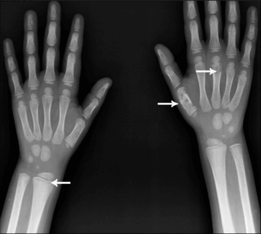
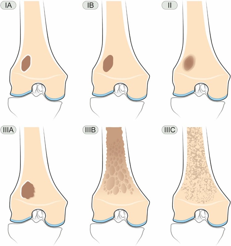
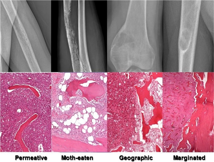
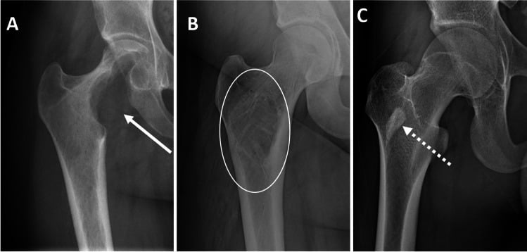
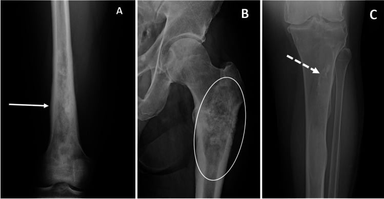
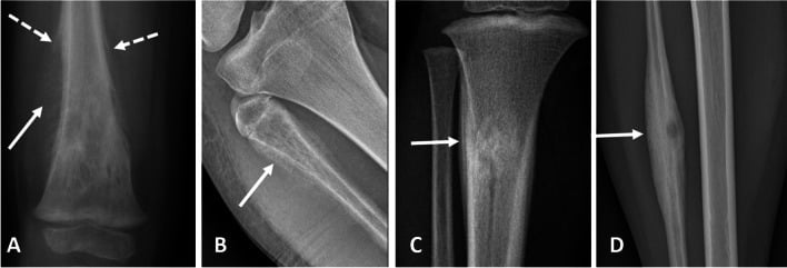
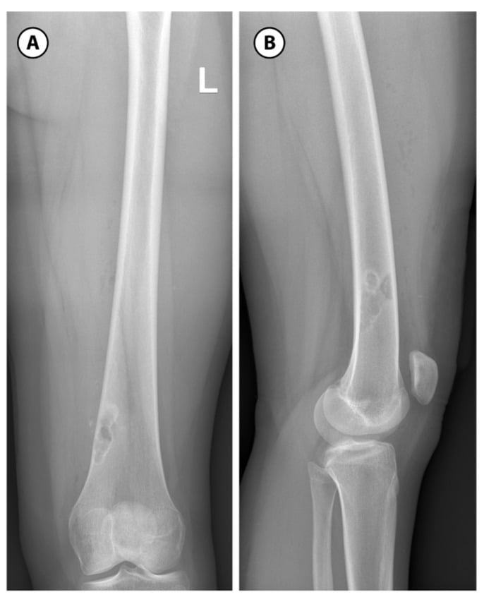
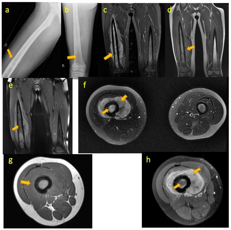
Next Investigation
The commonest correct answer to "what would you do next" in the viva for an aggressive lucent lesion is not "MRI" alone — it is "I would obtain orthogonal radiographs of the whole bone, arrange local staging MRI and a CT chest, send a myeloma screen if the patient is over 40, and refer to the regional sarcoma multidisciplinary team before any biopsy."
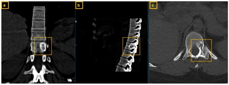
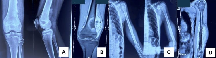
FEGNOMASHIC
Hook:The established mnemonic for the lucent bone lesion differential (Helms).
The Differential
- Typical age / setting
- Over 40; known or occult lung, breast, renal, thyroid, prostate primary
- Discriminating feature
- Multiple lesions in the axial skeleton and proximal long bones, distal to elbow and knee is rare (think lung, renal); pain at rest and at night
- What confirms it
- CT chest/abdomen/pelvis, whole-body bone scan or FDG-PET, myeloma screen; biopsy of the most accessible lesion
- Typical age / setting
- Over 50; back pain, anaemia, renal impairment
- Discriminating feature
- Punched-out lucencies with NO surrounding sclerosis and no reactive rim; skull vault involvement; often cold on bone scan
- What confirms it
- Serum and urine protein electrophoresis, serum free light chains, whole-body MRI or low-dose CT skeletal survey, marrow biopsy
- Typical age / setting
- 10-25 years; second peak in Paget disease and post-irradiation
- Discriminating feature
- Metaphysis about the knee, wide zone of transition, cortical destruction, aggressive periosteal reaction (Codman triangle, sunburst) with or without osteoid matrix
- What confirms it
- MRI whole bone, CT chest, alkaline phosphatase and LDH; image-guided biopsy planned by the sarcoma unit
- Typical age / setting
- 5-20 years; systemic upset, fever, raised inflammatory markers
- Discriminating feature
- Diaphyseal permeative lucency with lamellated 'onion-skin' periostitis and a soft-tissue mass disproportionately large for the bone change
- What confirms it
- MRI, CT chest, biopsy with molecular testing for the EWSR1 rearrangement
- Typical age / setting
- Over 40; pelvis, proximal femur, proximal humerus, scapula
- Discriminating feature
- Chondroid matrix with deep endosteal scalloping greater than two-thirds of cortical thickness, size greater than 5 cm, cortical thickening or breach, pain at rest — an enchondroma in the pelvis is a chondrosarcoma until proven otherwise
- What confirms it
- MRI for marrow and soft-tissue extension; sarcoma-unit biopsy — beware sampling error in low-grade disease
- Typical age / setting
- Any age; children commonest, plus diabetes, immunosuppression, sickle cell
- Discriminating feature
- Serpiginous lucency crossing the physis, a penumbra sign on T1 MRI, sequestrum within the lucency, and a clinical fever with raised CRP; can mimic any tumour
- What confirms it
- MRI with contrast, blood cultures, CRP/ESR, aspiration or biopsy for microbiology AND histology
- Typical age / setting
- 5-20 years; usually asymptomatic and incidental
- Discriminating feature
- Eccentric, cortically based metaphyseal lucency with a scalloped sclerotic rim and a bubbly septated interior; migrates away from the physis and sclerose with age
- What confirms it
- Radiograph alone — a pathognomonic 'leave me alone' lesion; no follow-up needed unless very large or painful
- Typical age / setting
- 5-15 years; proximal humerus and proximal femur; presents with pathological fracture
- Discriminating feature
- Central, metaphyseal, mildly expansile, abutting the physis, long axis parallel to the bone; a fallen fragment sign after fracture
- What confirms it
- Radiograph; MRI shows uniform fluid signal with no solid enhancing component
- Typical age / setting
- 10-20 years; rapid growth, pain
- Discriminating feature
- Eccentric, markedly expansile, 'blown-out' cortex with a thin shell, fluid-fluid levels on MRI; may be secondary to another lesion
- What confirms it
- MRI for fluid-fluid levels; biopsy to exclude telangiectatic osteosarcoma and an underlying primary lesion
- Typical age / setting
- 20-45 years, physes CLOSED; knee, distal radius, sacrum
- Discriminating feature
- Eccentric, subarticular lucency extending to within 1 cm of the articular surface, no matrix, no sclerotic rim, narrow but non-sclerotic margin
- What confirms it
- MRI to assess subchondral and soft-tissue extent; biopsy; CT chest for benign pulmonary metastases
- Typical age / setting
- 20-50 years; hands and feet commonest site
- Discriminating feature
- Central, mildly expansile lucency with punctate or rings-and-arcs chondroid calcification, shallow endosteal scalloping, painless unless fractured
- What confirms it
- Radiograph is usually sufficient in the hand; MRI plus surveillance if in a long bone or if painful
- Typical age / setting
- 10-25 years; night pain relieved dramatically by NSAIDs
- Discriminating feature
- Lucent nidus less than 1.5 cm with a central density, surrounded by dense reactive cortical sclerosis; intra-articular lesions cause an effusion with little sclerosis
- What confirms it
- Thin-slice CT to demonstrate the nidus; bone scan shows a double-density sign
- Typical age / setting
- Adolescents and young adults; monostotic or polyostotic
- Discriminating feature
- Ground-glass (hazy, not truly lucent) matrix with a thick sclerotic 'rind', long lesion in a long bone, shepherd's crook deformity of the proximal femur
- What confirms it
- Radiograph is usually diagnostic; CT confirms ground-glass density; no biopsy needed if classic
- Typical age / setting
- Adults with chronic kidney disease or a parathyroid adenoma
- Discriminating feature
- Multiple lucencies plus the background clues — subperiosteal resorption of the radial side of the middle phalanges, acro-osteolysis, rugger-jersey spine
- What confirms it
- Serum calcium, phosphate, parathyroid hormone, vitamin D, renal function — biochemistry, not biopsy
- Typical age / setting
- 5-15 years; skull, spine, femur
- Discriminating feature
- 'Great mimic' — bevelled-edge skull lucency, or vertebra plana with preserved disc heights; can look aggressive yet heal spontaneously
- What confirms it
- Skeletal survey, MRI; biopsy showing CD1a and langerin positive cells
- Typical age / setting
- Over 40; adjacent to a degenerate or inflamed joint
- Discriminating feature
- Subchondral lucency with a sclerotic rim in a joint that shows osteoarthritis, avascular necrosis or inflammatory arthritis — the joint disease is the clue
- What confirms it
- Radiograph of the joint; MRI if solitary and the joint appears normal
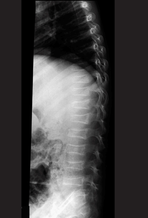
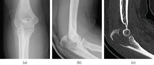
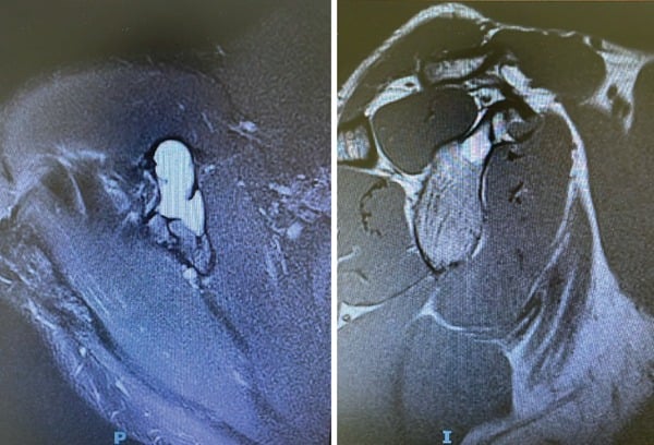
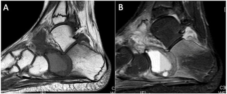
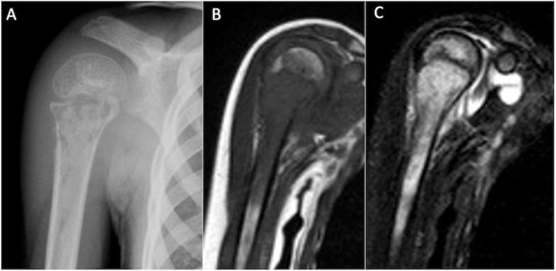
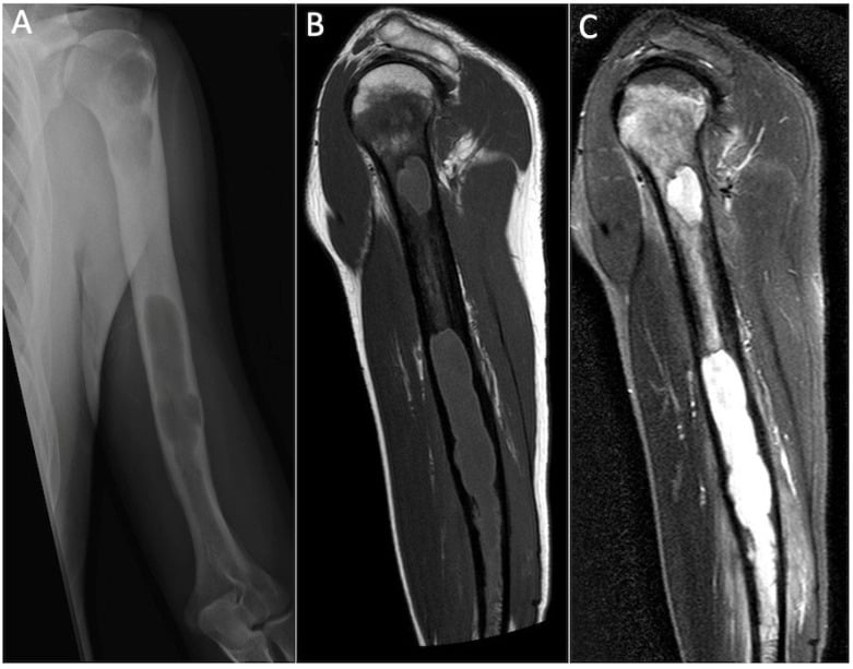
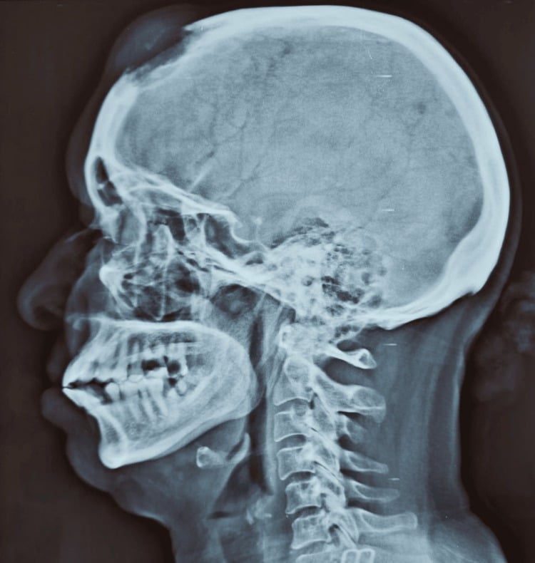
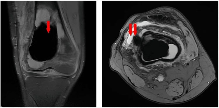
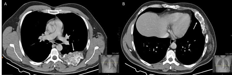
Narrowing It Down
- 1Step 1 — How old is the patient, and are the physes open?
Establish age and physeal status before looking at the lesion itself.
Under 30 with open or recently closed physes: simple bone cyst, non-ossifying fibroma, aneurysmal bone cyst, chondroblastoma, osteoid osteoma, Ewing sarcoma, osteosarcoma, infection, Langerhans cell histiocytosis. Age 20 to 45 with closed physes: giant cell tumour enters the list. Over 40: metastasis, myeloma and chondrosarcoma dominate, and that remains true even when the lesion looks benign. This single question discards more than half the differential.
- 2Step 2 — Solitary or multiple?
Image the whole bone and screen the rest of the skeleton before committing to a single-lesion diagnosis.
Multiple lucencies means metastases, myeloma, brown tumours of hyperparathyroidism, polyostotic fibrous dysplasia, enchondromatosis (Ollier, Maffucci), Langerhans cell histiocytosis, or multifocal infection. Solitary keeps the primary bone tumour list alive.
- 3Step 3 — Where in the bone, longitudinally?
Place the lesion in epiphysis, metaphysis or diaphysis, and note whether the physis is open.
Epiphysis or apophysis with open physes: chondroblastoma or infection. Subarticular with closed physes: giant cell tumour or geode. Metaphysis: the widest list - simple bone cyst, non-ossifying fibroma, aneurysmal bone cyst, osteosarcoma, infection. Diaphysis: Ewing sarcoma, Langerhans cell histiocytosis, fibrous dysplasia, adamantinoma in the anterior tibial cortex, osteoid osteoma.
- 4Step 4 — Where in the bone, transversely?
Decide whether the lesion is central, eccentric, intracortical or on the surface.
Central: simple bone cyst, enchondroma, fibrous dysplasia. Eccentric: non-ossifying fibroma, aneurysmal bone cyst, giant cell tumour, chondromyxoid fibroma. Intracortical: osteoid osteoma, fibrous cortical defect, intracortical abscess. Surface or juxtacortical: osteochondroma, periosteal chondroma, parosteal or periosteal osteosarcoma.
- 5Step 5 — Is there matrix?
Look inside the lucency for mineralised material and characterise its pattern.
Rings, arcs, popcorn or punctate densities mean CARTILAGE - enchondroma or chondrosarcoma, separated by pain, size, endosteal scalloping and location. Fluffy cloud-like density means OSTEOID - osteosarcoma, or the nidus of an osteoid osteoma. Hazy ground-glass means fibrous dysplasia. No matrix at all keeps the cystic and fibrous lesions and giant cell tumour in play.
- 6Step 6 — How aggressive does it look?
Grade the zone of transition, then check the cortex, the periosteal reaction and the soft tissues - and describe each finding explicitly rather than resting on the grade label.
Lodwick IA (sclerotic rim) and IB (sharp, no rim) with an intact cortex and no periosteal reaction mean indolent - offer observation. IC (ill-defined geographic), II (moth-eaten) and III (permeative) with cortical destruction, aggressive periosteal reaction or a soft-tissue mass mean aggressive - sarcoma, infection or metastasis, and the patient enters a staging pathway. Hold the grade loosely: inter-observer agreement for Lodwick is only 39 per cent (kappa 0.23) and is no better with seniority (Willenbring), so the FINDINGS you describe carry more weight than the grade you assign.
- 7Step 7 — What do the patient and the bloods say?
Take the systemic history and send calcium, phosphate, alkaline phosphatase, parathyroid hormone, full blood count, CRP, ESR and a myeloma screen.
Fever with raised CRP and ESR pushes towards infection, Ewing sarcoma or Langerhans cell histiocytosis. Night pain relieved by NSAIDs is osteoid osteoma. Weight loss, smoking history, a breast lump or haematuria is metastasis. Anaemia, hypercalcaemia and renal impairment is myeloma. Raised calcium with raised parathyroid hormone is a brown tumour - and no biopsy is needed.
MCQ Practice Points
Q: Which single radiographic feature best predicts the biological aggressiveness of a lucent bone lesion?
A: The zone of transition. A narrow, sclerotic margin (Lodwick IA) indicates slow growth that the bone has had time to wall off; a wide, imperceptible margin with moth-eaten or permeative change (Lodwick II–III) indicates rapid growth. Size and matrix are far less reliable. Say the feature rather than the grade if you can — inter-observer agreement on the Lodwick grade itself is only 39 per cent (Willenbring 2025), so the description carries more information than the label.
Q: A lucent epiphyseal lesion in a 13-year-old with open physes — what is the diagnosis?
A: Chondroblastoma. Typically under 4 cm, epiphyseal or apophyseal, with a thin sclerotic rim, fine internal chondroid calcification, and often striking surrounding marrow oedema and a joint effusion on MRI. Infection is the main alternative. Giant cell tumour requires closed physes and is therefore the answer only in the skeletally mature.
Q: Which features distinguish an enchondroma from a low-grade chondrosarcoma in the proximal femur?
A: Endosteal scalloping of more than two-thirds of cortical thickness, lesion length greater than 5 to 6 cm, cortical thickening, expansion or breach, a soft-tissue mass, axial or proximal appendicular location, and above all pain at rest unrelated to another cause. Matrix calcification is present in both and does not help.
Q: A 70-year-old has multiple punched-out skull lucencies without surrounding sclerosis and a normal bone scan. What is the diagnosis and the confirmatory test?
A: Multiple myeloma. Lesions are purely lytic with no osteoblastic response, so the isotope bone scan is typically negative or "cold" — which is precisely why a skeletal survey, whole-body MRI or low-dose CT is used instead. Confirm with serum and urine protein electrophoresis, serum free light chains and bone marrow biopsy.
Q: Which lucent lesions may be diagnosed on plain radiography alone and require no follow-up?
A: Non-ossifying fibroma and fibrous cortical defect, bone island, classic monostotic fibrous dysplasia, a small asymptomatic hand enchondroma, and a subchondral geode adjacent to an obviously arthritic joint — provided the patient is asymptomatic and the appearances are pathognomonic. Recognising these is the other half of Lodwick's purpose: his stated aim was deciding which lesions need tissue and which may safely be followed.
Q: A 22-year-old has a permeative diaphyseal femoral lesion with lamellated periosteal reaction and a soft-tissue mass. Your unit has an interventional radiology list tomorrow. Do you biopsy?
A: No — refer, do not biopsy. The biopsy tract becomes part of the definitive resection and must be planned by the surgeon who will perform it. Mankin's 597-patient Musculoskeletal Tumor Society series found a diagnostic error rate of 17.8 per cent, a biopsy problem forcing a more complex operation or unplanned adjuvant therapy in 19.3 per cent, an altered outcome — disability, local recurrence or death — in 10.1 per cent, and 18 unnecessary amputations. Errors and adverse outcomes were two to twelve times greater when the biopsy was done at a referring institution rather than a treatment centre, and those figures were unchanged from the 1982 study fourteen years earlier.
Exam Viva Scenarios
Practise clinical reasoning and management decisions out loud
“You are shown this radiograph of the knee in a 14-year-old who sustained a twisting injury at football. There is a well-defined lucent lesion in the distal femoral metadiaphysis. Describe what you see and give me your differential.”
“You are shown this radiograph of the humerus in a 62-year-old ex-smoker with three months of arm pain, now worse at night. There is a lucency in the proximal diaphysis with cortical thinning. How do you proceed?”
“You are shown this femoral radiograph in a 9-year-old with two weeks of thigh pain, fever and a CRP of 90. There is a permeative diaphyseal lucency with lamellated periosteal reaction and soft-tissue swelling.”
Describe in this order
- Age and skeletal maturity
- Bone and longitudinal site: epiphysis / metaphysis / diaphysis
- Transverse site: central / eccentric / cortical / surface
- Size in centimetres, margin and zone of transition
- Matrix, cortex, periosteal reaction, soft-tissue mass
Lodwick grading
- IA — geographic with sclerotic rim: indolent
- IB — geographic, sharp, no rim
- IC — geographic, ill-defined margin
- II — moth-eaten: aggressive
- III — permeative: most aggressive
Site shortcuts
- Epiphysis, physes open — chondroblastoma or infection
- Subarticular, physes closed — giant cell tumour or geode
- Metaphysis, eccentric — non-ossifying fibroma, aneurysmal bone cyst
- Metaphysis, central — simple bone cyst
- Diaphysis, permeative — Ewing sarcoma, Langerhans cell histiocytosis, lymphoma, infection
- Anterior tibial cortex — adamantinoma or osteofibrous dysplasia
Age shortcuts
- Under 30 — cystic and fibrous lesions, Ewing sarcoma, osteosarcoma, infection
- 20-45 with closed physes — giant cell tumour
- Over 40 — metastasis, myeloma, chondrosarcoma until proven otherwise
Red flags demanding staging
- Wide zone of transition or permeative pattern
- Cortical destruction or a soft-tissue mass
- Aggressive periosteal reaction: Codman triangle, sunburst, onion-skin
- Night pain, rest pain, systemic symptoms
- Any lytic lesion in a patient over 40
Never do this
- Never biopsy before local and systemic staging and sarcoma-unit discussion
- Never nail or curette an undiagnosed aggressive lesion
- Never send tissue for histology alone — always microbiology too
- Never call a lesion solitary on a single view
Evidence Base
Determining Growth Rates of Focal Lesions of Bone from Radiographs
- The source of the grading used on this page: rate of growth divides focal bone lesions into two largely mutually exclusive classes
- Five grades are defined by rules applied to the radiographic signs of bone destruction - the zone of transition, cortical behaviour and periosteal response
- The stated purpose is triage, in the authors' own words: 'Not all focal lesions require biopsy, and grading is especially helpful in deciding which should be biopsied and which may be safely followed'
Classifications for Radiographic Evaluation of Radiolucent Bone Lesions Have Poor Inter- and Intra-observer Agreement
- 48 case sets of radiolucent lesions, each with two orthogonal views, classified TWICE by 20 observers at least two weeks apart, using the original Lodwick, modified Lodwick and Enneking systems
- INTER-OBSERVER RELIABILITY WAS POOR for all three: agreement 39 per cent for Lodwick (kappa 0.23), 39 per cent for modified Lodwick (kappa 0.25) and 53 per cent for Enneking (kappa 0.28)
- Intra-observer reproducibility also lacked strong agreement (kappa 0.42 to 0.45); individual self-agreement ranged from 39 to 78 per cent
- TRAINING LEVEL HAD NO EFFECT on reproducibility (R squared under 0.2, p over 0.05 for all three systems)
- On the question that actually matters - benign versus malignant - lesions were correctly classified in only 73.3 per cent of cases using Lodwick, 59.0 per cent with modified Lodwick and 62 per cent with Enneking
- Reproducibility was best at the extremes: the highest and lowest grades were the most consistently assigned