A permeative diaphyseal lesion in a child with fever and a raised ESR — the differential that must be recited perfectly
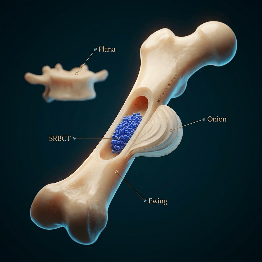
- Small round blue cell tumour (SRBCT) is a HISTOLOGICAL descriptor, not a radiological one — but the radiological pattern it produces is stereotyped: a permeative or moth-eaten lesion with a disproportionately large soft tissue mass and little bone destruction visible on radiograph.
- The core bone differential in a child: Ewing sarcoma, metastatic neuroblastoma, lymphoma of bone, leukaemia/chloroma, rhabdomyosarcoma metastasis, Langerhans cell histiocytosis and osteomyelitis.
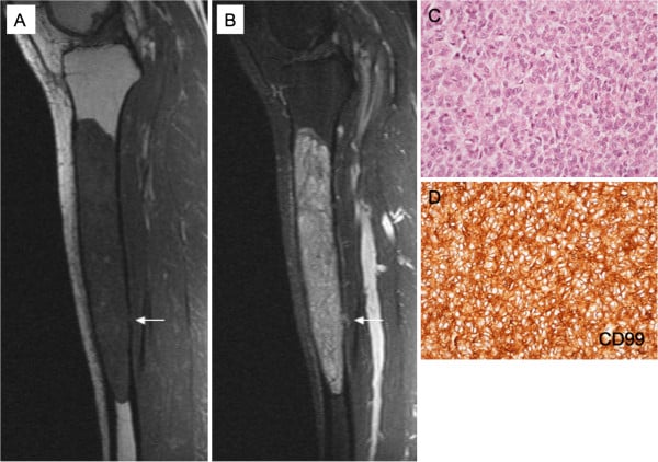
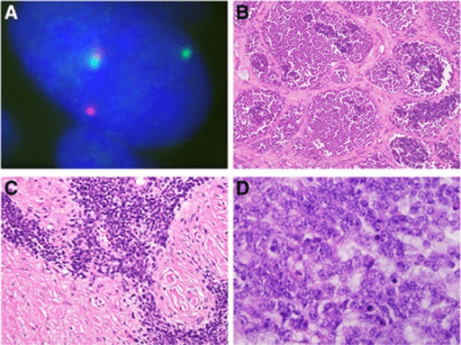
- Ewing sarcoma is defined by EWSR1 rearrangement, most often t(11;22)(q24;q12) EWSR1::FLI1, with CD99 membranous positivity — but CD99 alone is NOT specific (lymphoblastic lymphoma, synovial sarcoma, some rhabdomyosarcomas). Add NKX2.2: alone it is only 84 per cent specific, but CD99 AND NKX2.2 together reach 98 per cent, fooled in Shibuya's 131 tumours by only one mesenchymal chondrosarcoma and one small cell carcinoma.
- 'Ewing-like sarcoma' and 'PNET' are retired terms. WHO now names FOUR categories: Ewing sarcoma, round cell sarcoma with EWSR1-non-ETS fusion (NFATc2, PATZ1), CIC-rearranged sarcoma, and sarcoma with BCOR genetic alteration.
- Those categories are NOT a naming exercise: three-year overall survival in the 148-patient global series was 92.2 per cent for BCOR::CCNB3 and 39.6 per cent for CIC-rearranged (p less than 0.0001). Identical down the microscope, opposite in prognosis — so no family can be counselled before the molecular result returns.
- EWSR1 break-apart FISH alone is not enough: a positive result also occurs with EWSR1-non-ETS fusions such as NFATc2 and PATZ1, which are a different WHO entity. Sequencing identifies the partner.
- Fever, malaise, raised ESR/CRP and leucocytosis occur in BOTH Ewing sarcoma and osteomyelitis. Inflammatory markers never discriminate.
- Age under 5 years with multiple bone lesions and periorbital ecchymosis means neuroblastoma metastases until urinary catecholamines say otherwise.
- A lesion that looks worse on MRI than on radiograph, with soft tissue mass out of proportion to cortical destruction, is lymphoma of bone until proven otherwise in the adult.
- Biopsy must be planned by the treating sarcoma unit, along the future resection axis, with fresh tissue sent for cytogenetics and a separate sample for microbiology.
- “Never say 'Ewing sarcoma' without in the same breath saying 'and I would send tissue for culture because osteomyelitis is the mimic'.
- “Diaphyseal permeative lesion in a child = Ewing, LCH, osteomyelitis, leukaemia. Metaphyseal = osteosarcoma, osteomyelitis.
- “Onion-skin periosteal reaction is not pathognomonic of Ewing — osteomyelitis and LCH both produce it.
- “In an adult over 40 years with a permeative lesion, myeloma and metastasis outrank all round cell tumours.
Subacute osteomyelitis reproduces every feature of Ewing sarcoma: permeative diaphyseal destruction, lamellated periosteal reaction, soft tissue oedema, fever, raised ESR, and even histological small round cells if a reactive lymphoid infiltrate is sampled. Always send a microbiology sample at biopsy. The reverse error — treating Ewing as osteomyelitis and doing an open washout through a non-oncological approach — contaminates compartments and may cost the limb.
Biopsy tract haematoma and post-biopsy oedema make the MRI uninterpretable for skip lesions and true marrow extent. The whole-bone T1 sequence defines resection length. Image first, then biopsy — always through the sarcoma unit that will resect.
CD99 membranous positivity is the classic Ewing marker but is also expressed by lymphoblastic lymphoma, synovial sarcoma and some rhabdomyosarcomas. Diagnosis requires FISH or RT-PCR for EWSR1 rearrangement. Say this out loud — it separates a pass from a distinction.
Ewing sarcoma is rare under 5 years. In a toddler with multiple lytic bone lesions, the answer is metastatic neuroblastoma, leukaemia or LCH. Ask for urinary vanillylmandelic and homovanillic acid, a blood film and an abdominal ultrasound before anything else.
Recognising the Pattern
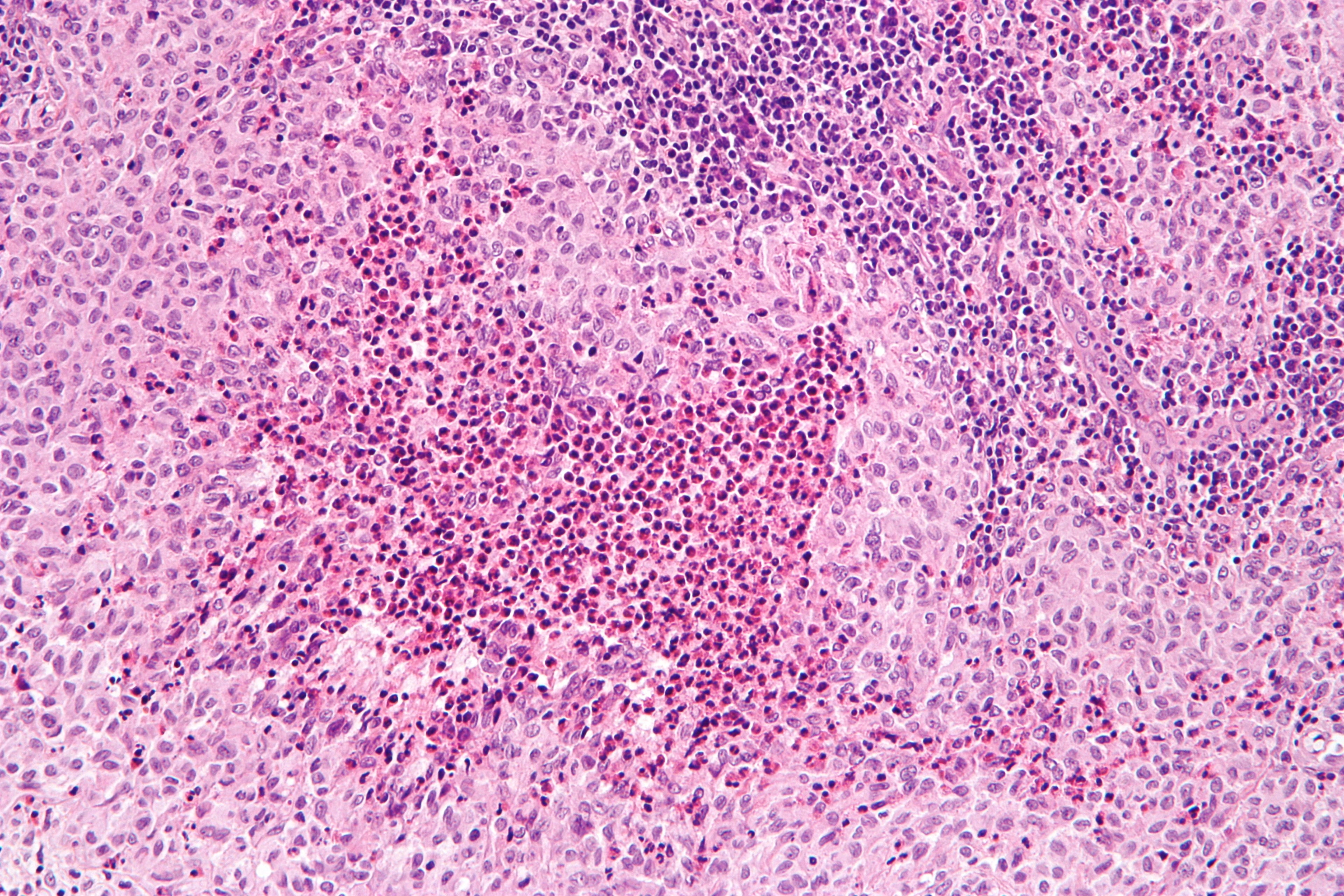
What the pattern actually is. The small round blue cell tumours are a histological family — tumours composed of monotonous cells with scant cytoplasm, round hyperchromatic nuclei and a high nuclear-to-cytoplasmic ratio, staining densely blue on haematoxylin and eosin. Radiologically they converge on a single stereotyped appearance in bone, and that appearance is what you are being shown.
The radiographic signature, in order of importance:
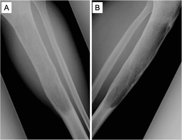
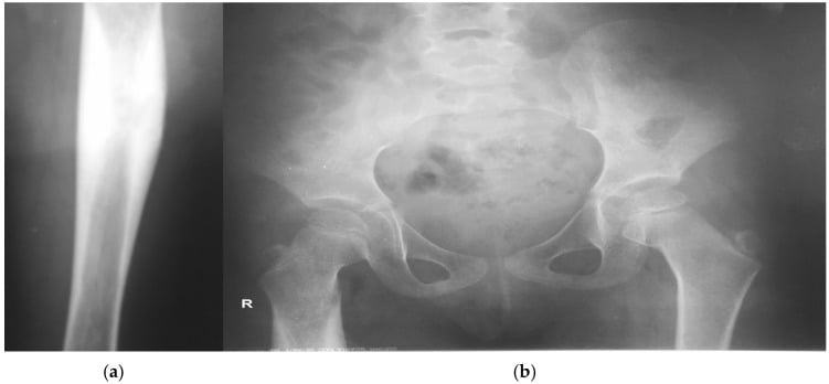
- Permeative or moth-eaten bone destruction — multiple tiny ill-defined lucencies, no discernible margin, no sclerotic rim. This is Lodwick grade III, a wide zone of transition. The lesion cannot be outlined with a pencil.
- Aggressive periosteal reaction — lamellated ("onion-skin"), spiculated ("sunburst"), or a Codman triangle. It signals rapid, interrupted periosteal elevation.
- A soft tissue mass out of proportion to bone destruction — the single most useful discriminator of this family. The tumour permeates Haversian canals and escapes into soft tissue before it destroys enough bone to be radiographically obvious.
- Diaphyseal or metadiaphyseal location in a long bone, or flat bone involvement (ilium, scapula, rib, vertebral body).
- No tumour matrix — no chondroid rings-and-arcs, no osteoid cloud. Any reactive sclerosis is host bone, not tumour bone.
Confirming it is genuinely present. Ask three questions before committing. Is the zone of transition truly wide, or is the radiograph underpenetrated? Is the periosteal reaction interrupted or continuous — continuous solid periosteal reaction suggests a slower process. Is there a soft tissue mass, or only oedema? A mass displaces fat planes; oedema blurs them.
The words to use in the viva. "This is a radiograph of the left femur of a skeletally immature patient. There is an ill-defined permeative lesion centred on the diaphysis with a wide zone of transition, associated lamellated periosteal reaction and an adjacent soft tissue mass. There is no visible tumour matrix. This is an aggressive lesion, Lodwick grade III. My differential in a patient of this age is Ewing sarcoma, osteomyelitis, lymphoma of bone, leukaemic infiltration, Langerhans cell histiocytosis and metastatic neuroblastoma. I would want the full history, examination, inflammatory markers and a whole-bone MRI before biopsy."
Mimics — the false positives:
- Underexposed or rotated film producing apparent trabecular loss. Repeat the view before diagnosing permeation.
- Osteopenia of disuse — regional, symmetric, spares the cortex focally and lacks a soft tissue mass.
- Healing stress fracture with periosteal callus — the periosteum is thick, solid and continuous, and there is a linear sclerotic band.
- Normal physeal or nutrient foramen anatomy, and the physiological cortical tunnelling of rapid infantile bone turnover.
- Post-radiotherapy or post-surgical bone, which can appear permeative for months.
Next Investigation
The commonest technical error in this pathway is a biopsy performed locally before staging MRI, through a transverse incision or across an uninvolved compartment. This can convert a limb-salvageable Ewing sarcoma into an amputation. In the viva, state the referral before you state the biopsy.
L-E-M-O-NLEMON — small round blue cell tumours of bone
The Differential
- Typical age / setting
- 10-20 years; diaphysis of femur/tibia, pelvis, rib, scapula; Caucasian predominance
- Discriminating feature
- Large soft tissue mass with minimal cortical destruction, lamellated periosteum, systemic fever and raised ESR; saucerisation of cortex
- What confirms it
- EWSR1 rearrangement on FISH (t(11;22) EWSR1-FLI1 in about 85 per cent); membranous CD99 with NKX2.2
- Typical age / setting
- Any age; metaphysis in children, spine in adults; sickle cell, immunosuppression, penetrating injury
- Discriminating feature
- Sequestrum, involucrum or a rim-enhancing abscess on MRI; pain often improves on antibiotics; joint effusion sympathetic
- What confirms it
- Culture from bone biopsy or aspirate; penumbra sign on T1 MRI supports Brodie abscess
- Typical age / setting
- Under 5 years; multiple skull, orbit, long bone metaphyses; periorbital ecchymosis, abdominal mass
- Discriminating feature
- Multiplicity plus sutural diastasis and 'hair-on-end' skull; abdominal or posterior mediastinal primary
- What confirms it
- Elevated urinary VMA and HVA, MIBG scan avidity, abdominal imaging showing adrenal primary
- Typical age / setting
- Peak 40-60 years but occurs in adolescence; femur, pelvis, spine
- Discriminating feature
- Patient looks well despite huge lesion; soft tissue mass with preserved cortical outline on MRI ('cortex intact, tumour through it')
- What confirms it
- Core biopsy with CD20 positivity and flow cytometry; staging PET-CT and bone marrow
- Typical age / setting
- 2-10 years; symmetrical, multiple bones, metaphyseal
- Discriminating feature
- Metaphyseal lucent bands, diffuse osteopenia, symmetric bilateral involvement, cytopenias on full blood count
- What confirms it
- Peripheral blood film and bone marrow aspirate with immunophenotyping
- Typical age / setting
- 5-15 years; skull, femur, spine, rib
- Discriminating feature
- Beveled edge in skull, vertebra plana in spine; lesion can look aggressive yet the child is systemically well; may heal spontaneously
- What confirms it
- Biopsy: CD1a and langerin (CD207) positive with Birbeck granules; skeletal survey for further lesions
- Typical age / setting
- 10-25 years; metaphysis around the knee and proximal humerus
- Discriminating feature
- Any recognisable osteoid matrix or cloud-like mineralisation within the soft tissue mass; markedly raised alkaline phosphatase
- What confirms it
- Biopsy showing malignant osteoid produced directly by tumour cells
- Typical age / setting
- 2-15 years; head and neck, genitourinary or extremity primary
- Discriminating feature
- Bone lesions accompany a demonstrable soft tissue primary rather than arising within bone
- What confirms it
- Biopsy with desmin and myogenin/MyoD1 positivity; PAX3/PAX7-FOXO1 fusion in alveolar subtype
- Typical age / setting
- Over 50 years; skull, spine, pelvis, proximal long bones
- Discriminating feature
- Punched-out lucencies without periosteal reaction, and a normal or low alkaline phosphatase despite extensive lysis
- What confirms it
- Serum and urine electrophoresis, serum free light chains, whole-body low-dose CT or MRI, marrow aspirate
- Typical age / setting
- Over 40 years; axial skeleton, proximal femur, humerus
- Discriminating feature
- Multiplicity above the elbow and knee, known primary, cortical 'blow-out' from renal or thyroid primaries
- What confirms it
- CT chest/abdomen/pelvis, bone scan or PET, biopsy of the most accessible lesion
- Typical age / setting
- Adolescent and young adult males; abdominal/pelvic serosal masses with bone involvement secondary
- Discriminating feature
- Bulky peritoneal masses dominate the picture; bone disease is metastatic not primary
- What confirms it
- EWSR1-WT1 fusion; polyphenotypic staining (desmin dot-like, keratin, WT1)
- Typical age / setting
- 20-30 years; jaw, ribs, spine, and extraskeletal sites
- Discriminating feature
- Biphasic pattern — round cell areas plus islands of recognisable hyaline cartilage, giving punctate chondroid calcification
- What confirms it
- HEY1-NCOA2 fusion; biopsy showing the biphasic pattern
- Typical age / setting
- 7-14 years, girls more often; clavicle, metaphyses of tibia/femur, spine
- Discriminating feature
- Multifocal, symmetrical, culture-negative, sterile; medial clavicle involvement is near-characteristic; associated psoriasis or palmoplantar pustulosis
- What confirms it
- Whole-body MRI showing multifocal marrow oedema plus negative cultures on biopsy
- Typical age / setting
- Children and young adults; more often soft tissue than bone
- Discriminating feature
- Morphologically Ewing-like but EWSR1 FISH negative
- What confirms it
- Molecular testing showing CIC-DUX4 or BCOR-CCNB3 fusion
Narrowing It Down
- 1Step 1 — How old is the patient?
Fix the age before proposing anything, and let it set the prior.
Under 5: neuroblastoma metastases, leukaemia, Langerhans cell histiocytosis and osteomyelitis dominate — Ewing is rare. 5 to 25: Ewing sarcoma, osteosarcoma, Langerhans cell histiocytosis, osteomyelitis. Over 40: metastasis, myeloma and lymphoma outrank everything, and a permeative lesion in this group is a carcinoma or plasma cell disorder until proven otherwise. Age alone removes half the list.
- 2Step 2 — Solitary or multiple?
Obtain a skeletal survey in the young child and whole-body imaging in the adult before characterising the index lesion.
Multiple lesions in a toddler point to neuroblastoma, leukaemia or multifocal Langerhans cell histiocytosis. Multiple lesions in an adult point to metastasis or myeloma. A truly solitary aggressive diaphyseal lesion in a teenager keeps Ewing at the top.
- 3Step 3 — Where in the bone?
Name the segment — diaphysis, metaphysis, epiphysis, flat bone, vertebral body — before naming a lesion.
Diaphysis or metadiaphysis of a long bone, or a flat bone (ilium, scapula, rib): Ewing, lymphoma, Langerhans cell histiocytosis, osteomyelitis. Metaphysis: osteosarcoma, osteomyelitis, leukaemia. Epiphysis: chondroblastoma, infection, Langerhans cell histiocytosis. Vertebral body with complete collapse and preserved discs: vertebra plana of Langerhans cell histiocytosis in a child, metastasis or myeloma in an adult.
- 4Step 4 — Is there matrix?
Search the SOFT TISSUE MASS for mineralisation, not just the bone — this is the highest-yield look on the whole study.
Cloud-like or fluffy mineralisation means tumour osteoid — osteosarcoma. Rings, arcs or punctate calcification means chondroid — mesenchymal chondrosarcoma, which is also one of only two tumours that fooled the CD99-plus-NKX2.2 pair. No matrix at all keeps the round cell family and infection in play.
- 5Step 5 — How aggressive, and does MRI match the radiograph?
Compare the size of the soft tissue component against the visible bone destruction, and look specifically for a collection, sequestrum or sinus.
A soft tissue mass far larger than the radiographic destruction, with the cortex still traceable, favours lymphoma or Ewing. A rim-enhancing collection, sequestrum, sinus tract or the penumbra sign favours osteomyelitis. Fluid-fluid levels raise telangiectatic osteosarcoma or aneurysmal bone cyst.
- 6Step 6 — What do the systemic features and simple bloods say?
Send a full blood count and film, ESR, CRP, LDH, alkaline phosphatase, urinary VMA and HVA in the young child, and serum electrophoresis with light chains over 50. Examine the abdomen, testes, nodes and skin.
Blasts and cytopenias mean leukaemia. ESR and CRP are raised in BOTH Ewing and infection and never discriminate. LDH is prognostic in Ewing and raised in lymphoma. Alkaline phosphatase rises in osteosarcoma. Catecholamine metabolites identify neuroblastoma.
- 7Step 7 — Refer BEFORE the needle, stage, then biopsy for molecules
Refer to the sarcoma unit before any biopsy if the mass is painful, progressively enlarging, over 5 cm, deep to deep fascia, or recurrent after an inadvertent excision. Complete whole-bone MRI with contrast, staging CT chest and PET-CT FIRST. Then let the unit that will resect plan the tract along the future resection axis.
Send THREE things from one biopsy: fresh tissue for cytogenetics, FISH and sequencing; formalin tissue for histology and immunohistochemistry; and a dedicated sample for microbiology including mycobacteria. The molecular sample is not optional — three-year survival across the WHO round cell categories ranges from 39.6 to 92.2 per cent, and EWSR1 break-apart FISH alone cannot separate Ewing sarcoma from an EWSR1-non-ETS fusion. Biopsy without the requisite expertise can compromise reconstruction and may make amputation necessary to obtain margins.
MCQ Practice Points
Q: Two round cell sarcomas of bone look identical down the microscope. How much can the fusion alone change the prognosis?
A: From 40 per cent to 92 per cent. In the 148-patient global GRACefUl series, three-year overall survival was 92.2 per cent for BCOR::CCNB3-rearranged sarcoma, 78.7 per cent for unclassified undifferentiated round cell sarcoma, and 39.6 per cent for CIC-rearranged sarcoma (p less than 0.0001). They also differ clinically: BCOR::CCNB3 patients are younger (median 17 versus 32), male-predominant, more often bone primaries, with fewer synchronous metastases. This is why no family can be counselled and no outcome quoted until the molecular result returns — and why fresh tissue must leave theatre for sequencing. Caveat: retrospective over 36 years with non-standardised treatment, so the survival gap is confounded by era and regimen as well as biology.
Q: A biopsy shows small round blue cells with strong membranous CD99. Is this diagnostic of Ewing sarcoma?
A: No. CD99 is sensitive but not specific — lymphoblastic lymphoma, synovial sarcoma and some rhabdomyosarcomas express it, and staining intensity also varies in CIC::DUX4 and BCOR::CCNB3 sarcomas. Add NKX2.2: alone it is 80 per cent sensitive and only 84 per cent specific, with false positives in olfactory neuroblastoma, extraskeletal myxoid chondrosarcoma, mesenchymal chondrosarcoma, small cell carcinoma and Merkel cell carcinoma. CD99 and NKX2.2 together reach 98 per cent specificity, fooled in Shibuya's 131 tumours by only one mesenchymal chondrosarcoma and one small cell carcinoma. Molecular demonstration of the fusion remains the reference standard.
Q: EWSR1 break-apart FISH is positive on a round cell sarcoma of bone. Does that settle the diagnosis?
A: No, and this is the current trap. WHO recognises a separate category — round cell sarcoma with EWSR1-non-ETS fusion, including NFATc2 and PATZ1 partners — in which the break-apart probe is also positive because EWSR1 is genuinely rearranged, just not with an ETS partner. Break-apart FISH detects the split; it does not name the partner. Sequencing does. The four WHO categories are Ewing sarcoma, EWSR1-non-ETS fusion sarcoma, CIC-rearranged sarcoma, and sarcoma with BCOR genetic alteration — and "Ewing-like sarcoma" and "PNET" are retired terms.
Q: The panel returns NKX2.2 negative on a round cell sarcoma. What does that tell you?
A: It argues against Ewing sarcoma. NKX2.2 was diffusely nuclear positive in 93 per cent of Ewing sarcomas but negative in both CIC::DUX4 and BCOR::CCNB3 sarcomas in Çetin's ten-year single-centre reclassification. DUX4 positivity then identifies CIC::DUX4 outright. Morphology helps while sequencing is pending: pleomorphism, prominent nucleoli, wide clear or eosinophilic cytoplasm, myxoid or pink hyaline stroma and spindle cell areas all point away from Ewing.
Q: Is it reasonable to give an Ewing-type regimen to any round cell sarcoma of bone while awaiting molecular results?
A: It is the pragmatic default, but know the signal against it. In Çetin's series, BCOR::CCNB3 sarcomas treated on an Ewing regimen showed a histopathological regression response of 30 per cent, against 70 per cent in those given non-Ewing regimens. Treat that as a reason to obtain the molecular diagnosis urgently rather than as established practice: it rests on eighteen patients split between non-randomised arms, with regression response as a surrogate rather than survival.
Q: Which single finding best distinguishes Ewing sarcoma from osteosarcoma on plain radiograph?
A: Tumour matrix. Osteosarcoma produces cloud-like or fluffy mineralised osteoid within the lesion and the soft tissue mass; Ewing produces no matrix, only reactive host sclerosis. Location helps secondarily (diaphysis for Ewing, metaphysis for osteosarcoma) but periosteal reaction patterns overlap completely — onion-skin is not pathognomonic of anything.
Q: A 4-year-old has multiple lytic skull and long bone lesions. What is the first-line non-invasive test?
A: Urinary vanillylmandelic acid and homovanillic acid, together with a full blood count and film. These identify metastatic neuroblastoma and acute leukaemia respectively, and can spare the child an open bone biopsy. Ewing sarcoma is rare under 5.
Q: Which MRI sign favours a Brodie abscess over a round cell tumour?
A: The penumbra sign — a thin rim of relatively high signal on unenhanced T1-weighted images at the margin of the abscess cavity, representing vascular granulation tissue. Supporting features are a sequestrum, sinus tract, involucrum and rim enhancement around a non-enhancing centre.
Q: Why must every biopsy of a suspected Ewing sarcoma include a microbiology sample?
A: Because subacute osteomyelitis reproduces the entire clinical and radiological picture — fever, raised ESR, permeative diaphyseal destruction, lamellated periosteal reaction — and reactive lymphoid infiltrate can be misread histologically. Culture is the only reliable way to exclude it, and missing infection delays curative antibiotic treatment. Inflammatory markers never discriminate between the two.
Exam Viva Scenarios
Practise clinical reasoning and management decisions out loud
“You are shown this radiograph of the left femur of a 14-year-old boy with six weeks of thigh pain, night pain, intermittent fevers to 38.2 degrees and a 3 kg weight loss. There is a permeative mid-diaphyseal lesion with lamellated periosteal reaction and a palpable tender mass. CRP is 78 and ESR 62.”
“You are shown radiographs of a 3-year-old girl with irritability, refusal to weight bear, bilateral periorbital ecchymosis and proptosis. There are multiple lytic metaphyseal lesions in both femora and lucencies with sutural widening in the skull.”
“You are shown a radiograph of the proximal humerus of a 58-year-old man with two months of dull shoulder ache. There is a permeative lesion with a subtle wide zone of transition; the cortex is thinned but traceable. MRI shows a marrow-replacing mass with a circumferential soft tissue component far larger than the bone destruction suggests. He is systemically well with a normal alkaline phosphatase.”
The pattern in one sentence
- Permeative or moth-eaten bone destruction, wide zone of transition, aggressive (lamellated or spiculated) periosteal reaction, no matrix, and a soft tissue mass out of proportion to the bone destruction.
Differential by age
- Under 5 years: metastatic neuroblastoma, leukaemia, LCH, osteomyelitis
- 5-25 years: Ewing sarcoma, osteosarcoma, LCH, osteomyelitis, lymphoma
- Over 40 years: metastasis, myeloma, primary lymphoma of bone
- Any age: osteomyelitis — always
One-line discriminators
- Ewing — teenager, diaphysis, huge soft tissue mass, EWSR1 rearrangement
- Osteomyelitis — sequestrum, penumbra sign, rim-enhancing abscess, positive culture
- Neuroblastoma metastases — under 5, multiple, raccoon eyes, raised urinary VMA/HVA
- Lymphoma — well patient, cortex traceable, CD20 positive
- Leukaemia — symmetric metaphyseal bands, cytopenias, blasts on film
- LCH — beveled skull edge, vertebra plana, CD1a and langerin positive
- Small cell osteosarcoma — visible osteoid matrix, raised alkaline phosphatase
- Myeloma — over 50, punched-out lucencies, monoclonal band
Investigation order
- Orthogonal radiographs of the whole bone with both joints
- Bloods: FBC and film, ESR, CRP, LDH, ALP, calcium; electrophoresis if over 50; urinary catecholamines if under 5
- Whole-bone MRI with contrast BEFORE biopsy
- Staging CT chest and PET-CT or bone scintigraphy
- Planned core biopsy by the sarcoma unit — histology plus cytogenetics plus microbiology
Traps to avoid
- Local biopsy or washout before referral — contaminates compartments
- Believing raised CRP or ESR excludes tumour
- Accepting CD99 positivity as diagnostic of Ewing
- Diagnosing Ewing in a child under 5 without excluding neuroblastoma
- Sending all tissue in formalin and losing flow cytometry and cytogenetics
- Fixing an impending fracture before a tissue diagnosis is established
Viva opening line
- 'This is an aggressive Lodwick grade III permeative lesion of the diaphysis with lamellated periosteal reaction, no matrix and an associated soft tissue mass. In a patient of this age my differential is Ewing sarcoma and osteomyelitis first, then lymphoma, leukaemia, LCH and small cell osteosarcoma.'
Evidence Base
A Global Collaborative Study of CIC-Rearranged, BCOR::CCNB3-Rearranged and Other Ultra-Rare Unclassified Undifferentiated Small Round Cell Sarcomas (GRACefUl)
- 148 patients with undifferentiated round cell sarcoma treated 1983-2019 at 21 worldwide sarcoma reference centres, classified molecularly
- 88 (60 per cent) CIC-rearranged, 33 (22 per cent) BCOR::CCNB3-rearranged, 27 (18 per cent) unclassified
- THREE-YEAR OVERALL SURVIVAL DIFFERED BY MORE THAN FIFTY PERCENTAGE POINTS: 92.2 per cent for BCOR::CCNB3, 78.7 per cent for unclassified, and 39.6 PER CENT for CIC-rearranged (p less than 0.0001)
- The subtypes differ clinically too: BCOR::CCNB3 patients were younger (median 17 versus 32 years), predominantly male, more often bone primaries, with fewer synchronous metastases
- Nearly a third (47 of 148, 31.8 per cent) had metastases at diagnosis; the authors conclude a full molecular assessment should be undertaken whenever this diagnosis is suspected
The Extensive Case Series of 21 CIC::DUX4, 18 BCOR::CCNB3 and 148 Ewing's Sarcomas From a Single Center
- Ten years of round cell sarcomas from one university department reclassified with an 8-antibody panel (CD99, NKX2.2, ETV4, WT1, DUX4, BCOR, CCNB3, SS18-SSX), FISH and next-generation sequencing
- NKX2.2 was diffusely nuclear positive in 93 per cent of Ewing sarcomas and NEGATIVE in both CIC::DUX4 and BCOR::CCNB3; DUX4 was positive in all CIC::DUX4 cases
- CD99 staining INTENSITY VARIED in the two non-Ewing groups - it does not separate them
- Morphology helps before molecular results return: pleomorphism, prominent nucleoli, wide clear or eosinophilic cytoplasm, myxoid or pink hyaline stroma and spindle cell areas all point AWAY from Ewing sarcoma
- Overall, histopathological regression response to neoadjuvant therapy on the Ewing regimen was SIMILAR across all three groups - the signal below sits inside that null, not against it
- TREATMENT SIGNAL: BCOR::CCNB3 sarcomas treated on an Ewing regimen showed a histopathological regression response of only 30 per cent, against 70 PER CENT for those given non-Ewing regimens. Tumour size 5 cm or above was a significant negative survival factor; age, sex and site were not
The Combination of CD99 and NKX2.2, a Transcriptional Target of EWSR1-FLI1, Is Highly Specific for the Diagnosis of Ewing Sarcoma
- 46 genetically confirmed Ewing sarcomas against 85 non-Ewing small round cell tumours - a proper comparison group, which most marker studies lack
- NKX2.2 alone: sensitivity 80 per cent, specificity 84 PER CENT. It was expressed in 14 of the 85 non-Ewing tumours
- Those false positives are named and are exactly the tumours in this differential: olfactory neuroblastoma, extraskeletal myxoid chondrosarcoma, mesenchymal chondrosarcoma, small cell carcinoma and Merkel cell carcinoma
- CD99 AND NKX2.2 TOGETHER gave a specificity of 98 per cent, with both markers co-expressed in only two non-Ewing tumours (one mesenchymal chondrosarcoma, one small cell carcinoma)
- NR0B1, E2F3 and EZH2 were less useful singly or in any combination
Updates on WHO Classification for Small Round Cell Tumors: Ewing Sarcoma vs. Everything Else
- The WHO Classification of Soft Tissue and Bone Tumours now recognises FOUR separate categories of undifferentiated small round cell sarcoma, not one
- They are: Ewing sarcoma; round cell sarcoma with EWSR1-non-ETS fusions (including NFATc2 and PATZ1); CIC-rearranged sarcoma; and sarcoma with BCOR genetic alterations
- The authors emphasise that these entities overlap substantially in both morphology and immunohistochemistry, so neither alone reliably separates them
- Molecular testing is frequently required to reach a definitive diagnosis - and carries its own pitfalls, which the review discusses explicitly
- Areas of genuine uncertainty and ongoing investigation remain within and between these categories
Biopsy and the Diagnostic Evaluation of Musculoskeletal Tumours: Critical but Often Missed in the 21st Century
- A review of biopsy practice for suspected bone and soft tissue sarcoma from a specialist Australian unit's surgical, radiological and pathological team
- Biopsy performed without the requisite expertise can compromise reconstruction and MAY MAKE AMPUTATION NECESSARY to achieve adequate margins
- Biopsy must always be preceded by imaging - never the reverse
- There must be NO attempt at biopsy or excision before referral if malignancy is suspected at all
- Explicit referral triggers before any biopsy: a mass that is painful, progressively enlarging, greater than 5 cm, deep to deep fascia, or recurrent after inadvertent excision