A small round cell sarcoma that happens to make cartilage — never manage it like a conventional chondrosarcoma, but know that the margin, not the chemotherapy, is what the evidence supports
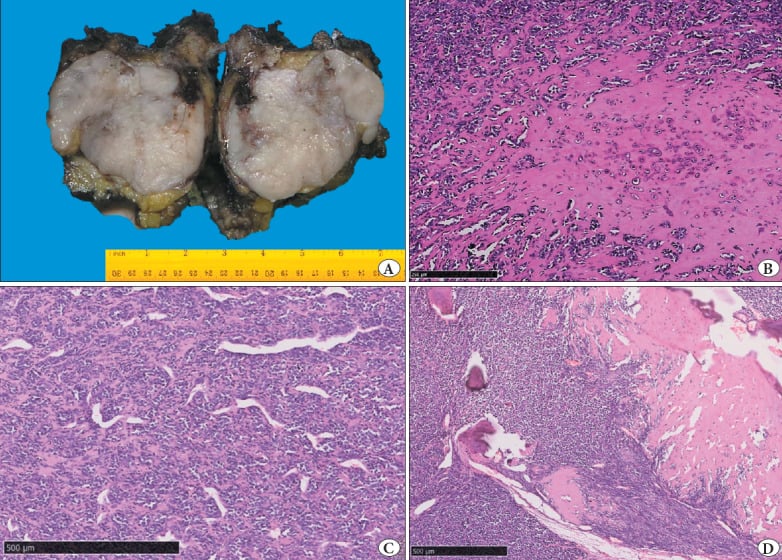
- Biphasic histology: sheets of primitive small round blue cells with a haemangiopericytoma-like branching vascular pattern, abruptly transitioning to islands of well-differentiated hyaline cartilage.
- HEY1-NCOA2 fusion is the diagnostic molecular hallmark, detected by RT-PCR or fusion-directed next-generation sequencing; it is not present in conventional chondrosarcoma or Ewing sarcoma.
- Unlike conventional chondrosarcoma, this tumour is routinely given multi-agent chemotherapy - but say WHY carefully: it is treated that way by extrapolation from its small round cell biology and by convention at sarcoma centres, NOT because a survival benefit has been shown. The largest systematic review (Xu, 107 patients) found anthracycline-based chemotherapy did NOT improve overall survival. Complete excision with negative margins is the intervention with evidence behind it.
- Craniofacial bones, ribs, spine, pelvis and femur are the classic sites; a jaw or rib lesion in a 20-year-old with stippled calcification should raise it.
- A small biopsy sampling only the small cell area is readily misdiagnosed as Ewing sarcoma, synovial sarcoma, small cell osteosarcoma or solitary fibrous tumour.
- Late relapse beyond 10 years is characteristic; surveillance must be lifelong, not five-year.
- “Radiologically it is an aggressive lytic destructive lesion with a soft-tissue mass containing stippled or ring-and-arc chondroid calcification — the mix of aggression and chondroid matrix is the clue.
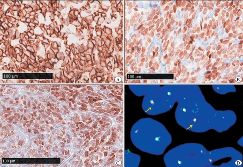
- “Immunohistochemistry: small cells are CD99 positive (a trap), SOX9 positive, and the cartilage islands are S100 positive; NKX2.2 and FLI1 help exclude Ewing sarcoma.
- “Grading is not applied — all mesenchymal chondrosarcoma is treated as high grade regardless of how mature the cartilage looks.
- “Discordance between a benign-looking cartilage island on frozen section and a clinically aggressive lesion should never downgrade the plan.
- “Local recurrence and pulmonary metastasis dominate failure; margin quality remains the strongest surgeon-controlled variable.
Conventional chondrosarcoma is managed by surgery alone. Mesenchymal chondrosarcoma is managed by wide resection plus multi-agent chemotherapy at essentially every sarcoma centre — so reading the pathology report as plain "chondrosarcoma" and discharging the patient without a sarcoma MDT discussion is a serious error. But be precise about what the systemic therapy is for. No study has shown it improves survival: the largest systematic review (Xu, 107 patients) found anthracycline-based chemotherapy did not benefit overall survival, and radiotherapy showed no survival association either. What that review did find is that surgery, and specifically a negative margin, drives local control and event-free survival. The defensible position is that chemotherapy is standard, reasonable and worth giving — by extrapolation from small round cell biology rather than from demonstrated benefit — while the operation is the part you know matters.
The small cell component is frequently CD99 membranous positive. Combined with a small round blue cell morphology on a needle core, this produces a false diagnosis of Ewing sarcoma. Demand molecular testing: HEY1-NCOA2 versus EWSR1 rearrangement.
A core through the cartilage island alone reads as low-grade cartilage tumour; a core through the small cell area alone reads as round cell sarcoma. Plan an image-guided multi-site biopsy through the enhancing soft-tissue mass, tract placed for later excision, at the treating sarcoma unit.
Relapse at 10, 15 and even 20 years is well documented. Discharging at five years is unsafe. Sustained chest and local imaging surveillance, with clear handover to the patient and primary physician, is part of the operation's aftercare.
Definition, Epidemiology and Biology
Mesenchymal chondrosarcoma is a rare high-grade malignant cartilage-forming tumour defined by a biphasic architecture of undifferentiated small round cells and islands of well-formed hyaline cartilage. It is biologically closer to the translocation-associated round cell sarcomas than to the enchondroma–chondrosarcoma continuum, and its clinical behaviour and treatment follow that biology.
Epidemiology
- Accounts for approximately 1 to 2 percent of chondrosarcomas; a genuinely rare tumour with worldwide incidence in the range of a few cases per ten million per year.
- Peak incidence in the second and third decades - approximately 60 per cent of Nakashima's 111 cases - i.e. a decade or two earlier than conventional chondrosarcoma. The full reported range runs from 5 to 74 years.
- Sex distribution is essentially equal. Nakashima found "no significant sex predilection" across 111 cases; a slight female excess is quoted in some smaller series but is not established.
- The skeletal-to-extraskeletal split depends entirely on where the series came from, and the difference is large enough to change what you expect in clinic. The classic bone-tumour archives report roughly two thirds skeletal (Nakashima: 72 of 111 in bone, 38 extraskeletal). Population-based SEER data report the opposite: 123 of 205, or 60 per cent, extraskeletal (Schneiderman 2017), whose authors concluded that extraskeletal tumours "are more common than previously reported". A bone-tumour referral registry is where bone tumours are sent - it undercounts a soft tissue mass that went to a different service. Expect an extraskeletal presentation to be at least as likely as a skeletal one.
- Skeletal sites: craniofacial bones (maxilla, mandible, skull base), ribs, vertebrae, ilium and other pelvic bones, femur and humerus. Craniofacial and axial predilection is a strong distinguishing feature from conventional chondrosarcoma.
- Extraskeletal sites: meninges and brain, orbit, head and neck soft tissue, lower limb musculature, retroperitoneum.
Molecular pathology
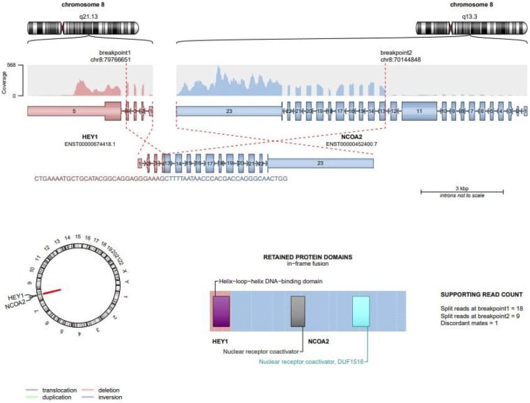
- The recurrent HEY1-NCOA2 fusion (an intrachromosomal deletion on 8q) is present in the great majority of cases and is the diagnostic molecular hallmark.
- A minority harbour IRF2BP2-CDX1.
- Neither fusion is found in conventional or dedifferentiated chondrosarcoma, Ewing sarcoma, or small cell osteosarcoma — hence its discriminating value.
- IDH1/IDH2 mutations, characteristic of conventional central chondrosarcoma, are absent, which is another useful negative discriminator.
Staging and Multidisciplinary Planning
Suspected primary bone sarcoma referred before biopsy to a sarcoma centre. Local radiograph, whole-bone MRI with contrast, CT of the lesion, staging chest CT, bloods including LDH and alkaline phosphatase.
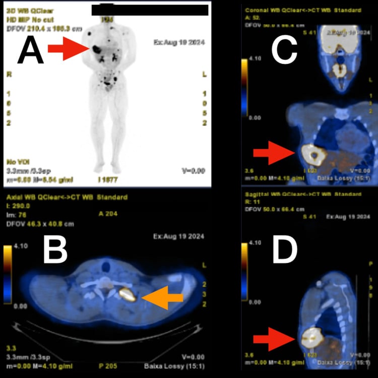
Image-guided core biopsy with tract planning; fresh tissue for RNA-based fusion testing. Whole-body staging (PET-CT or bone scintigraphy).
Radiologist, sarcoma pathologist, medical and radiation oncologist, orthopaedic and, where relevant, maxillofacial, neurosurgical or thoracic surgeon. Decide resectability, reconstruction, and whether radiotherapy is required. Fertility preservation counselling before chemotherapy in this young population.
Multi-agent regimen; baseline echocardiogram and audiometry as dictated by agents used. Restaging MRI and chest CT before surgery.
Definitive local control with reconstruction. Margins assessed by the pathologist on the intact specimen.
Completion of chemotherapy once the wound is sound; radiotherapy for close, marginal or intralesional margins or unresectable disease. Surveillance protocol initiated and explicitly stated to be lifelong.
Staging uses the standard bone sarcoma frameworks — the Enneking (MSTS) system (all mesenchymal chondrosarcoma is high grade, therefore stage IIA if intracompartmental, IIB if extracompartmental, III if metastatic) and the AJCC system. Grading of the cartilage component is not performed and must never be used to argue for de-escalation.
Clinical Presentation and Examination
- Pain and progressive swelling over weeks to months are the dominant symptoms; night pain and rest pain suggest a malignant process.
- Craniofacial disease: facial swelling, loosening teeth, malocclusion, nasal obstruction, epistaxis, proptosis or diplopia (orbital or skull base extension), cranial nerve palsies.
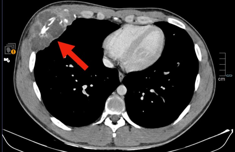
- Rib and chest wall: chest wall mass, pleuritic pain, occasionally an incidental finding on a chest radiograph obtained for another reason.
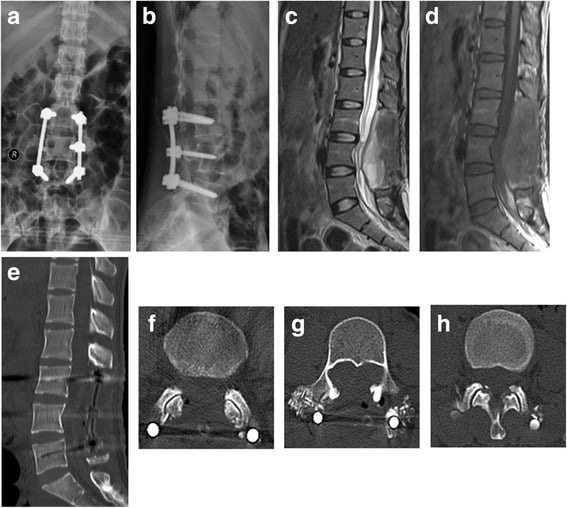
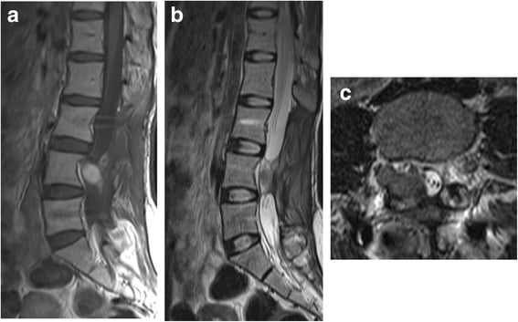
- Spine: axial pain progressing to radicular pain, then myelopathy or cauda equina symptoms — a fast tempo of neurological decline is characteristic of an aggressive extradural mass.
- Pelvis: deep dull pain, referred to the groin or buttock, often mistaken for hip pathology; large tumour volume before diagnosis because the pelvis accommodates growth silently.
- Ask specifically about duration, tempo of change, prior irradiation, constitutional symptoms and family history of syndromic cancer.
Imaging: What to Order and How to Read It
Plain radiograph (two orthogonal views, whole bone).
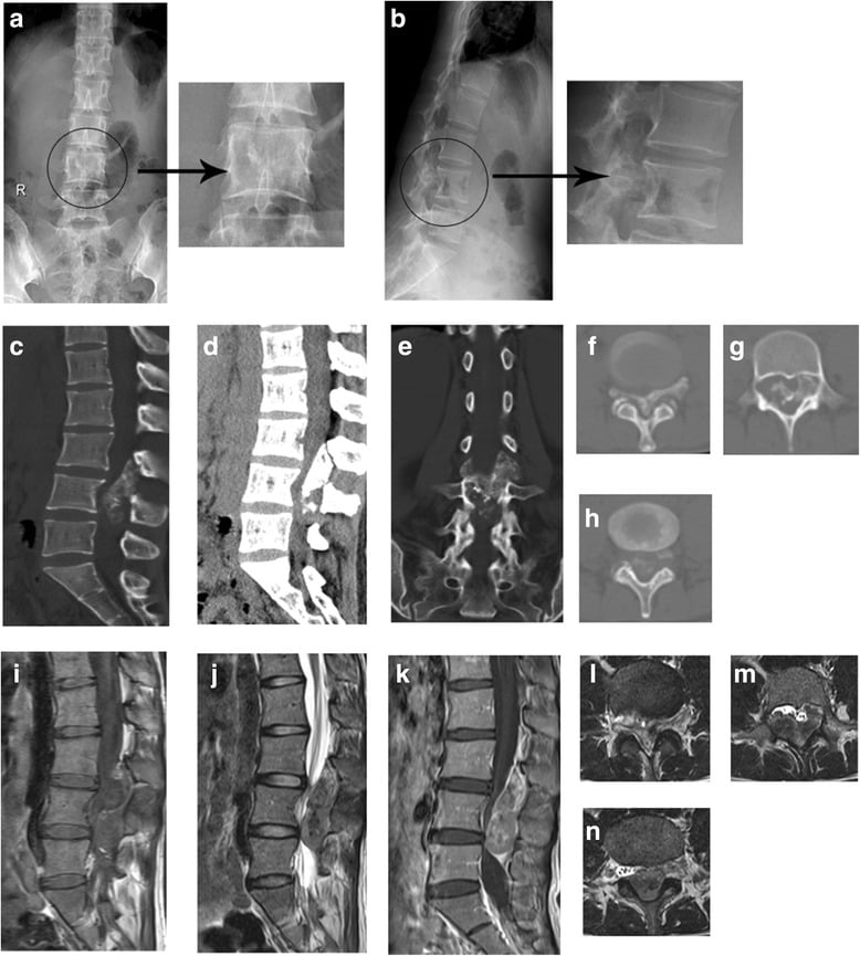
- Aggressive lytic destruction with a wide zone of transition and ill-defined margins (Lodwick grade III behaviour).
- Stippled, flocculent, punctate or ring-and-arc calcification within the lesion or the associated soft-tissue mass — the chondroid signature.
- Cortical destruction and an extraosseous soft-tissue mass; periosteal reaction is variable and may be interrupted.
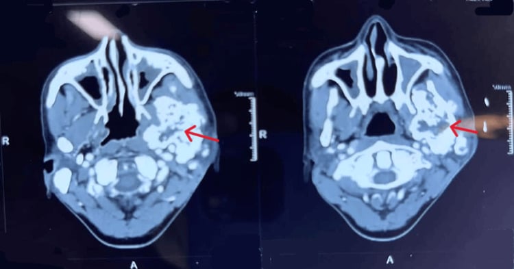
- In the mandible or maxilla, a lytic destructive lesion with root resorption and a soft-tissue component.
CT (with intravenous contrast; the matrix study).
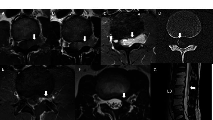
- Best modality for matrix characterisation — the punctate and ring-and-arc calcification that separates a chondroid tumour from Ewing sarcoma or lymphoma.
- Defines cortical breach, endosteal scalloping, pathological fracture risk and, in the pelvis or skull base, the exact osseous corridors for resection.
- Staging chest CT is mandatory at diagnosis: the lung is the dominant metastatic site and small nodules will be missed on radiograph.
- CT angiography where the mass abuts iliac, femoral, subclavian or vertebral vessels, to plan vascular control or reconstruction.
Biopsy: Principles and the Classic Pitfall
- 1Step 1 — Refer before biopsy
Aggressive lytic lesion with chondroid stippling in the second or third decade, often craniofacial, spine, rib, scapula or sternum
Unplanned or badly sited biopsy contaminates compartments, converts limb-sparing resection to amputation and complicates flap reconstruction
- 2Step 2 — Plan the tract
Post-contrast MRI or PET identifies the enhancing solid soft-tissue component versus necrotic centre and mineralised cartilage island
Skin entry marked with indelible ink dot or small tattoo and documented; tract excised en bloc; meticulous haemostasis as a haematoma disseminates tumour along fascial planes
- 3Step 3 — Sample both phases
The tumour is biphasic and a 1 mm core may capture only one component
Both the primitive small cell component and the hyaline cartilage islands available to the pathologist
- 4Branch A — Core samples the small cell component only
Sheets of primitive small round to spindled cells, scant cytoplasm, staghorn (haemangiopericytoma-like) branching vessels, mitoses and necrosis; CD99 membranous positive
Classic misdiagnosis: a 'Ewing sarcoma' of the mandible, rib or skull base in a young adult with chondroid stippling on imaging is mesenchymal chondrosarcoma until proven otherwise
- 5Branch B — Core samples the cartilage island only
Well-differentiated hyaline cartilage, S100 and SOX9 positive, possible endochondral ossification
Misdiagnosis as low-grade conventional chondrosarcoma or enchondroma, with under-treatment
- 6Step 4 — Immunohistochemical triage
Small cells CD99 positive and SOX9 positive, desmin negative; NKX2.2 typically negative (contrast Ewing sarcoma, NKX2.2 positive); STAT6 negative excludes solitary fibrous tumour
Differential narrowed but not settled by immunohistochemistry alone
- 7Step 5 — Molecular confirmation
RT-PCR or targeted RNA sequencing on fresh tissue
Definitive diagnosis of mesenchymal chondrosarcoma
- 8Step 6 — Multimodal planning
Managed as a small round cell sarcoma rather than a chondrosarcoma - but the negative margin is the part with evidence behind it
Lifelong chest surveillance for late pulmonary relapse beyond ten years
An unplanned or badly sited biopsy contaminates compartments, converts a limb-sparing resection into an amputation, and complicates flap reconstruction. Referral before biopsy is the standard of care worldwide for any suspected primary bone sarcoma.
Technique.
- Image-guided core needle biopsy (CT for deep osseous or pelvic lesions, ultrasound for a soft-tissue component) is first line; open incisional biopsy is reserved for non-diagnostic cores.
- Target the enhancing solid soft-tissue component identified on post-contrast MRI or the PET-avid region — avoid necrotic centre and avoid sampling only the mineralised cartilage island.
- Take multiple cores from more than one region of the mass to capture both phases. Send fresh tissue for molecular studies as well as formalin-fixed material.
- Longitudinal tract in line with the planned incision, shortest route through one compartment, no transgression of neurovascular bundles or an uninvolved joint. Mark the skin entry point with an indelible ink dot or a small tattoo and document it — the tract is excised en bloc.
- Achieve meticulous haemostasis; a haematoma disseminates tumour along fascial planes.
Histopathology.
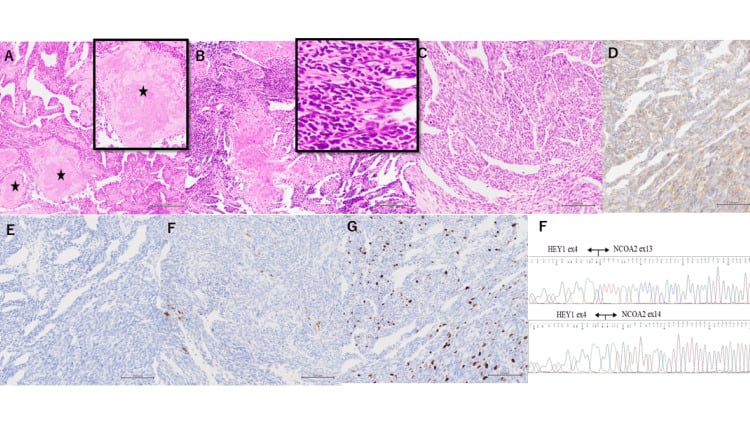
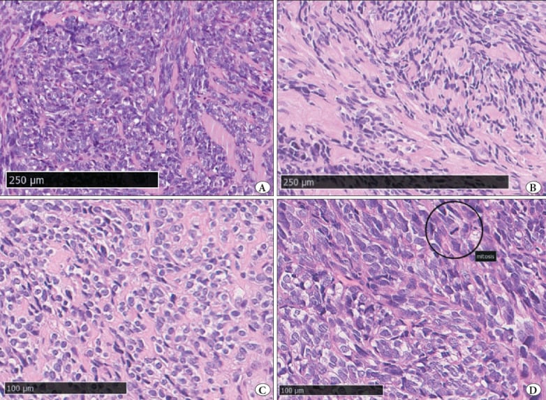
- Biphasic pattern: sheets and nests of primitive small round to spindled cells with scant cytoplasm and a haemangiopericytoma-like (staghorn) branching vascular pattern, abruptly interrupted by islands of well-differentiated hyaline cartilage that may show endochondral ossification.
- Mitoses are present in the small cell component; necrosis is common.
- Immunohistochemistry: small cells CD99 positive (membranous, frequently), SOX9 positive, desmin negative; cartilage islands S100 and SOX9 positive. NKX2.2 is typically negative (contrast Ewing sarcoma, which is NKX2.2 positive). STAT6 is negative (excludes solitary fibrous tumour). TLE1 may be weakly positive and must not be over-read as synovial sarcoma.
- Molecular confirmation: RT-PCR or targeted RNA sequencing for HEY1-NCOA2; EWSR1 and SS18 rearrangements absent.
A core biopsy that samples only the primitive small cell region reads as a small round blue cell tumour, CD99 positive — and is reported as Ewing sarcoma. The cartilage islands may be entirely absent from a 1 mm core. If a "Ewing sarcoma" arises in the mandible, rib or skull base of a young adult and the imaging shows chondroid stippling, insist on molecular testing before committing to a Ewing protocol.
BIPHASICRecognising Mesenchymal Chondrosarcoma
Hook:Two faces, one tumour — and it needs two treatments: the knife and the drip.
Treatment Principles
- Typical site
- Ribs, appendicular bone, favourable pelvis
- Margin feasibility
- Wide en bloc margin achievable
- Local control
- Wide resection is the definitive local step
- Systemic therapy
- Neoadjuvant multi-agent chemotherapy, then resection, then adjuvant chemotherapy
- Typical site
- Craniofacial and skull base, mobile spine, sacrum, deep pelvis
- Margin feasibility
- Margin close or unobtainable; cord and optic apparatus limit dose
- Local control
- Resection where feasible plus adjuvant radiotherapy; proton or carbon ion at skull base and mobile spine; definitive high-dose radiotherapy if unresectable, then reassess resectability after response
- Systemic therapy
- Chemotherapy in all cases, including as cytoreduction before reassessment
- Typical site
- Meninges, orbit, limb and trunk soft tissue; 60 per cent of cases in population data - the commoner presentation, not the exception
- Margin feasibility
- Wide margin usually achievable if compartmental anatomy permits
- Local control
- Wide excision with radiotherapy for close or planned marginal clearance
- Systemic therapy
- Same anthracycline and alkylating agent based multi-agent regimens
- Typical site
- Lung is the dominant site; relapse characteristically very late
- Margin feasibility
- Primary margin planned only after systemic response is demonstrated
- Local control
- In good responders with limited pulmonary disease, metastasectomy plus primary resection offers meaningful long-term survival
- Systemic therapy
- Systemic therapy first, then reassess for complete surgical clearance
- Typical site
- Any involved skeletal site
- Margin feasibility
- Fracture haematoma threatens the planned margin
- Local control
- Definitive resection remains the aim; internal fixation through an unresected sarcoma contaminates the whole bone and is a reconstructive disaster
- Systemic therapy
- Stabilise the biology with chemotherapy first where possible
- Typical site
- Previous operative field
- Margin feasibility
- Re-resection where feasible
- Local control
- Strong adverse prognostic marker; trigger re-staging and multidisciplinary re-planning rather than a reflex further local procedure
- Systemic therapy
- Reconsider systemic regimen at re-planning
Decision thresholds
- Resectable disease, no metastases: neoadjuvant multi-agent chemotherapy, then wide en bloc resection, then adjuvant chemotherapy. This is the standard pathway in contemporary sarcoma practice.
- Margin achievable but predicted close (spine, pelvis, skull base): add adjuvant radiotherapy; consider proton or carbon ion therapy at the skull base and mobile spine where dose to cord and optic apparatus limits photons.
- Unresectable disease: chemotherapy plus definitive high-dose radiotherapy; reassess resectability after response.
- Pathological fracture or impending fracture: stabilise the biology first where possible; definitive resection remains the aim, and internal fixation through an unresected sarcoma contaminates the whole bone and is a reconstructive disaster.
- Metastatic at presentation: systemic therapy first. In good responders with limited pulmonary disease, metastasectomy plus primary resection offers meaningful long-term survival and is worth pursuing.
- Local recurrence: re-resection where feasible; recurrence is a strong adverse prognostic marker and should trigger re-staging and multidisciplinary re-planning rather than a reflex further local procedure.
The primitive small round cell component behaves like other translocation-driven round cell sarcomas and responds to anthracycline and alkylating agent based regimens. Registry and multi-institutional series consistently report better survival in chemotherapy-treated patients, particularly those with localised disease and those undergoing complete resection. The cartilage islands themselves are not chemosensitive — which is why systemic therapy never replaces the operation.
Surgery: Wide En Bloc Resection
Multimodal Management Timeline: Mesenchymal Chondrosarcoma
Indication. Localised or oligometastatic mesenchymal chondrosarcoma where a wide margin is achievable with acceptable function.
Contraindications. Unreconstructable vital structure involvement (e.g. circumferential encasement of brainstem, bilateral carotid, or aorta), widely disseminated disease without response to systemic therapy, or a patient unfit for major resection. Relative: an anatomical site where wide margin would demand unacceptable neurological sacrifice — here plan planned close margin plus radiotherapy.
Why this operation over alternatives. Intralesional curettage, adequate for low-grade central cartilage tumours of the appendicular skeleton, is never acceptable here: the small cell component is high grade and seeds locally. Radiotherapy alone gives inferior local control where resection is feasible.
PIPADRAW sequence (worked for an iliac wing or periacetabular resection, the commonest orthopaedic scenario):
- Position: floppy lateral or supine on a radiolucent table with a bump; prepare the whole hemipelvis, ipsilateral limb free-draped to allow intraoperative manipulation, abdomen prepared to the costal margin. Urinary catheter; consider preoperative ureteric stents for large pelvic masses.
- Imaging and equipment: image intensifier, navigation or patient-specific instrumentation where available for the osteotomy planes, cell salvage not used, vascular clamps and grafts available, oscillating and Gigli saws, high-speed burr, allograft or endoprosthetic implants on the shelf, plastic surgery and vascular surgery on standby.
- Preparation: cross-matched blood, tranexamic acid, antibiotic prophylaxis, normothermia, considered preoperative embolisation of a hypervascular mass, marking of the biopsy tract with an ellipse of skin.
- Approach: ilioinguinal or utilitarian pelvic approach tailored to the tumour; incision incorporates the biopsy tract ellipse.
- Dissection: stay outside the reactive zone at all times — dissect through normal tissue planes, taking a cuff of muscle over the tumour. Ligate and divide internal iliac branches as required. Never enter the pseudocapsule.
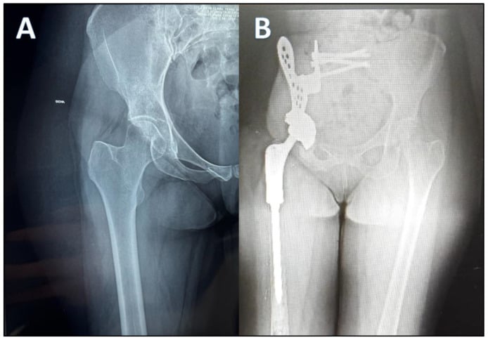
- Reduction/Resection/Reconstruction: perform planned osteotomies with a margin measured from preoperative MRI (aim for at least 1 to 2 cm of bone beyond marrow signal abnormality, adjusted for anatomical constraint). Deliver the specimen intact and orient it for the pathologist with sutures and a diagram. Reconstruct according to zone — Enneking zone I resections often need no reconstruction; periacetabular (zone II) resections require pelvic endoprosthesis, allograft-prosthetic composite, hip transposition or ischiofemoral arthrodesis depending on resource and surgeon preference.
- At-risk structures: external and internal iliac vessels, femoral and obturator nerves, lumbosacral trunk and sciatic nerve, ureter, bladder, rectum and superior gluteal vessels (their sacrifice compromises flap viability).
- Fixation: implants placed only after the tumour is out and instruments changed; separate clean set.
- Closure: meticulous dead space obliteration with muscle flaps, drains brought out in line with the incision so the tract can be included in any future re-resection, layered tension-free closure.
- Aftercare: thromboprophylaxis, protected weight bearing dictated by reconstruction, early physiotherapy, chemotherapy resumed once the wound is healed (commonly at 2 to 3 weeks).
- Pitfalls: tumour spillage, unrecognised skip lesion, biopsy tract not excised, drain sited remotely, resuming chemotherapy over a compromised wound.
- Salvage of failure: re-resection for positive margin where anatomically possible; otherwise adjuvant radiotherapy. Deep infection of a megaprosthesis is managed by debridement with component retention early, or two-stage revision or amputation late.
Prognosis and Surveillance
- Overall survival is 51 per cent at 5 years and 43 per cent at 10 years in the 205-patient SEER series (Schneiderman 2017). The older Mayo figures - 54.6 per cent at 5 years falling to 27.3 per cent at 10 - come from only 23 patients treated before modern chemotherapy, so treat the 1986 ten-year figure as the pessimistic bound rather than the expected one. Both agree on the point that matters: a substantial number of deaths occur after the fifth year.
- What independently predicts death, on multivariable analysis: metastasis at presentation (HR 12.38) and tumour size (HR 1.16 for every additional centimetre). Nothing else in the SEER model came close.
- Extraskeletal origin is NOT an independent prognostic factor. SEER found no survival difference between extraskeletal and skeletal disease. The impression that soft tissue cases do better is confounded by resectability and size, not by the tissue of origin.
- Location changes its prognostic meaning across the age range - one of the few genuinely counterintuitive findings in this tumour. Taking cranial site as the reference: at age 20, axial (HR 5.56) and appendicular (HR 6.26) disease carry a markedly worse outlook, so the young patient with a skull base lesion is comparatively fortunate. At age 60 the same hazard ratios are 0.10 and 0.14 - the relationship has inverted, and the cranial site is now much the worst place to have this tumour. Do not quote "cranial tumours do better" without saying in whom.
- Other factors reported as favourable, but not independently validated: complete resection with negative margins, receipt of chemotherapy, good histological response to neoadjuvant therapy. SEER records none of these, so they rest on retrospective institutional series that cannot separate them from selection.
- Failure pattern: local recurrence and pulmonary metastasis, with bone and lymph node metastasis less common. Relapse at 10 to 20 years is well documented and is the single most distinctive prognostic feature of this tumour.
Surveillance schedule (pragmatic, adapted to resources):
- Clinical
- Examination of site and regional nodes
- Local imaging
- MRI or CT of operative site
- Chest imaging
- CT chest
- Clinical
- Examination plus function assessment
- Local imaging
- MRI or CT of site
- Chest imaging
- CT chest, radiograph alternating where CT access limited
- Clinical
- Examination, implant review
- Local imaging
- Site imaging annually
- Chest imaging
- Chest imaging annually
- Clinical
- Annual review; survivorship, cardiac and endocrine late effects
- Local imaging
- Site imaging for new symptoms plus periodic
- Chest imaging
- Annual chest imaging
Counsel the patient explicitly that this tumour can return more than a decade later, and that new chest symptoms, local swelling or pain must trigger immediate review. Hand over a written surveillance plan to the patient and their primary care physician — essential where patients relocate across borders or health systems.
Guidelines, Registries & Global Practice
Global epidemiology and access. Mesenchymal chondrosarcoma is uniformly rare across all populations with no established geographic clustering. The dominant global variable is access to a sarcoma centre, molecular diagnostics and multi-agent chemotherapy. In many low- and middle-income settings, fusion testing is unavailable and diagnosis rests on morphology and immunohistochemistry alone — an acceptable pragmatic pathway provided the biphasic pattern is recognised and CD99 positivity is not over-interpreted. Telepathology and regional reference laboratories are increasingly used to bridge this gap.
Society guidance that genuinely addresses this tumour:
- Relevant guidance
- Bone sarcomas should be diagnosed and treated in reference centres or networks; mesenchymal chondrosarcoma is treated with chemotherapy in addition to surgery, unlike conventional chondrosarcoma
- Practical consequence
- Refer before biopsy; do not extrapolate conventional chondrosarcoma management
- Relevant guidance
- Recognises mesenchymal chondrosarcoma as a distinct chemosensitive subtype; recommends wide excision plus consideration of chemotherapy and radiotherapy
- Practical consequence
- Supports neoadjuvant and adjuvant systemic therapy in localised disease
- Relevant guidance
- Centralised sarcoma diagnostic and treatment services with mandatory multidisciplinary team review and defined referral pathways
- Practical consequence
- Structural argument for centralisation applicable to any health system
- Relevant guidance
- Any suspected primary bone tumour should be referred to a specialist centre before biopsy
- Practical consequence
- Prevents the unplanned-biopsy disaster that most commonly harms these patients
- Relevant guidance
- Defines mesenchymal chondrosarcoma as a distinct entity with HEY1-NCOA2 fusion; no grading applied
- Practical consequence
- All cases are high grade; grading language should not appear in the report
Registry evidence. There is no implant registry role here, but national cancer registries and sarcoma-specific databases (national cancer registries in Europe and North America, the Scandinavian Sarcoma Group registry, and cooperative group databases) supply nearly all outcome data for this tumour. Their consistent messages: rarity mandates pooled data, chemotherapy is associated with better survival, and late relapse is real. Where megaprostheses are used for reconstruction, national bone tumour endoprosthesis registries inform expected implant survivorship and infection rates.
Practice variation by resource setting.
- High-resource: neoadjuvant chemotherapy, navigation-assisted or patient-specific-instrument resection, particle therapy for skull base disease, custom endoprosthetic reconstruction.
- Intermediate-resource: chemotherapy with standard agents, conventional wide resection with allograft or standard modular endoprosthesis, photon radiotherapy.
- Low-resource: priority is accurate diagnosis and achieving a wide margin, accepting more ablative surgery (e.g. amputation or hindquarter resection) where reconstruction is unavailable; chemotherapy where deliverable safely. An oncologically sound amputation is superior to a marginal limb-sparing resection with no adjuvant therapy.
Controversies & Areas of Uncertainty
- Mechanism
- The cartilage islands dominate the sampled field, so a biphasic tumour is signed out as a low-grade cartilage lesion and treated by surgery alone
- Prevention
- Insist on the biphasic pattern being actively excluded; confirm HEY1-NCOA2 fusion by FISH or RNA sequencing in any young patient with a cartilage tumour
- Salvage
- Re-review the archival blocks at a sarcoma reference centre and restage; offer systemic therapy even if resection has already occurred
- Mechanism
- The primitive round cell component is CD99 positive, prompting a diagnosis of Ewing sarcoma and loss of the cartilage-based diagnosis
- Prevention
- Correlate with the cartilage islands and SOX9 positivity; test for HEY1-NCOA2 rather than accepting a negative EWSR1 result as indeterminate
- Salvage
- Molecular re-testing; note that a Ewing-type regimen overlaps therapeutically, so the systemic error is smaller than the surgical one
- Mechanism
- The tumour is heterogeneous; a needle passing only through the chondroid zone misses the round cell component entirely
- Prevention
- Multiple cores across radiologically distinct zones, guided by the most cellular and least mineralised area on MRI
- Salvage
- Repeat targeted biopsy of the non-mineralised component before committing to any definitive resection
- Mechanism
- A tract sited outside the planned resection field or crossing compartments forces a wider or amputative resection later
- Prevention
- Biopsy performed or directed by the operating sarcoma surgeon, in line with the planned incision and excisable en bloc
- Salvage
- Excise the tract with the specimen; accept a larger soft tissue reconstruction or consider adjuvant radiotherapy to the contaminated field
- Mechanism
- Frozen artefact obscures the round cell population and margins are called clear on inadequate sampling
- Prevention
- Do not use frozen section to grade this tumour; reserve it for margin orientation and rely on permanent sections and molecular confirmation
- Salvage
- Formal permanent section review of all margins; re-resect a close or positive margin, or consider radiotherapy where re-resection is not feasible
- Mechanism
- Late recurrence and late metastasis are characteristic; the survival curve does not plateau at five years
- Prevention
- Counsel and schedule surveillance well beyond five years, including chest imaging and imaging of the primary site
- Salvage
- Restage fully at relapse; isolated resectable pulmonary or local recurrence still merits aggressive metastasectomy or re-resection
- Optimal chemotherapy regimen. No randomised evidence exists. Practice ranges from Ewing-type regimens (vincristine, doxorubicin, cyclophosphamide alternating with ifosfamide and etoposide) to osteosarcoma-style or soft tissue sarcoma-style doxorubicin and ifosfamide combinations. Choice is centre-dependent; the argument for a Ewing-type regimen rests on the shared primitive round cell biology.
- Magnitude of chemotherapy benefit. Retrospective series are subject to selection bias — fitter, younger, localised patients receive chemotherapy. Some analyses show no significant survival gain. Most centres nonetheless offer it given the alternative is surgery alone for a high-grade sarcoma in a young patient.
- Role and timing of radiotherapy. Clearly indicated for unresectable and close-margin disease, but whether routine adjuvant radiotherapy after a genuinely wide resection reduces recurrence is unproven, and the long-term costs (growth disturbance, secondary malignancy) are substantial in adolescents.
- Particle versus photon therapy at the skull base and spine: physically attractive dose distributions, but access is limited to a small number of centres worldwide and comparative outcome data are scarce.
- Response assessment. Histological necrosis thresholds validated for osteosarcoma are not validated here; the cartilage islands do not necrose and can distort percentage estimates.
- Extraskeletal versus skeletal prognosis - largely settled, in the negative. The largest population series (205 patients, SEER) found no survival difference between the two, and identified metastasis and size as the independent predictors instead. Institutional series reporting better outcomes for extraskeletal disease are describing resectability, not biology. What is not settled is the reversal of the location effect with age, which rests on a single Cox model and needs replication.
- Targeted therapy. The HEY1-NCOA2 fusion drives Notch pathway and PTHrP-related signalling; targeting has not yet translated into clinical benefit. Enrolment in sarcoma trials and tissue banking should be offered to every patient.
CHOP UPManaging the Confirmed Case
Hook:Cut wide, drip hard, watch forever.
MCQ Practice Points
Q: Which molecular alteration defines mesenchymal chondrosarcoma, and how confidently can a positive result be used?
A: The HEY1-NCOA2 fusion — an in-frame join of HEY1 exon 4 to NCOA2 exon 13, arising from an intrachromosomal deletion on 8q. In the discovery study it was present in 9 of 9 further mesenchymal chondrosarcomas and 0 of 15 other chondrosarcoma subtypes, which is why the authors called it "the defining and diagnostic gene fusion". A minority carry IRF2BP2-CDX1 instead. EWSR1-FLI1 defines Ewing sarcoma, SS18-SSX synovial sarcoma, and IDH1/IDH2 mutation conventional central chondrosarcoma — all absent here. Note the asymmetry: those denominators make a positive result close to decisive, but nine and fifteen cases say little about sensitivity, so a negative result on a crushed core does not exclude the diagnosis.
Q: How does treatment differ from conventional chondrosarcoma, and how good is the evidence?
A: Multi-agent chemotherapy is added to wide resection. Conventional chondrosarcoma is chemoresistant and treated surgically; mesenchymal chondrosarcoma carries a primitive round cell component, and every sarcoma centre treats it systemically. Then say the harder half, because this is where candidates overreach: no study has shown a survival benefit. Nakashima, reviewing 111 cases, would only write that "the value of irradiation or chemotherapy was difficult to assess". SEER, the largest series, does not record chemotherapy at all. And Xu's systematic review of 107 patients — the only synthesis that actually tested it — found anthracycline-based chemotherapy did NOT improve overall survival, with radiotherapy showing no survival association either. What that review did find was that surgery, and a negative margin specifically, improved local control and event-free survival. So the defensible answer is: chemotherapy is standard of care on biological reasoning and convention, there is no randomised trial and at this incidence there never will be, and the margin is the part of the plan supported by evidence. Do not claim a consistent retrospective survival signal — the pooled data do not show one.
Q: Which anatomical sites are most characteristic, and what proportion is extraskeletal?
A: Craniofacial bones (maxilla, mandible, skull base), ribs, spine and pelvis — an axial and craniofacial predilection quite unlike conventional chondrosarcoma. But do not answer "one third extraskeletal". That figure comes from bone-tumour archives, which are where bone tumours get sent. In population-based SEER data 123 of 205 cases (60 per cent) were extraskeletal — meninges, orbit, head and neck, limb and trunk soft tissue — and the authors state plainly that extraskeletal disease is "more common than previously reported". It also carries no independent survival penalty.
Q: What is the classic biopsy pitfall?
A: Sampling only the small round cell component, finding it CD99 positive, and reporting Ewing sarcoma. The cartilage islands are patchy, so a single core through the cellular zone shows a CD99-positive small blue round cell tumour and nothing else. Prevention is multi-site targeted core biopsy of the enhancing soft-tissue mass, deeper levels, NKX2.2 and S100 immunostaining, and molecular fusion testing. This is the one situation where the fusion assay earns its cost outright.
Q: What is the typical survival and the dominant relapse pattern?
A: About 51 per cent at five years and 43 per cent at ten in 205 SEER patients; the older 23-patient Mayo series gives 54.6 and 27.3 per cent. The examiner's point is the gap between the two timepoints, not either figure. Failure is by local recurrence and pulmonary metastasis, characteristically late, with relapse documented at 10 to 20 years. On multivariable analysis only metastasis at presentation (HR 12.38) and tumour size (HR 1.16 per additional centimetre) independently predict death. Surveillance is therefore indefinite, and a five-year discharge is a real error rather than a theoretical one.
Q: What histological features confirm the diagnosis, and is it graded?
A: A bimorphic pattern — sheets of primitive small round to spindled cells with haemangiopericytoma-like staghorn branching vasculature, interrupted abruptly by islands of well-differentiated hyaline cartilage that may undergo endochondral ossification. Nakashima described this combination as "virtually pathognomonic in most cases". The abruptness is the diagnostic feature: conventional chondrosarcoma does not make that transition. Grading is not applied — every case is treated as high grade, which is why the tumour is stage IIA or IIB in Enneking terms unless metastatic.
Exam Viva Scenarios
Practise clinical reasoning and management decisions out loud
“A 19-year-old presents with three months of right-sided chest wall pain and a firm fixed mass over the sixth rib. Radiograph shows an expansile destructive rib lesion with a soft-tissue component containing stippled calcification. How do you proceed?”
“A 22-year-old has a destructive lesion of the mandible. Core biopsy is reported as a small round blue cell tumour, CD99 positive, and the referring team has started a Ewing sarcoma protocol. The CT shows fine stippled calcification within the associated soft-tissue mass. What is your concern?”
“A 34-year-old woman treated twelve years ago for mesenchymal chondrosarcoma of the ilium, with wide resection and chemotherapy, presents with a solitary 1.5 cm peripheral right lower lobe nodule found on a chest CT arranged for a cough. How do you manage this?”
“A 25-year-old man has two months of thoracic back pain and one week of progressive lower limb weakness. MRI shows a T7 vertebral body and posterior element lesion with a circumferential epidural mass compressing the cord. CT shows lysis with punctate calcification. What is your immediate and definitive management?”
Identity
- Rare high-grade biphasic cartilage-forming sarcoma; 1 to 2 percent of chondrosarcomas
- Second and third decades (about 60 per cent); no significant sex predilection
- Craniofacial, ribs, spine, pelvis - but 60 per cent are extraskeletal in SEER (meninges, orbit, soft tissue)
- HEY1-NCOA2 fusion defining; IRF2BP2-CDX1 in a minority; IDH wild type
Imaging
- Radiograph and CT: aggressive lysis, wide zone of transition, stippled or ring-and-arc chondroid matrix, soft-tissue mass
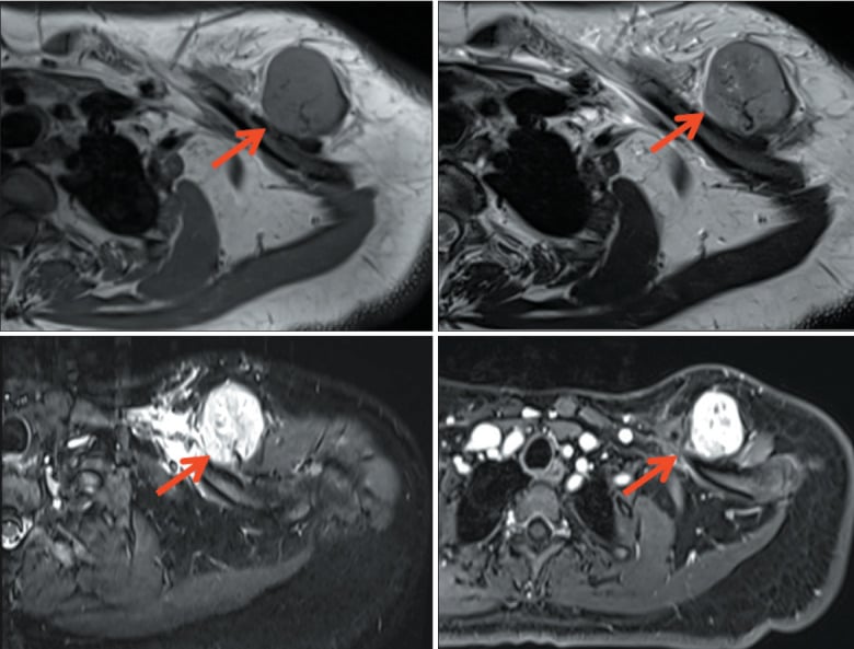
- MRI: heterogeneous — bright lobulated cartilage islands plus intermediate signal cellular areas, avid enhancement, flow voids
- Whole-bone T1 for intramedullary extent and skip lesions; measure from a fixed bony landmark
- Staging chest CT mandatory; PET-CT to direct biopsy and assess response
Biopsy
- Refer before biopsy; performed at the resecting unit
- Image-guided cores from multiple regions of the enhancing solid component; fresh tissue for RNA testing
- Longitudinal tract in line with planned incision, one compartment, marked and documented
- Pitfall: small cell only sample, CD99 positive, misread as Ewing sarcoma
Histology and immunohistochemistry
- Primitive small round cells plus staghorn vessels plus abrupt hyaline cartilage islands
- Small cells CD99 and SOX9 positive; cartilage S100 positive
- NKX2.2 negative (excludes Ewing sarcoma); STAT6 negative (excludes solitary fibrous tumour)
- No grading applied — always high grade
Treatment
- Neoadjuvant multi-agent chemotherapy, wide en bloc resection, adjuvant chemotherapy
- Radiotherapy for close or intralesional margins and unresectable axial or skull base disease; consider particle therapy
- Excise the biopsy tract en bloc; deliver the specimen intact and orient it for pathology
- Metastatic disease: systemic therapy first, then consider metastasectomy plus primary resection in responders
Complications
- Positive margin and local recurrence — the strongest surgeon-controlled variables
- Deep infection and wound breakdown, amplified by chemotherapy; delay resumption until wound sound
- Nerve injury, haemorrhage; consider preoperative embolisation for hypervascular masses
- Late effects: cardiotoxicity, infertility, growth arrest, secondary malignancy
Prognosis and follow-up
- Overall survival 51 per cent at 5 years, 43 per cent at 10 (205 SEER patients) - the curve keeps falling
- Independent predictors of death: metastasis (HR 12.38) and size (HR 1.16 per cm). Extraskeletal origin is NOT one
- Failure by local recurrence and lung metastasis, characteristically late
- Surveillance indefinite — chest and local imaging beyond ten years, with a written plan handed to patient and primary physician
Evidence Base
Identification of a Novel, Recurrent HEY1-NCOA2 Fusion in Mesenchymal Chondrosarcoma Based on a Genome-Wide Screen of Exon-Level Expression Data
- An in-frame fusion of HEY1 exon 4 to NCOA2 exon 13, found by an unbiased genome-wide screen for the intragenic expression discontinuity a fusion creates, then confirmed by 5' RACE
- Present in ALL nine further mesenchymal chondrosarcomas with a definitive histological diagnosis and adequate material - 9 of 9
- Absent in all 15 samples of other chondrosarcoma subtypes
- The authors' own conclusion: HEY1-NCOA2 is 'the defining and diagnostic gene fusion in mesenchymal chondrosarcomas'