Living autograft for segmental defects greater than 6 cm, hostile beds and biological intercalary reconstruction
- The fibula is a living bone flap: it unites like a fracture (6-12 weeks) rather than by creeping substitution, tolerates infected and irradiated beds, and hypertrophies under load.
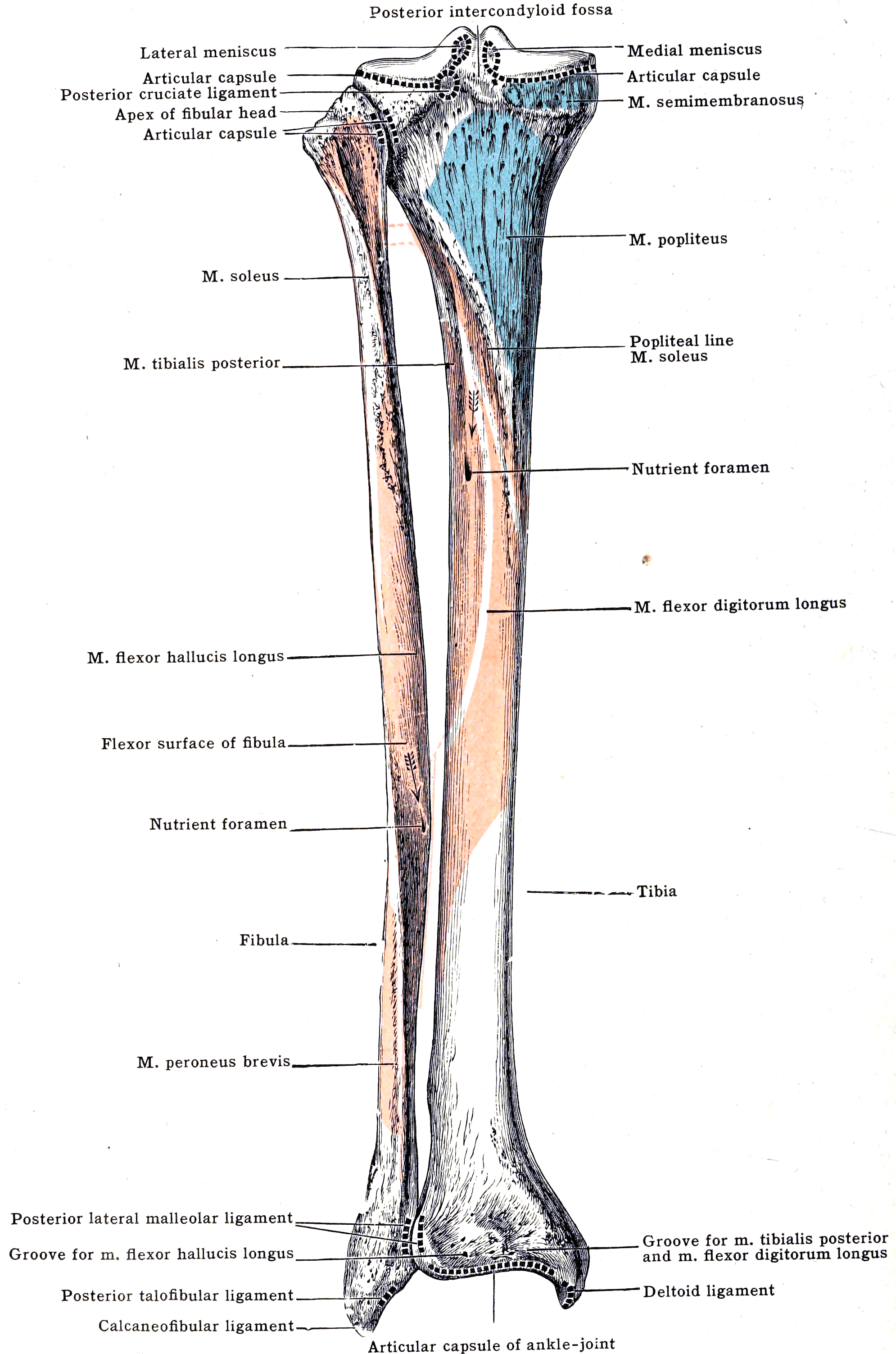
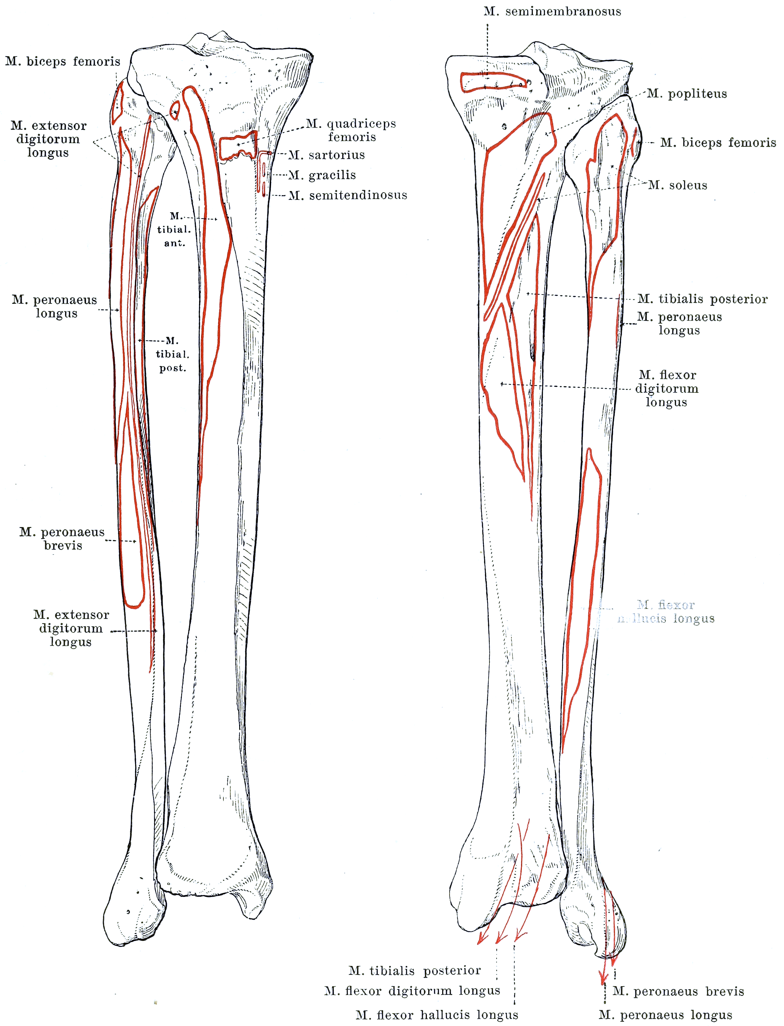
- Nutrient artery enters the middle third of the fibula posterior to the interosseous membrane — the middle third MUST be included in the harvested segment.
- Periosteal supply from the peroneal artery is segmental — closing-wedge osteotomies (double-barrel, contouring) are safe if periosteum and pedicle are preserved.
- Preserve the distal 6 cm of fibula for ankle mortise stability; in skeletally immature patients add a distal tibiofibular synostosis (Langenskiöld) to prevent progressive ankle valgus.
- Rule out peronea arteria magna (dominant peroneal artery with hypoplastic tibial vessels, roughly 0.2-5 percent) — harvesting the pedicle here devascularises the foot.
- Capanna technique: fibula telescoped inside a massive allograft combines immediate mechanical strength with biological healing and revascularisation potential.
- “Skin paddle survives on septocutaneous perforators in the posterior crural septum — it is the monitoring window for the buried bone flap.
- “Fix the fibula so the pedicle lies free of plate, bone and tourniquet of soft tissue — pedicle compression is the leading technical cause of flap loss.
- “Claw hallux from flexor hallucis longus scarring/contracture is the classic donor-site complication asked in exams.
A dominant peroneal artery supplying the foot with absent/hypoplastic anterior and posterior tibial arteries. Harvest here causes foot ischaemia. Screen with pulses and Doppler; image (CT/MR angiography or catheter angiography) if pulses are abnormal, previous trauma or vascular disease.
Winds around the fibular neck. Proximal harvest and pedicle exposure endanger it — identify and protect early. Leave the proximal fibula (head and neck) unless the physis/epiphysis is deliberately transferred on the anterior tibial vessels for growing reconstructions.
Enters posterior to the interosseous membrane in the middle third. Segments excluding the middle third rely on periosteal supply alone — plan the osteotomy levels so the nutrient foramen sits within the graft.
Retain at least 6 cm of distal fibula above the lateral malleolus. In children, even with 6 cm retained, proximal migration of the remnant causes valgus — perform a distal tibiofibular synostosis or screw fixation at harvest.
Rationale: Why a Vascularised Graft
- Best for
- Defects less than 5 to 6 cm in a healthy, well-vascularised bed
- Healing biology
- Dead scaffold incorporating by creeping substitution; transient weakening at 6 to 18 months
- Strengths
- Simple, no microsurgery, no donor vessel dissection
- Weaknesses
- Unpredictable failure in defects greater than 6 cm, infected beds and irradiated tissue; late fracture and non-union
- Best for
- Defects of 5 to 10 cm, especially trauma or infection after staged debridement
- Healing biology
- Cement spacer induces a vascular, osteoinductive membrane; stage two graft consolidates within it
- Strengths
- No microsurgery, familiar technique, tolerates staged debridement
- Weaknesses
- Two stages, large graft volume demand, less reliable beyond about 10 cm and in irradiated beds
- Best for
- Tibial defects with an intact soft tissue envelope
- Healing biology
- Regenerate forms in the distraction gap; native bone, native diameter
- Strengths
- Regenerates true host bone and corrects concurrent deformity
- Weaknesses
- Frame time roughly 1 month per cm, docking site problems, pin site infection, patient burden
- Best for
- Defects greater than 6 cm, infected or irradiated beds, congenital pseudarthrosis, oncologic intercalary defects
- Healing biology
- Living bone: osteocytes survive on the peroneal pedicle, heals as a two-level fracture with no creeping substitution
- Strengths
- Works in hostile beds, delivers antibiotics and immune cells, hypertrophies under load over 1 to 2 years, straight tubular cortical bone up to about 26 cm, double-barrelled for tibia and femur, epiphyseal transfer on the anterior tibial artery can reconstruct a growing physis
- Weaknesses
- Microsurgical expertise and monitoring, donor site morbidity, initial mechanical weakness with stress fracture before hypertrophy
- Best for
- Large-diameter intercalary defects of femur or tibia after tumour resection
- Healing biology
- Allograft gives immediate structure while the intramedullary fibula supplies living biology
- Strengths
- Immediate strength plus biology; allograft shields the fibula during the hypertrophy phase
- Weaknesses
- Complex and long operation; allograft non-union, fracture and infection still possible
- Best for
- Elderly patients, poor oncological prognosis, need for rapid weight-bearing
- Healing biology
- No biological healing; fixation is mechanical only
- Strengths
- Immediate function and early full weight-bearing
- Weaknesses
- Aseptic loosening, periprosthetic infection, not durable in the young or active
A non-vascularised cortical graft is dead scaffold: it incorporates by creeping substitution, transiently weakens at 6 to 18 months, and fails unpredictably in defects greater than 6 cm, infected beds and irradiated tissue. The vascularised fibula is living bone.
- Union biology — heals to host bone like a two-level fracture; osteocytes survive, no creeping substitution phase of weakness.
- Hostile beds — carries its own blood supply into irradiated, scarred or previously infected fields; delivers antibiotics and immune cells.
- Adaptation — hypertrophies under cyclical load over 1 to 2 years, eventually approaching host-bone diameter in the tibia and forearm.
- Growth — proximal fibular epiphyseal transfer (on the anterior tibial artery) can reconstruct a growing physis (e.g. proximal humerus in children).
- Geometry — straight tubular cortical bone up to about 26 cm; ideal cross-section for radius, ulna, humerus; double-barrelled for tibia/femur.
- Best For
- Defects less than 5-6 cm, healthy bed
- Strengths
- Simple, no microsurgery
- Weaknesses
- Creeping substitution, fracture, non-union in long/hostile defects
- Best For
- 5-10 cm, especially trauma/infection after staged debridement
- Strengths
- No microsurgery, familiar technique
- Weaknesses
- Two stages, graft volume demand, less reliable beyond about 10 cm and in irradiated beds
- Best For
- Tibial defects with intact soft tissue envelope
- Strengths
- Regenerates native bone, addresses deformity
- Weaknesses
- Long frame time (roughly 1 month per cm), docking problems, pin sites
- Best For
- Defects greater than 6 cm, infected/irradiated beds, congenital pseudarthrosis, oncologic intercalary defects
- Strengths
- Living bone, hypertrophy, works in hostile beds
- Weaknesses
- Microsurgical expertise, donor morbidity, initial mechanical weakness, stress fracture
- Best For
- Large-diameter intercalary defects (femur, tibia) after tumour resection
- Strengths
- Immediate strength plus biology; allograft protects fibula during hypertrophy
- Weaknesses
- Complex, long operation, allograft complications still possible
- Best For
- Elderly, poor prognosis, need for rapid weight-bearing
- Strengths
- Immediate function
- Weaknesses
- Aseptic loosening, infection, not durable in the young
Indications and Decision Thresholds
- 1Step 1 — Define the defect
Segmental long-bone loss after trauma, infection debridement or tumour resection. Measure the true gap after radical excision to healthy bleeding bone, and characterise the bed as clean or hostile (chronic infection, prior irradiation, scarred soft tissues, failed grafting).
Length and bed hostility become the two branch points.
- 2Step 2 — Less than 5 to 6 cm, clean bed
Non-vascularised autograft and the induced membrane (Masquelet) technique are reliable at this length.
Avoids donor fibula morbidity and microsurgical risk.
- 3Step 3 — 6 to 12 cm
Non-vascularised graft and induced membrane become less reliable; free vascularised fibular graft and distraction osteogenesis by bone transport are the competing options.
FVFG favoured where a flap is also required or frames are not tolerated; transport favoured where microsurgery is unavailable.
- 4Step 4 — Greater than 12 cm or large-diameter weight-bearing bone
A lone fibula is mechanically outmatched in the femur and proximal tibia.
Immediate structural support from allograft or the doubled strut, with living bone for long-term remodelling.
- 5Step 5 — Hostile bed override
Irradiated bone, chronic osteomyelitis, congenital pseudarthrosis of the tibia in NF1, or previously failed conventional grafting.
Living, independently perfused bone tolerates a bed that will not heal an avascular graft.
- 6Step 6 — Screen the donor limb
Assess donor leg vascular anatomy before committing; peronea arteria magna (dominant peroneal artery supplying the foot) is an absolute contraindication to fibular harvest.
Abnormal anatomy diverts to bone transport, allograft or the contralateral fibula.
- 7Step 7 — Screen the recipient site
Suitable recipient artery and vein must exist; radical dissection or radiation vasculitis may leave none.
No recipient vessels means no free flap — reassess before theatre, not on the table.
- 8Step 8 — Patient-level check and special sites
Elderly or low-demand patients are better served by endoprosthesis or acute shortening. In osteonecrosis of the femoral head in the young (Ficat II to III, pre-collapse or early post-collapse) the Urbaniak technique applies; collapse greater than 2 mm with acetabular involvement does poorly and favours arthroplasty.
Final choice balances biological need against donor morbidity and the results of modern arthroplasty.
- Segmental long-bone defects greater than 6 cm (trauma, infection, tumour) where non-vascularised graft and induced membrane are less reliable
- Infected or irradiated beds at any defect length where conventional grafting has failed or is expected to fail
- Congenital pseudarthrosis of the tibia (NF1-associated) after failed conventional surgery — FVFG and Ilizarov are the two workhorse salvage options
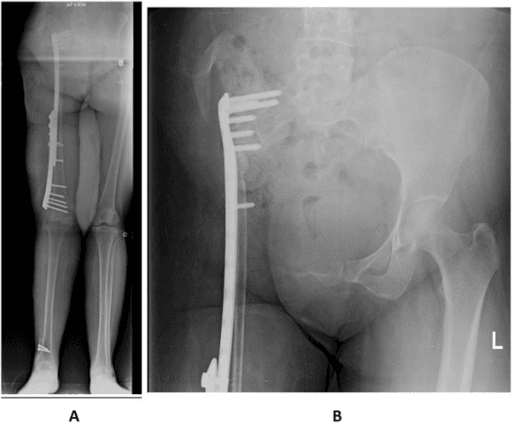
- Oncologic intercalary reconstruction — alone (forearm, humerus, fibula-for-radius) or as the Capanna hybrid inside a massive allograft (femur, tibia)
- Forearm and humeral reconstruction — fibular diameter closely matches radius/ulna; can arthrodese or reconstruct the distal radius after giant cell tumour resection (proximal fibula recreates the radiocarpal surface)
- Osteonecrosis of the femoral head in the young, pre-collapse or early post-collapse (Ficat II-III) — Urbaniak technique: core decompression plus vascularised fibula strut with anastomosis to ascending branch of lateral femoral circumflex vessels
- Peronea arteria magna in the donor limb (absolute)
- No suitable recipient vessels (previous radical dissection, radiation vasculitis) — reassess with angiography; consider arteriovenous loop
- Severe peripheral vascular disease; heavy smoking is a relative contraindication (flap thrombosis, non-union)
- Elderly/low-demand patients better served by endoprosthesis or acute shortening
- Femoral-head FVFG: collapse greater than 2 mm with acetabular involvement — outcomes poor, arthroplasty preferred; the whole technique is debated given modern arthroplasty results and its own donor morbidity
Below 5 to 6 cm: autograft or induced membrane. 6 to 12 cm: FVFG or bone transport depending on soft tissues, subspecialty availability and patient tolerance of frames. Greater than 12 cm, or large-diameter weight-bearing bone: Capanna hybrid, double-barrel FVFG, or transport-plus-flap combinations. Hostile bed (radiation, chronic infection) shifts the choice towards vascularised bone at any length.
Applied Vascular and Surgical Anatomy
- Where
- Fibular neck, winding under peroneus longus origin
- Mechanism
- Traction or direct division during proximal exposure and proximal osteotomy
- Avoid by
- Identify and protect at the neck; keep proximal osteotomy at least 6 cm distal to the head; subperiosteal dissection at the neck
- If injured
- Foot drop; explore and repair or graft, ankle-foot orthosis pending recovery
- Where
- Within peroneus longus and brevis, piercing fascia in the distal third of the lateral approach
- Mechanism
- Cut during lateral intermuscular septum dissection or skin paddle raising
- Avoid by
- Stay on the septum; raise the skin paddle including the septocutaneous perforators at the middle-distal third junction
- If injured
- Dorsal foot numbness; neuroma excision and burial if painful
- Where
- Immediately anterior to the interosseous membrane
- Mechanism
- Divided when the membrane is incised blindly from behind
- Avoid by
- Divide the membrane under direct vision close to the fibula, retracting anterior compartment muscle anteriorly
- If injured
- Foot ischaemia risk if posterior tibial compromised; repair vessel, repair nerve, monitor for compartment syndrome
- Where
- Peroneal artery is dominant or sole supply to the foot, 0.2 to 5 percent of limbs
- Mechanism
- Harvest devascularises the foot
- Avoid by
- Absolute contraindication; assess dorsalis pedis and posterior tibial pulses with triphasic Doppler, CTA or MRA if abnormal, previous trauma, PVD or congenital anomaly
- If injured
- Abandon harvest; if divided, immediate revascularisation and consider vein graft
- Where
- Medial to the peroneal pedicle in the deep posterior compartment
- Mechanism
- Injured tracing the pedicle proximally to the tibioperoneal trunk
- Avoid by
- Dissect along tibialis posterior in a defined plane; ligate only branches confirmed to run to the fibula
- If injured
- Plantar sensory loss and foot ischaemia; microvascular repair
- Where
- Enters posteromedial cortex of the middle third, posterior to the interosseous membrane
- Mechanism
- Avulsed by stripping the medial periosteum or by osteotomy through the middle third
- Avoid by
- Preserve a 1 to 2 mm cuff of tibialis posterior and periosteum on the medial fibula; keep osteotomies away from the nutrient foramen
- If injured
- Loss of endosteal (inner two-thirds) supply; graft survives on periosteal arcades if the cuff and pedicle are intact
- Where
- Segmental branches along the length of the bone supplying the outer cortex
- Mechanism
- Circumferential stripping at contouring or double-barrel osteotomy sites
- Avoid by
- Subperiosteal-sparing osteotomies leaving each segment attached to its periosteal sleeve and pedicle
- If injured
- Segmental avascular bone with delayed union; consider bone graft at that junction
- Where
- Distal 6 cm above the lateral malleolus and the syndesmosis
- Mechanism
- Over-long harvest destabilises the mortise and causes valgus ankle drift, especially in children
- Avoid by
- Retain at least 6 cm distally; syndesmotic screw or tibiofibular synostosis in the skeletally immature
- If injured
- Ankle valgus and instability; screw fixation, distal tibiofibular synostosis or corrective osteotomy
- Where
- Origin from the posterior fibula, elevated as a cuff with the graft
- Mechanism
- Excessive muscle harvest and postoperative scarring or compartment tightness
- Avoid by
- Take only a thin muscle cuff; avoid tight fascial closure; early hallux mobilisation
- If injured
- Claw hallux and great toe stiffness; splinting, FHL release or lengthening
SIX-SIXFibula Harvest Safety
Hook:Two sixes: six centimetres retained distally, six centimetres of defect as the indication threshold.
Preoperative Planning
- History and examination — donor leg trauma/surgery, claudication, smoking; palpate dorsalis pedis and posterior tibial pulses; handheld Doppler all three vessels.
- Imaging the donor leg — selective CTA/MRA as above; mandatory in congenital limb anomalies and abnormal pulses.
- Imaging the recipient site — full-length radiographs, CT for defect geometry, MRI for tumour margins; measure defect and add 2 to 3 cm for intramedullary docking or step-cuts.
- Recipient vessels — plan artery (end-to-side to a major axial vessel or end-to-end to a branch) and two venous options; in irradiated necks/limbs consider preoperative angiography of the recipient bed.
- Team and logistics — two-team approach (resection/recipient preparation simultaneous with harvest) shortens ischaemia time; microscope, microinstruments, couplers, implantable Doppler available.
- Consent — flap failure (roughly 5 percent in experienced hands), re-exploration, stress fracture, donor morbidity (great-toe clawing, ankle discomfort, weakness, sensory change), prolonged protected weight-bearing.
Operative Technique
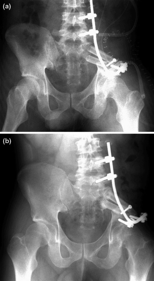
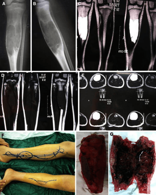
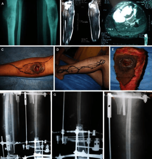
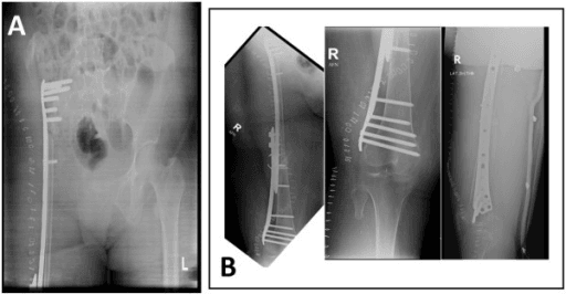
Position — supine with a bump or lateral decubitus; hip and knee flexed, thigh tourniquet (exsanguinate by elevation, not Esmarch, to preserve venous filling for perforator identification).
Landmarks and incision — line from fibular head to lateral malleolus along the posterior border of the fibula; centre the skin paddle over Doppler-marked perforators at the middle-distal third junction.
Sequence (Gilbert-type lateral approach):
- Incise anterior to the marked paddle; raise the anterior skin flap and identify septocutaneous perforators in the posterior crural septum; include a septal cuff with the paddle.
- Elevate peroneus longus and brevis off the fibula anteriorly, leaving a 1 to 2 mm muscle cuff on bone to protect periosteal vessels; protect the superficial peroneal nerve; proximally identify and protect the common peroneal nerve at the neck.
- Release the anterior compartment muscles (EDL, EHL) from the fibula; identify and protect the anterior tibial vessels and deep peroneal nerve on the interosseous membrane.
- Mark osteotomy levels — preserve 6 cm distally and stay below the neck proximally; confirm the middle third (nutrient foramen) lies within the graft. Perform osteotomies with an oscillating saw under retractor protection.
- Divide the interosseous membrane; rotate the fibula externally to expose the deep posterior compartment.
- Ligate and divide the distal peroneal vessels below the graft; dissect the pedicle proximally between tibialis posterior and FHL, taking a cuff of FHL/tibialis posterior with the bone, up to the tibioperoneal trunk for maximal length and calibre.
- Deflate the tourniquet; confirm perfusion of bone (bleeding osteotomy ends, punctate periosteal bleeding) and paddle; achieve haemostasis; divide the pedicle only when the recipient site is ready.
Closure — loose fascial closure (do not close the crural fascia tightly — compartment syndrome risk), drain, direct skin closure if paddle less than about 4 cm wide, otherwise split-skin graft; below-knee backslab in ankle neutral.
Paediatric addition — distal tibiofibular synostosis or transfixion screw to prevent proximal migration and ankle valgus.
The commonest avoidable technical failure is pedicle compromise at inset: a screw through the vessel groove, plate edge against the pedicle, kinking within a Capanna allograft window, or tension after limb lengthening. Always visualise the pedicle lying free after final fixation and in the final limb position, and recheck flow before closing.
SPONDeeHarvest Order
Hook:Skin, muscles, bone cuts, membrane, then pedicle — superficial to deep, distal to proximal.
Graft Biology: Hypertrophy and Stress Fracture
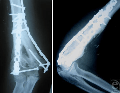
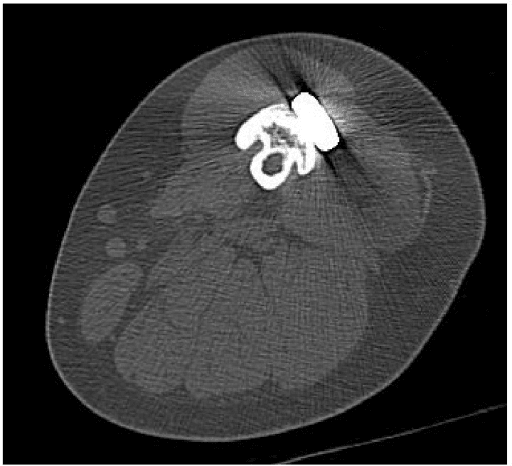
Vascularised fibula: union, hypertrophy and the stress-fracture window
Osteotomy and inset with microvascular anastomosis. Ischaemia time is kept as short as possible — the graft's value lies entirely in remaining a living, perfused bone with surviving osteocytes rather than a creeping-substitution scaffold.
The highest-risk period for pedicle thrombosis. Monitor the skin paddle where one is raised; suspected failure demands urgent re-exploration, since salvage is time-critical and a failed flap converts the reconstruction into a non-vascularised graft.
The fibula-host junctions heal like fractures, by callus, not by creeping substitution. Union typically at 3 to 6 months in a favourable bed. Protect fully: the fibula alone carries only a fraction of femoral or tibial load.
Loading drives periosteal apposition, but this is the period of peak stress-fracture risk — roughly 10 to 25 percent of lower-limb reconstructions, highest where a single-barrel fibula reconstructs femur or tibia without allograft or intramedullary nail protection.
Most unite with immobilisation and protected loading, because the bone is vascularised and retains intrinsic healing capacity. Persistent instability warrants supplementary fixation with bone graft.
Cortical thickening and diameter increase toward host dimensions. Greater and faster in children and in weight-bearing bones. The trap is to treat protection as a steady state. de Boer and Wood found hypertrophy was enhanced precisely where the graft was not bypassed by internal fixation - so a rigid construct that carries the load indefinitely prevents the remodelling that would eventually make the graft safe. Protection and hypertrophy pull in opposite directions, and the plan must be staged: shield through the fatigue window of the first year, then deliberately transfer load to the graft by graduated weight-bearing and, where appropriate, dynamisation or hardware removal. Note also what de Boer found made no difference - graft length and the use of additional bone graft affected neither stress fracture nor hypertrophy. For large-diameter bones the durable answer is more bone, not more metal: double-barrel or Capanna constructs.
The fibula-host junctions heal like fractures: callus and union typically by 3 to 6 months in favourable beds. Protect fully — the fibula alone carries a fraction of femoral or tibial load.
Graduated loading drives periosteal apposition. This is the window of stress fracture — reported in roughly 10 to 25 percent of lower-limb reconstructions, highest where a single-barrel fibula reconstructs the femur or tibia without allograft or nail protection.
Cortical thickening and diameter increase toward host dimensions (quantifiable by the de Boer and Wood hypertrophy index). Greater and faster in children and in weight-bearing bones.
Stress fracture management — most unite with immobilisation/protected loading because the bone is vascularised; persistent instability warrants supplementary fixation and bone graft. Prevention: double-barrel or Capanna constructs for large-diameter bones, spanning fixation retained until hypertrophy, graduated loading.
Complications
- Approximate Rate
- Roughly 5 percent (less in high-volume units)
- Prevention
- Recipient vessels outside zone of injury, tension-free anastomosis, paddle/Doppler monitoring
- Management
- Emergency re-exploration; thrombectomy, revision anastomosis; failed flap: debride and revise strategy
- Approximate Rate
- 10-25 percent (lower limb)
- Prevention
- Double-barrel/Capanna, protected loading until hypertrophy, retained spanning fixation
- Management
- Immobilise — most unite; fix and graft if persistent
- Approximate Rate
- 10-20 percent of junctions (higher with chemotherapy/radiation)
- Prevention
- Bleeding host bone, compression, stable fixation, intramedullary docking
- Management
- Revision fixation plus autograft; usually salvageable
- Approximate Rate
- Common (often subclinical); symptomatic in a minority
- Prevention
- Minimal FHL cuff, early toe mobilisation
- Management
- Stretching; FHL release or lengthening at the ankle if fixed
- Approximate Rate
- Significant if distal fibula inadequate or no synostosis
- Prevention
- Retain 6 cm; paediatric distal tibiofibular synostosis
- Management
- Synostosis/screw, supramalleolar osteotomy for established valgus
- Approximate Rate
- Weakness of eversion/great-toe flexion common early; compartment syndrome rare
- Prevention
- Loose fascial closure, drain, haemostasis
- Management
- Fasciotomy if compartment syndrome; physiotherapy for weakness — most recover to near-normal gait
Guidelines, Registries & Global Practice
- Global epidemiology of use — FVFG is practised worldwide wherever microsurgical expertise exists; the dominant indications differ by region: oncologic intercalary reconstruction and congenital pseudarthrosis in high-income centres; post-traumatic and post-infective defects dominate in many lower-resource settings, where bone transport is often preferred because it needs no microsurgical infrastructure.
- Society guidance — no society issues an FVFG-specific guideline. Relevant framework documents: BOA/BAPRAS BOAST on open fractures (early combined orthoplastic decision-making for segmental bone loss, reconstruction within a definitive soft-tissue plan); AO principles for segmental defect management (defect classification, staged strategies including Masquelet, transport and vascularised graft); musculoskeletal tumour society consensus favours biological reconstruction (allograft, FVFG, Capanna) for intercalary defects in the young where expertise allows.
- Registries — no implant-style registry captures FVFG; evidence rests on institutional series and systematic reviews, which consistently report union above 80 to 90 percent after all secondary procedures, with reoperation common (junctional grafting, stress-fracture management).
- Resource-setting variation — where microsurgery is unavailable: bone transport, Masquelet, ipsilateral pedicled fibula transfer for tibial defects (Huntington procedure — no microsurgery needed), or non-vascularised fibula strut for smaller defects. Selective rather than routine angiography is now the majority international practice for donor-limb assessment.
Controversies & Areas of Uncertainty
- Routine versus selective donor angiography — routine imaging detects rare variants but adds cost and (for catheter studies) risk; most units now image selectively on clinical/Doppler criteria, with a lower threshold in congenital deformity.
- FVFG for femoral head osteonecrosis — Urbaniak's results are difficult to reproduce; the operation is long, donor morbidity real, and modern arthroplasty in the young performs well. Many centres have abandoned it; others reserve it for pre-collapse disease in patients under 40.
- FVFG versus Ilizarov transport for tibial defects — comparable union in experienced hands; transport avoids microsurgery and donor morbidity but demands months in frame; choice is driven by defect length, soft tissues, patient tolerance and local expertise.
- Capanna versus fibula alone versus allograft alone — the hybrid adds operative complexity; whether it is required for defects under about 10 cm in the upper limb (where fibula alone performs well) is debated.
- Osteocutaneous paddle versus buried flap with implantable Doppler — paddle gives the most reliable monitoring but adds donor scarring and occasionally skin-graft morbidity.
MCQ Practice Points
Q: What is the pedicle of the standard free fibula flap?
A: The peroneal (fibular) artery with its paired venae comitantes, dissected proximally to the tibioperoneal trunk. The proximal fibular epiphysis, in contrast, travels on the anterior tibial vessels.
Q: Where does the nutrient artery enter the fibula?
A: The middle third, posterior to the interosseous membrane - which is why the middle third must be included in the graft to retain endosteal supply.
Q: How much distal fibula must be preserved, and what extra step is needed in children?
A: At least 6 cm above the lateral malleolus. In skeletally immature patients add a distal tibiofibular synostosis, or a transfixion screw, to prevent proximal migration of the remnant and progressive ankle valgus.
Q: Which classic donor-site complication affects the foot?
A: Claw hallux, from flexor hallucis longus scarring and contracture; eversion weakness and ankle discomfort also occur. Early great-toe mobilisation reduces the risk.
Q: Why is a closing-wedge osteotomy of a vascularised fibula safe?
A: Because segmental periosteal branches from the peroneal artery supply the outer cortex along the whole length. Provided the periosteal sleeve and pedicle are preserved, both limbs of a double-barrel construct remain perfused even if one lacks the nutrient artery.
Q: What is the Capanna technique, and what problem does it solve?
A: A vascularised fibula telescoped through the canal of a massive intercalary allograft, with the pedicle exiting through a trough. It solves the failure mode of each component: the allograft gives immediate size-matched mechanical strength that a slender fibula cannot, while the living fibula drives junctional union and salvages the construct if the allograft resorbs or fractures. Capanna's own paper reports no union rate - quote the concept to him and the numbers to a named cohort.
Exam Viva Scenarios
Practise clinical reasoning and management decisions out loud
“A 16-year-old has completed neoadjuvant chemotherapy for a mid-diaphyseal femoral osteosarcoma. Planned resection will leave a 14 cm intercalary defect with good soft tissues. How will you reconstruct, and why?”
“You plan a free fibula for a 10 cm post-infective radial and ulnar-side forearm defect. The anaesthetist asks whether the patient needs leg angiography first. How do you respond?”
“A 5-year-old with neurofibromatosis type 1 has an established congenital pseudarthrosis of the tibia that has refractured after intramedullary rodding and grafting. Discuss the role of free vascularised fibula transfer.”
“Twelve hours after a free fibula reconstruction of the humerus, the nurse reports the skin paddle is dusky and swollen with brisk capillary refill. What is happening and what do you do?”
Anatomy
- Pedicle: peroneal artery (1.5-2.5 mm) plus paired venae comitantes; dissect to tibioperoneal trunk; pedicle 4-8 cm
- Nutrient artery: middle third, posterior to interosseous membrane — include middle third in the graft
- Segmental periosteal supply permits careful osteotomy (double-barrel, contouring) with periosteum and pedicle intact
- Skin paddle: septocutaneous perforators in the posterior crural septum, middle-distal third junction
- Variant to exclude: peronea arteria magna — absolute contraindication to harvest
Indications and Thresholds
- Segmental defects greater than 6 cm; infected/irradiated beds at any length
- Congenital pseudarthrosis of the tibia (with rod protection to maturity)
- Oncologic intercalary reconstruction — alone (forearm/humerus) or Capanna hybrid (femur/tibia)
- Femoral head osteonecrosis in the young, pre-collapse (Urbaniak) — debated
- Greater than 12 cm or large-diameter bone: double-barrel or Capanna, not single fibula
Technique Essentials
- Lateral approach; muscle cuff on bone; protect common and superficial peroneal nerves, anterior tibial bundle
- Preserve 6 cm distal fibula; paediatric distal tibiofibular synostosis
- Ligate distal peroneal vessels; pedicle dissection distal to proximal; divide pedicle last
- Inset: intramedullary docking or step-cuts, spanning fixation, pedicle free of compression
- Selective donor angiography: image only if pulses/Doppler abnormal, prior trauma, vascular disease, congenital anomaly
Aftercare and Complications
- Monitor via paddle or implantable Doppler; venous congestion commonest — immediate re-exploration
- Flap loss roughly 5 percent; junctional non-union 10-20 percent per junction — revise and graft
- Stress fracture 10-25 percent during remodelling — most heal with immobilisation
- Hypertrophy over 1-2 years (de Boer and Wood index) governs return to unprotected loading
- Donor morbidity: claw hallux (FHL), eversion weakness, ankle valgus in children without synostosis
Evidence Base
The Free Vascularized Bone Graft - a Clinical Extension of Microvascular Techniques
- The founding paper. A free vascularised bone graft was integrated with an appropriate soft-tissue flap repair for lower limb injury with extensive loss of BOTH skin and bone
- The technique was developed to salvage TWO legs that would otherwise have been amputated - the entire report is those two cases
- The authors claim Case 2 as 'the first successful distant transfer of a composite fibular graft by microvascular anastomoses to be reported in man'
- The result in Case 2 is described by the authors themselves as 'preliminary' and 'encouraging' - not as an established outcome
A New Reconstructive Technique for Intercalary Defects of Long Bones - Massive Allograft with Vascularized Fibular Autograft
- The technique arose in 1988 out of experience using a vascularised fibular autograft as a SALVAGE procedure for massive allograft failures caused by non-union or massive resorption
- That salvage success prompted the authors to combine the two grafts PRIMARILY rather than waiting for the allograft to fail - the allograft gives immediate structural bulk, the fibula gives living bone that can unite, remodel and hypertrophy inside it
- The authors state that the excellent final results and the avoidance of further salvage procedures justify the primary use of this more complicated technique
- They acknowledge the trade-off explicitly: longer surgical times and greater complexity
Treatment of Osteonecrosis of the Femoral Head with Free Vascularized Fibular Grafting - 103 Hips
- 103 consecutive hips in 89 patients, followed prospectively for at least five years. Aetiology: alcohol 30%, steroids 17%, trauma 13%, Perthes 3%, idiopathic 38%
- Kaplan-Meier probability of conversion to total hip arthroplasty within five years, BY STAGE (Marcus): stage II 11%, stage III 23%, stage IV 29%, stage V 27%
- Harris hip scores improved in every stage (p less than 0.001): II 56 to 80, III 52 to 85, IV 41 to 76, V 36 to 75 - so even hips that were not salvaged structurally improved symptomatically
- A trend to lower conversion in patients under 30 did NOT reach significance (p = 0.06), and no causative factor predicted conversion
Bone Changes in the Vascularised Fibular Graft
- Retrospective review of 62 consecutive patients who had a vascularised fibular transfer for a large skeletal defect
- FRACTURES OCCURRED IN 25 PER CENT, at an average of EIGHT MONTHS after surgery - the fatigue window is the first year, not the first weeks
- Hypertrophy was more common when the limb was mechanically LOADED, and was enhanced where the graft was NOT bypassed by internal fixation - a plate that shields the graft prevents the remodelling that makes it strong
- Graft length and the use of additional bone graft material had NO influence on either stress fracture or hypertrophy
- The authors conclude that the graft should be protected against fatigue fracture for the first year, and that GRADUAL increases in loading enhance remodelling