The epiphyseal cartilage malignancy that masquerades as chondroblastoma for years
- Epiphyseal lytic lesion in a patient older than 25 years is clear cell chondrosarcoma until proven otherwise — not chondroblastoma.
- Years-long, low-grade hip or shoulder pain with a slowly enlarging epiphyseal lesion is the classic history.
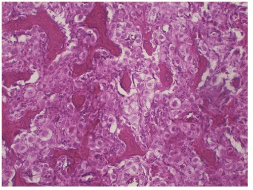
- Histology: sheets of large cells with clear, glycogen-rich cytoplasm and distinct membranes, scattered osteoclast-type giant cells, and intralesional woven bone — a cartilage tumour that makes bone.
- Intralesional curettage is the classic and avoidable error: high local recurrence, and recurrence can be higher grade.
- Definitive treatment is wide en-bloc resection with endoprosthetic or osteoarticular allograft reconstruction.
- Chemotherapy and radiotherapy are ineffective — surgery is the treatment.
- Metastasis to lung and bone can appear more than 10 to 20 years later; surveillance is lifelong.
- “Age over 25, size greater than 4 cm and symptom duration in years favour clear cell chondrosarcoma over chondroblastoma.
- “Both lesions can contain giant cells and both are epiphyseal — the discriminator is clear cytoplasm, woven bone, absence of chicken-wire calcification, and the patient's age.
- “S100 positive; the tumour cells are cytokeratin negative, which separates it from metastatic clear cell renal carcinoma.
- “Never curette an epiphyseal lesion in an adult before a definitive biopsy diagnosis.
- “Approximately one third show matrix mineralisation on radiographs — absence of visible chondroid matrix does not exclude the diagnosis.
Chondroblastoma is a lesion of the open physis — typically 10 to 20 years. An epiphyseal lesion in a skeletally mature adult, particularly if larger than 3 to 4 cm with a years-long history, must be assumed malignant until biopsy proves otherwise.
Intralesional curettage of clear cell chondrosarcoma contaminates the joint and soft tissues, produces local recurrence rates several-fold higher than wide resection, and can convert a curable low-grade tumour into an unsalvageable one.
The biopsy tract must be excisable in the definitive resection and planned by the operating surgeon. A transgluteal or multi-compartment hip tract can force a hemipelvectomy that a correctly placed anterolateral tract would have avoided.
Recurrence and pulmonary or skeletal metastasis are documented 10 to 20 years and beyond after resection. Five-year clearance means nothing here — commit the patient to lifelong imaging surveillance.
Definition, Epidemiology and Behaviour
Clear cell chondrosarcoma is a rare, low-grade malignant cartilage-forming tumour defined by sheets of tumour cells with abundant clear, glycogen-rich cytoplasm, admixed osteoclast-type giant cells and intralesional woven bone formation. It was delineated as a distinct entity by Unni and colleagues at the Mayo Clinic in 1976 and remains recognised as a separate variant in the WHO classification of bone tumours.
Key epidemiological features
- Accounts for roughly 1 to 2 percent of chondrosarcomas — most units see a handful of cases in a career.
- Peak incidence in the third to fifth decades; a male-to-female ratio of about 2 to 3 : 1.
- Site distribution: proximal femur (femoral head/neck) is the commonest by a wide margin, then proximal humerus; proximal tibia, distal femur, spine, ribs and craniofacial bones are described but uncommon.
- Almost always epiphyseal or epiphyseal-equivalent (apophyses, small round bones); extension into the metaphysis occurs with growth.
Biological behaviour. It is conventionally regarded as grade 1 in behaviour: slow growing, locally destructive, low metastatic propensity. But it is not benign — untreated or inadequately treated disease recurs locally, and metastases to lung and to other bones are well documented, sometimes after very long latency. Recurrent tumours may show dedifferentiation to high-grade sarcoma, which carries a poor prognosis.
Two unrelated malignancies share the words "clear cell", and both are in this corpus. The name is the only thing they have in common.
- Clear cell CHONDROSARCOMA (this page)
- Bone - cartilage-forming
- Clear cell SARCOMA of soft tissue
- Soft tissue - tendons and aponeuroses
- Clear cell CHONDROSARCOMA (this page)
- Epiphysis: femoral head and neck, then proximal humerus
- Clear cell SARCOMA of soft tissue
- Distal extremity, especially foot and ankle
- Clear cell CHONDROSARCOMA (this page)
- S100 positive, HMB-45 and Melan-A NEGATIVE
- Clear cell SARCOMA of soft tissue
- S100 and HMB-45, Melan-A, SOX10 all positive
- Clear cell CHONDROSARCOMA (this page)
- No EWSR1 rearrangement
- Clear cell SARCOMA of soft tissue
- EWSR1::ATF1 t(12;22) - the defining lesion
- Clear cell CHONDROSARCOMA (this page)
- Chondroblastoma
- Clear cell SARCOMA of soft tissue
- Malignant melanoma ("melanoma of soft parts")
- Clear cell CHONDROSARCOMA (this page)
- Low grade; over 85% survival after wide resection
- Clear cell SARCOMA of soft tissue
- Aggressive; nodal spread; 5-year survival around 42%
The practical discriminator at the bench is straightforward: a melanocytic marker panel separates them outright. HMB-45 and Melan-A are positive in clear cell sarcoma of soft tissue and negative here, and EWSR1 break-apart FISH is positive there and negative here. What the two genuinely share is the clinical trap - both are slow-growing, both look benign, and in both the unplanned intralesional excision is the single event that ruins the outcome.
Pathology
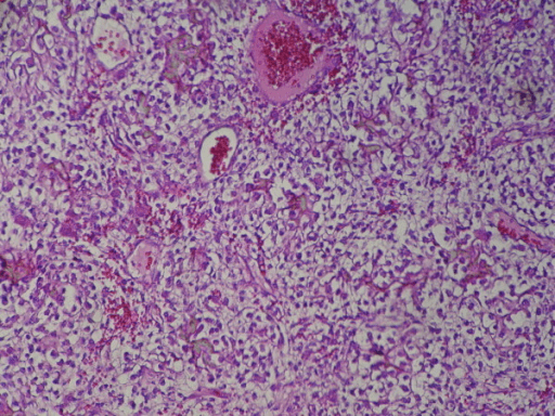
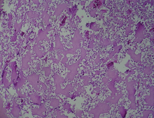
Macroscopic. Soft to gritty, grey-white to reddish-brown tissue; cystic haemorrhagic areas are common; gritty foci correspond to woven bone and mineralised cartilage. The lesion abuts and may breach the subchondral plate.
Microscopic — the diagnostic triad
- Clear cells: sheets and lobules of large, round to polygonal cells with abundant, empty-appearing (glycogen-rich) cytoplasm, sharply defined cell membranes, and central rounded nuclei with fine chromatin and small nucleoli. Mitoses are scarce.
- Osteoclast-type multinucleated giant cells: scattered throughout — this is why it is misread as chondroblastoma or giant cell tumour.
- Intralesional woven bone: irregular trabeculae of reactive and tumour-derived woven bone within the lesion. A malignant cartilage tumour that forms bone — this is the feature that clinches it and also the feature that leads to the erroneous diagnosis of osteosarcoma if the cartilage component is under-sampled.
Additional features: low-grade hyaline cartilage lobules (present in most cases, sometimes focal), secondary aneurysmal bone cyst change, and areas indistinguishable from conventional grade 1 chondrosarcoma.
Special stains and immunohistochemistry
- PAS positive, diastase sensitive cytoplasm (glycogen).
- S100 positive — confirms chondroid lineage.
- Cytokeratin, EMA, PAX8, CD10 negative — excludes metastatic clear cell renal carcinoma.
- H3K36M immunostain negative — chondroblastoma is positive (H3F3B K36M mutation); this is a genuinely useful modern discriminator.
- IDH1/IDH2 mutations, characteristic of conventional chondrosarcoma and enchondroma, are generally not a feature of clear cell chondrosarcoma.
"Clear cell chondrosarcoma is a low-grade cartilage malignancy defined by clear glycogen-rich tumour cells, osteoclast-type giant cells and intralesional woven bone, arising in the epiphysis of a skeletally mature adult. S100 positive, cytokeratin negative, H3K36M negative."
Staging and Multidisciplinary Assessment
- Stage using the Enneking (MSTS) system for surgical staging: this is a low-grade (G1) lesion, usually intracompartmental (T1) unless there is soft tissue extension or joint contamination, and M0 — hence Stage IA or IB. Stage III denotes metastatic disease.
- The AJCC staging for bone sarcoma may also be quoted (grade, size, and presence of skip or distant metastasis).
- Full staging package: whole-bone MRI with contrast, CT of the lesion, CT chest, bone scan or whole-body MRI, baseline bloods.
- Discuss every case at a specialist bone and soft tissue sarcoma MDT with a bone tumour pathologist, musculoskeletal radiologist, sarcoma oncologist and orthopaedic oncology surgeon before any definitive procedure.
- Consent discussion should include limb salvage versus amputation, implant survivorship, function expectations and the requirement for lifelong follow-up.
Clinical Presentation
- Duration measured in years, not weeks. Median symptom duration commonly exceeds 12 to 24 months; histories of 5 to 10 years of "groin strain" or "shoulder impingement" are typical and highly characteristic.
- Deep, aching, activity-related joint pain — groin pain radiating to the anterior thigh or knee for proximal femoral lesions; deep shoulder pain with night ache for humeral lesions.
- Pain is often initially mechanical and intermittent, then becomes constant and nocturnal as the lesion enlarges.
- Pathological fracture through the femoral neck is a recognised presentation and may be the first event, in perhaps 20 to 25 percent of femoral cases.
- Systemic upset, weight loss and fever are absent — their presence should redirect towards infection or a different diagnosis.
- Ask specifically about a previous curettage or "cyst removal" elsewhere; recurrent clear cell chondrosarcoma after mislabelled chondroblastoma is a classic referral.
Imaging: What to Order and How to Read It
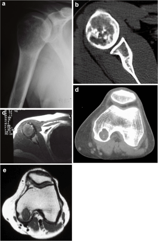
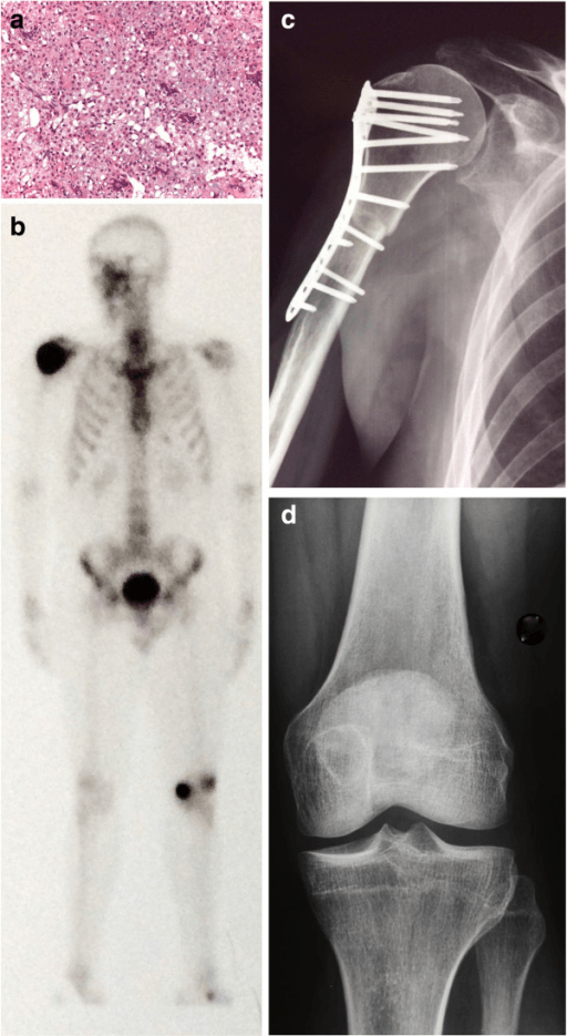
Sequenced imaging and staging workup in clear cell chondrosarcoma
Views. AP pelvis plus a cross-table lateral or frog-lateral of the affected hip; AP and axillary lateral of the shoulder. Always image the whole bone.
Findings
- Typical appearance
- Epiphyseal, often eccentric, may cross the closed physeal scar into the metaphysis
- Typical appearance
- Well-defined lytic (radiolucent) lesion, geographic, often lobulated
- Typical appearance
- Variable — a sclerotic rim may be present (mimicking benignity) or the margin may be indistinct
- Typical appearance
- Frequently greater than 3 to 4 cm at diagnosis, reflecting the long history
- Typical appearance
- Punctate or stippled mineralisation in roughly one third; the majority show no visible chondroid matrix
- Typical appearance
- Thinning, endosteal scalloping, expansion; frank cortical breach and soft tissue mass are late
- Typical appearance
- Pathological femoral neck fracture through the lesion is common
Reading discipline. Draw the lesion's boundary, state whether it is geographic type IA (sclerotic rim), IB (well defined, no sclerosis) or IC (ill defined) using the Lodwick descriptors, and state whether it crosses the joint surface. A geographic lesion with a sclerotic rim in an adult epiphysis is not reassuring — clear cell chondrosarcoma frequently looks benign.
Only about a third of clear cell chondrosarcomas show visible matrix mineralisation. The absence of rings-and-arcs calcification in an adult epiphyseal lytic lesion therefore does not push you away from a cartilage malignancy.
Differential Diagnosis — and How to Win the Chondroblastoma Argument
- Chondroblastoma (benign)
- 10 to 20 years, physis still open
- Clear cell chondrosarcoma (malignant)
- 25 to 50 years, skeletally mature
- Metastatic clear cell renal carcinoma
- Older adult, typically 50 years and above
- Chondroblastoma (benign)
- Weeks to months
- Clear cell chondrosarcoma (malignant)
- Years — often greater than 12 to 24 months
- Metastatic clear cell renal carcinoma
- Short history, often pathological fracture or known renal primary
- Chondroblastoma (benign)
- Usually less than 3 cm
- Clear cell chondrosarcoma (malignant)
- Often greater than 4 cm
- Metastatic clear cell renal carcinoma
- Variable, often destructive and rapidly enlarging
- Chondroblastoma (benign)
- Epiphysis: proximal humerus, distal femur, proximal tibia
- Clear cell chondrosarcoma (malignant)
- Epiphysis: proximal femur first, then proximal humerus
- Metastatic clear cell renal carcinoma
- Any site; frequently multiple lesions
- Chondroblastoma (benign)
- Small lytic epiphyseal lesion, thin sclerotic rim, chicken-wire calcification
- Clear cell chondrosarcoma (malignant)
- Larger lytic lesion, variable rim, mineralisation in about one third
- Metastatic clear cell renal carcinoma
- Aggressive lytic destruction, no rim, no chondroid matrix, hypervascular
- Chondroblastoma (benign)
- Extensive surrounding marrow and soft-tissue oedema typical
- Clear cell chondrosarcoma (malignant)
- Oedema less marked; lobulated cartilage-type tumour signal
- Metastatic clear cell renal carcinoma
- Solid enhancing vascular mass, soft-tissue extension
- Chondroblastoma (benign)
- Polygonal chondroblasts, pericellular chicken-wire calcification, giant cells, chondroid islands
- Clear cell chondrosarcoma (malignant)
- Large clear glycogen-rich cells with distinct membranes, giant cells, intralesional woven bone
- Metastatic clear cell renal carcinoma
- Clear cells in delicate sinusoidal vascular network, no chondroid matrix
- Chondroblastoma (benign)
- S100 positive; DOG1 positive; H3K36M positive (H3F3B mutation)
- Clear cell chondrosarcoma (malignant)
- S100 positive; H3K36M negative; cytokeratin negative
- Metastatic clear cell renal carcinoma
- Cytokeratin, PAX8 and CD10 positive; S100 negative
- Chondroblastoma (benign)
- Intralesional curettage and bone grafting
- Clear cell chondrosarcoma (malignant)
- Wide en-bloc resection and reconstruction
- Metastatic clear cell renal carcinoma
- Systemic therapy, embolisation, stabilisation or excision of solitary metastasis
- Chondroblastoma (benign)
- Over-treatment: needless joint sacrifice
- Clear cell chondrosarcoma (malignant)
- Under-treatment: local recurrence, dedifferentiation, death
- Metastatic clear cell renal carcinoma
- Missed primary: uncontrolled bleeding at curettage, missed systemic disease
- Chondroblastoma (benign)
- 10 to 20 years, open physis
- Clear cell chondrosarcoma (malignant)
- 25 to 50 years, skeletally mature
- Chondroblastoma (benign)
- Weeks to months
- Clear cell chondrosarcoma (malignant)
- Years — often greater than 12 to 24 months
- Chondroblastoma (benign)
- Usually less than 3 cm
- Clear cell chondrosarcoma (malignant)
- Often greater than 4 cm
- Chondroblastoma (benign)
- Epiphysis: proximal humerus, distal femur, proximal tibia
- Clear cell chondrosarcoma (malignant)
- Epiphysis: proximal femur first, then proximal humerus
- Chondroblastoma (benign)
- Small lytic epiphyseal lesion, thin sclerotic rim, chicken-wire calcification
- Clear cell chondrosarcoma (malignant)
- Larger lytic lesion, variable rim, mineralisation in one third
- Chondroblastoma (benign)
- Extensive surrounding oedema typical
- Clear cell chondrosarcoma (malignant)
- Oedema less marked; lobulated tumour signal
- Chondroblastoma (benign)
- Polygonal chondroblasts, pericellular chicken-wire calcification, giant cells, chondroid islands
- Clear cell chondrosarcoma (malignant)
- Large clear glycogen-rich cells, distinct membranes, giant cells, intralesional woven bone
- Chondroblastoma (benign)
- S100 positive; DOG1 and H3K36M (H3F3B mutation) positive
- Clear cell chondrosarcoma (malignant)
- S100 positive; H3K36M negative; cytokeratin negative
- Chondroblastoma (benign)
- Intralesional curettage and grafting
- Clear cell chondrosarcoma (malignant)
- Wide en-bloc resection and reconstruction
- Chondroblastoma (benign)
- Over-treatment: needless joint sacrifice
- Clear cell chondrosarcoma (malignant)
- Under-treatment: recurrence, dedifferentiation, death
Other differentials for an adult epiphyseal lytic lesion
- Discriminating features
- Subarticular, no sclerotic rim, no matrix; mononuclear stroma with evenly distributed giant cells; H3G34W positive
- Discriminating features
- Clear cells, but cytokeratin/PAX8/CD10 positive, S100 negative; vascular, often multiple lesions; renal primary
- Discriminating features
- Metaphyseal/diaphyseal epicentre, hyaline lobules, deep endosteal scalloping, rings-and-arcs
- Discriminating features
- Metaphyseal, eccentric, lobulated sclerotic margin; myxoid lobules
- Discriminating features
- Small, adjacent to degenerate joint, no enhancing solid component
- Discriminating features
- Penumbra sign, systemic markers, clinical sepsis
CLEARDistinguishing clear cell chondrosarcoma from chondroblastoma
Hook:A CLEAR cell tumour is CLEARly not a chondroblastoma when the patient is old, the lesion is big and the story is long.
Biopsy: Principles and Pitfalls
The biopsy must be performed or directed by the surgeon who will do the definitive resection, in the unit that will do it. Poorly planned biopsies alter management, necessitate more extensive resection or amputation, and worsen outcomes — the principle established by Mankin's landmark musculoskeletal tumour society studies.
Technique
- Preferred: image-guided (CT or ultrasound) core needle biopsy, multiple cores from the solid enhancing component, avoiding cystic or necrotic areas identified on post-contrast MRI.
- Tract: longitudinal, in line with the planned incision, through a single compartment. For the proximal femur, an anterolateral tract in line with a lateral or anterolateral approach; mark the entry point with ink or a suture and record it in the operation note and imaging report so it can be excised en bloc.
- Avoid: transgluteal tracts, crossing the hip joint capsule unnecessarily, traversing neurovascular bundles, and traversing the deltoid in multiple planes at the shoulder.
- Open incisional biopsy if core sampling is non-diagnostic; meticulous haemostasis, no drains through separate stab incisions (or place the drain exit in line with the incision so it can be excised).
- Send tissue for histology, microbiology and fresh tissue for molecular studies where available.
Common pitfalls
- Sampling only the secondary aneurysmal bone cyst component and reporting "aneurysmal bone cyst" — always correlate with the enhancing solid nodule on MRI.
- A frozen section report of "chondroblastoma-like lesion with giant cells" leading directly to curettage on the same list. Never proceed to definitive treatment on frozen section for an adult epiphyseal cartilage lesion.
- Failure to obtain a second opinion from a specialist bone tumour pathologist — this variant is rare and the misdiagnosis rate outside referral centres is meaningful.
Management: Decision Thresholds
- 1Step 1 — Index suspicion
Lytic epiphyseal or apophyseal lesion in a skeletally mature adult, most often proximal femur or proximal humerus
Referral to a specialist bone sarcoma unit before any instrumentation
- 2Step 2 — Full staging first
Local MRI of the whole bone plus radiographs, and CT chest for pulmonary metastases
Resection and reconstruction planned before tissue is ever taken
- 3Step 3 — Surgeon-planned biopsy
Core biopsy performed or directed by the surgeon who will do the definitive resection
Histology confirms clear cells with abundant glycogen, vacuolated cytoplasm and lattice-like woven bone
- 4Step 4 — Curettage is prohibited
Intralesional treatment is inadequate once the diagnosis is made
Intralesional treatment predicts local recurrence and risks dedifferentiation
- 5Step 5 — Wide en-bloc resection
Localised, resectable disease with feasible limb salvage
Generally curative; margin quality is the dominant prognostic variable
- 6Step 6 — Escalate when the joint is breached
Joint breached, effusion containing tumour cells, capsular involvement or prior intralesional surgery
Amputation reserved for neurovascular encasement, extensive contamination, fungating tumour or failed salvage; proximal femur may need hip disarticulation, proximal humerus a forequarter amputation
- 7Step 7 — Adjuvants do not rescue a poor margin
Chemotherapy is ineffective in low-grade cartilage malignancy; radiotherapy is ineffective as primary treatment
Surgery remains the only reliably curative modality
- 8Step 8 — Lifelong surveillance
Late local recurrence and late pulmonary metastasis are characteristic of this tumour
Widespread metastatic disease is managed palliatively with symptomatic surgery and radiotherapy for pain; no reliable systemic option
Once clear cell chondrosarcoma is diagnosed, intralesional treatment is inadequate. Wide en-bloc resection with a cuff of normal tissue on all surfaces is the treatment of choice and is generally curative.
Surgical Technique
Indication. Clear cell chondrosarcoma of the femoral head/neck with intact neurovascular structures and a resectable margin.
Contraindication. Encasement of the femoral or external iliac vessels, extensive pelvic extension not amenable to en-bloc pelvic resection, uncontrolled sepsis, patient unfit for major reconstruction.
Why this over alternatives. Curettage has an unacceptable recurrence rate. Osteoarticular allograft of the proximal femur has high rates of fracture, non-union and joint degeneration; a modular tumour endoprosthesis gives immediate, durable, weight-bearing reconstruction in a young adult and is the usual choice, with allograft-prosthetic composite used where abductor reattachment to allograft trochanter is favoured.
PIPADRAW sequence
- Position: lateral decubitus with the pelvis squarely supported, or supine on a radiolucent table for an anterolateral approach; whole limb and hemipelvis prepped free, image intensifier available.
- Imaging / equipment: preoperative MRI-based resection level marked; navigation or patient-specific guides optional; modular proximal femoral replacement trays with a bipolar or constrained/dual-mobility acetabular option; cell salvage is not used in tumour surgery.
- Preparation: cross-match; tranexamic acid; antibiotic prophylaxis; plan for a synthetic mesh or attachment tube for soft tissue reconstruction; consent for possible acetabular resection and possible amputation.
- Approach: extended lateral or anterolateral approach incorporating and excising the biopsy tract as an ellipse of skin in continuity with the specimen.
- Dissection: develop planes outside the tumour pseudocapsule at all times. Detach the abductors with a cuff of tissue (or take the greater trochanter with the specimen if involved). Divide the vastus lateralis origin. Identify and protect the femoral neurovascular bundle anteriorly and the sciatic nerve posteriorly. Ligate perforators sequentially.
- Resection / reconstruction: perform a capsulotomy well outside the joint if the joint is uninvolved, or an extra-articular resection (osteotomy through the pubis/ischium or acetabular roof, joint kept sealed) if the joint is breached or has been previously operated. Osteotomise the femur at the pre-planned level, 2 to 3 cm distal to the marrow margin on T1 MRI. Send the distal marrow margin for frozen section. Reconstruct with a modular proximal femoral replacement, cemented or press-fit stem, and bipolar head or dual-mobility construct to reduce dislocation risk.
- At-risk structures: sciatic nerve (posterior retraction), femoral nerve and vessels (anterior dissection and acetabular retractors), obturator vessels in extra-articular resections, superior gluteal nerve.
- Fixation and soft tissue: reattach the abductors to the prosthesis using an attachment tube or synthetic mesh under appropriate tension; repair the capsule/pseudocapsule around the head to reduce dislocation; secure iliopsoas where possible.
- Closure: layered over deep drains brought out in line with the incision; meticulous haemostasis.
- Aftercare: hip abduction brace or knee immobiliser for 6 weeks; touch or partial weight-bearing progressing per implant fixation; abductor rehabilitation deferred until soft tissue healing; thromboprophylaxis.
- Pitfalls: inadequate distal margin from misreading the T1 marrow edge; violating the pseudocapsule when mobilising posteriorly; failure to excise the biopsy tract; abductor failure producing a permanent Trendelenburg gait; dislocation from under-tensioned soft tissues.
- Salvage of failure: revision of modular components for loosening or fracture; hip disarticulation for uncontrolled local recurrence or infected unreconstructable failure.
Complications
- Where and how often
- Adult epiphysis or apophysis mistaken for chondroblastoma or a cyst; the dominant avoidable complication
- Mechanism
- Intralesional entry leaves tumour behind and contaminates the whole cavity, so local recurrence is substantially higher than after wide resection
- Avoid by
- Correct diagnosis before any surgery; never curette an adult epiphyseal cartilage lesion
- If it happens
- Re-stage, re-biopsy looking for dedifferentiation, then wide re-resection or amputation
- Where and how often
- Proximal femur, proximal humerus; any tract not in line with the definitive incision
- Mechanism
- Tract seeding through uninvolved compartments makes an en bloc excision of the tract impossible and compromises the margin
- Avoid by
- Biopsy planned and ideally performed by the resecting surgeon, in line with the definitive approach; excise the tract en bloc at resection
- If it happens
- Widen the resection to include the tract; amputation if a clear margin is otherwise unachievable
- Where and how often
- Low, single figures, with clear margins
- Mechanism
- Marginal or intralesional margin at a critical structure, or unexcised tract
- Avoid by
- Frozen-section margin control; excise the biopsy tract; plan the approach around the neurovascular bundle
- If it happens
- Re-resection; amputation if a clear margin cannot be regained
- Where and how often
- Uncommon but devastating; typically in repeatedly treated recurrent tumour
- Mechanism
- A low-grade cartilage tumour acquires a high-grade non-cartilaginous sarcoma component
- Avoid by
- Adequate primary surgery the first time — this is the single strongest protection
- If it happens
- Treat as high-grade sarcoma with chemotherapy and radical surgery; prognosis is poor
- Where and how often
- Around a fifth of femoral cases, classically the femoral neck
- Mechanism
- Epiphyseal lytic destruction of load-bearing bone in a young adult
- Avoid by
- Recognise the lesion and protect weight-bearing before definitive surgery; biopsy and stage first
- If it happens
- Do not internally fix — reaming and nailing seeds the entire femoral canal; proceed to staging and resection
- Where and how often
- Lung is the commonest site and may present decades later; skeletal spread is characteristically over-represented for a low-grade cartilage tumour
- Mechanism
- Indolent haematogenous dissemination that outlives conventional five-year follow-up
- Avoid by
- Lifelong CT chest surveillance plus baseline and interval bone imaging — never discharge at five years
- If it happens
- Metastasectomy for isolated resectable pulmonary lesions; local treatment, radiotherapy for painful bone deposits, prophylactic stabilisation
- Where and how often
- Periprosthetic infection is among the leading causes of endoprosthetic failure, roughly 10 percent in large series
- Mechanism
- Large implant surface, long operative time, extensive dead space and compromised soft tissue envelope
- Avoid by
- Antibiotic prophylaxis, silver-coated implants in selected centres, meticulous soft tissue closure and drain management
- If it happens
- DAIR if early; two-stage revision for established infection; amputation for intractable infection
- Where and how often
- Aseptic loosening and stem fracture rise with time and in young active patients; dislocation after proximal femoral replacement and inferior subluxation after proximal humeral replacement
- Mechanism
- Long lever arms, absent capsule and muscle detachment leave the implant unsupported
- Avoid by
- Adequate stem length and fixation with an appropriate bearing; bipolar or dual-mobility hip bearing with capsular and abductor reconstruction; mesh suspension and tendon transfers at the shoulder
- If it happens
- Revision of modular components; closed reduction and bracing then revision to constrained or dual mobility; revision suspension at the shoulder
- Where and how often
- Abductor insufficiency with Trendelenburg gait is very common after proximal femoral replacement; sciatic, femoral or axillary nerve injury is uncommon but functionally decisive
- Mechanism
- Abductor detachment without secure reattachment; traction or excessive retraction on a nerve during a wide exposure
- Avoid by
- Secure abductor reattachment to an attachment tube or mesh with delayed rehabilitation; formal identification and protection of named nerves
- If it happens
- Prolonged rehabilitation and a stick — some abductor lag is usually permanent; observe neurapraxia, splint, tendon transfer for a permanent deficit
- Approximate frequency / context
- Substantially higher than after wide resection — the dominant avoidable complication
- Prevention
- Correct diagnosis before surgery; never curette an adult epiphyseal lesion
- Management
- Re-stage, re-biopsy for dedifferentiation, wide re-resection or amputation
- Approximate frequency / context
- Low (single figures) with clear margins
- Prevention
- Excise biopsy tract; frozen-section margins
- Management
- Re-resection; amputation if unachievable
- Approximate frequency / context
- Uncommon but devastating
- Prevention
- Adequate primary surgery
- Management
- High-grade sarcoma protocol; prognosis poor
- Approximate frequency / context
- The commonest metastatic site; may present decades later
- Prevention
- Lifelong CT surveillance
- Management
- Metastasectomy for isolated resectable lesions
- Approximate frequency / context
- Characteristically over-represented for a low-grade cartilage tumour
- Prevention
- Baseline and interval bone imaging
- Management
- Local treatment, radiotherapy for pain, stabilisation
- Approximate frequency / context
- Among the leading causes of endoprosthetic failure after tumour resection (roughly 10 percent in large series)
- Prevention
- Antibiotic prophylaxis, silver-coated implants in selected centres, meticulous soft tissue closure, drain management
- Management
- DAIR early; two-stage revision; amputation for intractable infection
- Approximate frequency / context
- Increases with time and young active patients
- Prevention
- Adequate stem length and fixation, appropriate bearing
- Management
- Revision of modular components
- Approximate frequency / context
- Recognised early complication
- Prevention
- Bipolar or dual-mobility bearing, capsular and abductor reconstruction, brace
- Management
- Closed reduction, bracing; revision to constrained/dual mobility
- Approximate frequency / context
- Very common after proximal femoral replacement
- Prevention
- Secure abductor reattachment with attachment tube or mesh, delayed rehabilitation
- Management
- Prolonged rehabilitation; stick; usually permanent to some degree
- Approximate frequency / context
- Uncommon but functionally decisive
- Prevention
- Formal identification and protection; avoid excessive retraction
- Management
- Observation for neurapraxia; splinting; tendon transfer for permanent deficit
- Approximate frequency / context
- Common after proximal humeral replacement
- Prevention
- Mesh suspension, capsular reconstruction, tendon transfers
- Management
- Bracing; revision suspension
- Approximate frequency / context
- Around a fifth of femoral cases
- Prevention
- Recognise the lesion and protect weight-bearing before definitive surgery
- Management
- Do not internally fix — this disseminates tumour; proceed to staging and resection
Reaming and nailing a femoral neck fracture through an undiagnosed clear cell chondrosarcoma seeds the entire femoral canal and converts a curable tumour into one requiring total femoral resection or amputation. Any pathological fracture in a young adult with no known primary must be biopsied and staged first.
Outcomes, Prognosis and Surveillance
Prognostic determinants
- Surgical margin is the dominant modifiable factor. Wide resection yields long-term disease-free survival in the great majority; intralesional treatment produces high recurrence.
- Metastasis at presentation, large tumour size, axial location and dedifferentiation in recurrent disease predict worse survival.
- Overall survival after adequate wide resection is commonly reported in excess of 85 percent at 5 to 10 years in collected series, but the survival curve does not plateau — late events occur.
Surveillance schedule (representative; adapt to unit protocol)
Clinical review with local radiographs and CT chest every 3 to 4 months. MRI of the operated site at 6 and 12 months (metal-suppression sequences) or CT where the implant precludes MRI.
Review every 6 months with plain radiographs of the reconstruction and CT chest. Assess implant fixation, abductor function, and rehabilitation goals.
Annual clinical review, radiographs and chest imaging. Continue to counsel the patient explicitly that late relapse occurs.
Annual or biennial review indefinitely, combining oncological surveillance with arthroplasty surveillance for loosening, wear and periprosthetic fracture. Provide the patient with written information so relapse is recognised if they move care.
WIDEManagement principles
Hook:Go WIDE first time; there is no effective rescue from a drug or a beam.
Guidelines, Registries & Global Practice
Global epidemiology and access. Clear cell chondrosarcoma is rare and geographically uniform in distribution; there is no established ethnic or regional predilection. What varies enormously is time to diagnosis and access to a sarcoma centre. In high-income systems with established referral pathways, most patients reach a bone tumour unit before any surgical intervention. In settings with limited MRI access and no centralised sarcoma service, the most common pathway is inadvertent curettage of a presumed benign cyst, followed by late referral with contaminated tissue planes.
Society guidance that genuinely addresses this tumour
- Relevant guidance
- All suspected primary bone sarcomas should be referred to a reference centre before biopsy; biopsy by the treating surgical team; chondrosarcoma treated surgically with adequate margins; chemotherapy and radiotherapy have no established role in low-grade chondrosarcoma.
- Relevant guidance
- Centralised diagnosis and treatment in designated sarcoma centres with a specialist MDT including a bone tumour pathologist; defined referral criteria for any bone lesion suspected of malignancy.
- Relevant guidance
- Do not biopsy or operate on a suspected primary bone tumour in a non-specialist unit; unexplained bone pain, particularly night pain, in a young adult warrants radiographs and prompt referral.
- Relevant guidance
- Wide excision for chondrosarcoma; radiotherapy considered only where wide excision is not achievable; particle therapy for unresectable axial disease; long-term surveillance with imaging of the primary site and chest.
- Relevant guidance
- Defines clear cell chondrosarcoma as a distinct malignant cartilage-forming entity with its own morphology and behaviour.
Registry and reconstruction evidence. Because tumour endoprostheses are implanted in small numbers, the informative datasets are national and international bone tumour registries and implant registries (for example the Australian Orthopaedic Association National Joint Replacement Registry and the National Joint Registry, both of which capture tumour prostheses, alongside dedicated sarcoma registries such as those of the Rizzoli Institute and the Netherlands Committee on Bone Tumours). Consistent messages: infection and aseptic loosening dominate late failure; revision burden accumulates in young patients over decades; and cumulative implant survivorship falls steadily beyond ten years, which is precisely the horizon relevant to a 30-year-old with a curable low-grade tumour. Consent must reflect a lifetime of revision surgery, not just the index operation.
Practice variation by resource setting
- Where modular tumour endoprostheses are unavailable, osteoarticular allograft, allograft-prosthetic composite, resection-arthrodesis or resection without reconstruction are legitimate reconstructions; the oncological principle (wide margin) is not negotiable, the reconstruction is.
- Where MRI is limited, whole-bone plain radiography plus CT with careful clinical correlation should still identify the epiphyseal lesion; the imperative is referral before intervention.
- Telepathology and international second-opinion networks materially reduce misdiagnosis of this rare variant and should be used where local specialist bone pathology is unavailable.
Controversies & Areas of Uncertainty
1. Is intralesional treatment ever acceptable? A minority argue that a very small, entirely intraosseous, radiologically contained lesion could be treated by meticulous extended curettage with adjuvants, preserving a native hip in a young adult. The counter-argument is that recurrence rates after intralesional treatment are unequivocally higher in every published series, recurrences may dedifferentiate, and the tumour is curable with wide resection. Current consensus: wide resection is standard; intralesional treatment should be an exceptional, MDT-documented decision.
2. Endoprosthesis versus osteoarticular allograft in the proximal femur and humerus. Endoprostheses give immediate stability and reliable early function but accrue infection, loosening and revision over decades. Allografts offer soft tissue reattachment and bone stock but carry fracture, non-union, resorption and joint degeneration. Allograft-prosthetic composites attempt to combine both. No randomised data exist; choice is centre- and patient-dependent.
3. Reverse total shoulder after proximal humeral resection. Improves active elevation compared with a hemiarthroplasty-type endoprosthesis, but requires an intact deltoid, functioning axillary nerve and preserved glenoid — conditions frequently not met after an oncological resection. Selection, not technique, determines the result.
4. Duration and intensity of surveillance. There is no evidence-based schedule. Late metastasis is well documented, but the yield of annual chest CT at 15 or 20 years and the cumulative radiation burden in a young patient are unquantified. Many units transition to lower-dose or lower-frequency imaging combined with a strong safety-netting message.
5. Role of particle (proton/carbon-ion) radiotherapy. Increasingly used for unresectable axial chondrosarcoma. Its role in clear cell chondrosarcoma specifically is essentially undefined given the rarity and the usually resectable appendicular location.
6. Molecular characterisation. Unlike conventional chondrosarcoma (IDH-mutant in a large proportion), clear cell chondrosarcoma lacks a defining driver. Whether it is a distinct entity biologically or a morphological pattern remains an open question, and there is currently no targeted therapy.
MCQ Practice Points
Q: Which single feature best distinguishes clear cell chondrosarcoma from chondroblastoma?
A: Skeletal maturity with a closed physis in a patient older than 25 years, supported by intralesional woven bone and clear glycogen-rich cytoplasm on histology and a negative H3K36M immunostain. Both lesions are epiphyseal and both contain osteoclast-type giant cells, so neither of those features discriminates. Note the limit of the stain: 5 per cent of genuine chondroblastomas are mutation-negative, so it supports a morphological conclusion rather than overturning one.
Q: What proportion of clear cell chondrosarcomas show radiographic matrix mineralisation?
A: Approximately one third. The majority appear as purely lytic epiphyseal lesions, frequently with a partial sclerotic rim, which is why they are so often mistaken for benign lesions.
Q: What is the treatment of choice for a localised clear cell chondrosarcoma of the femoral head?
A: Wide en-bloc resection of the proximal femur with endoprosthetic (or allograft-composite) reconstruction, excising the biopsy tract in continuity. Intralesional curettage is inadequate — Unni's founding series already recorded that excision usually recurred while resection appeared curative. Chemotherapy and radiotherapy have no established role.
Q: Which immunohistochemical panel excludes metastatic clear cell renal cell carcinoma?
A: Clear cell chondrosarcoma is S100 positive and cytokeratin, EMA, PAX8 and CD10 negative. Metastatic clear cell renal carcinoma shows the reverse pattern.
Q: A patient is 12 years post wide resection and disease free. Can surveillance be stopped?
A: No. Local recurrence and pulmonary or skeletal metastasis are documented beyond 10 to 20 years. Surveillance should continue indefinitely, combining oncological imaging with monitoring of the endoprosthetic reconstruction for loosening, wear and periprosthetic fracture.
Q: What is the most common site of clear cell chondrosarcoma, and the second?
A: Proximal femur (femoral head and neck) is by far the commonest, followed by the proximal humerus. Both are epiphyseal — the location is the diagnostic signature. Note the qualifier: the Hamburg register series also recorded rib, ilium and even the distal phalanx of the hand, so an epiphyseal site is typical rather than obligatory.
Exam Viva Scenarios
Practise clinical reasoning and management decisions out loud
“A 34-year-old male labourer has had right groin pain for five years, treated as femoroacetabular impingement with physiotherapy and a hip arthroscopy two years ago with no benefit. Pain is now constant and wakes him. An AP pelvis shows a 4.5 cm well-defined lytic lesion in the femoral head with a partial sclerotic rim and faint stippled densities. What is your diagnosis and what do you do next?”
“A 41-year-old woman is referred to you two years after curettage and grafting of a proximal humeral epiphyseal lesion reported at the time as chondroblastoma. She now has recurrent pain and a radiograph shows lysis around the graft with cortical thinning and a small soft tissue shadow. Review of the original slides by your bone pathologist reports clear cell chondrosarcoma. How do you proceed?”
“A 29-year-old man attends the emergency department after a trip, with a displaced femoral neck fracture. The trauma registrar has consented him for cannulated screw fixation. On review of the film you note a lucency in the femoral head with an ill-defined margin, and he admits to 18 months of groin pain preceding the fall. What do you do?”
“A pathologist telephones you intraoperatively during a planned curettage of an epiphyseal lesion in a 27-year-old. The frozen section shows a giant-cell-rich cartilage lesion with polygonal cells. She is uncertain between chondroblastoma and clear cell chondrosarcoma. What is your decision?”
Identity
- Rare low-grade malignant cartilage tumour, 1 to 2 percent of chondrosarcomas
- Epiphyseal: proximal femur first, proximal humerus second
- Age 25 to 50, male predominance, skeletally mature
- Delineated by Unni and colleagues, Mayo Clinic, 1976
Presentation
- Deep joint pain for YEARS — often mislabelled FAI or impingement
- Night pain, progressive, unrelieved by physiotherapy or arthroscopy
- Pathological femoral neck fracture in around a fifth of femoral cases
- Examination is bland: painful FADIR, painful log roll, reduced internal rotation
Imaging
- Lytic geographic epiphyseal lesion, variable sclerotic rim, often greater than 4 cm
- Matrix mineralisation in only about one third
- MRI: low to intermediate T1, high lobulated T2, septal and nodular enhancement
- T1 marrow margin defines the resection level; add 2 to 3 cm
- Mandatory CT chest plus bone scan at baseline
Pathology
- Clear, glycogen-rich cells with sharp membranes (PAS positive, diastase sensitive)
- Osteoclast-type giant cells scattered throughout
- Intralesional woven bone — a cartilage tumour that makes bone
- S100 positive; cytokeratin/PAX8 negative; H3K36M negative
- IDH mutations generally absent
Differential
- Chondroblastoma — younger, smaller, shorter history, chicken-wire calcification, H3K36M positive
- Giant cell tumour — subarticular, no matrix, H3G34W positive
- Metastatic clear cell renal carcinoma — cytokeratin/PAX8 positive, S100 negative
- Conventional chondrosarcoma — metaphyseal/diaphyseal, rings and arcs
- Intraosseous ganglion, Brodie abscess
Management
- Stage fully, then biopsy by the definitive treating team along an excisable tract
- Sarcoma MDT decision in every case
- Wide en-bloc resection with endoprosthetic or allograft-composite reconstruction
- Extra-articular resection if the joint is breached or previously operated
- Chemotherapy and radiotherapy ineffective; surgery is curative
- Metastasectomy for isolated resectable pulmonary metastases
Errors to avoid
- Curetting an adult epiphyseal lesion
- Internally fixing an undiagnosed pathological fracture in a young adult
- Proceeding on an equivocal frozen section
- Biopsy through a non-excisable or multi-compartment tract
- Discharging the patient at five years
Outcome
- Long-term survival commonly greater than 85 percent after adequate wide resection
- Margin quality is the dominant modifiable prognostic factor
- Recurrence may dedifferentiate to high-grade sarcoma
- Late lung and bone metastasis beyond 10 to 20 years — lifelong surveillance
- Endoprosthetic revision burden accumulates over decades in young patients
Evidence Base
Chondrosarcoma: Clear-Cell Variant - a Report of Sixteen Cases
- Sixteen adult patients, ten of them men, with the tumour usually involving the proximal femur or humerus - the series that established the entity
- RADIOGRAPHICALLY 'WELL DEFINED AND INDISTINGUISHABLE FROM CHONDROBLASTOMA' in the authors' own words, which is the whole diagnostic problem stated in the founding paper
- Benign giant cells and cells with clear cytoplasm were ALWAYS present; areas of conventional chondrosarcoma were seen in about half the cases
- The operative lesson, verbatim: EXCISION USUALLY RESULTED IN RECURRENCE, RESECTION SEEMED TO BE CURATIVE - the curettage-versus-wide-resection distinction dates from here
- Four of the sixteen patients (25 per cent) died as a result of the tumour, and serial radiographs occasionally showed malignant progression of the lesion
Clear-Cell Chondrosarcoma: the Hamburg Bone Tumour Register Series
- Sixteen cases from a national bone tumour register, analysed for age distribution, location and radiological and pathomorphological criteria
- Confirms the classic profile: adults in the third or fourth decade, predilection for the epiphyses of long tubular bones and particularly the femoral head, with a sharply defined radiolucent lesion
- BUT ALSO RECORDS UNUSUAL SITES that break the rule - rib, ilium and the DISTAL PHALANX OF THE HAND - so an epiphyseal location is typical rather than obligatory
- THE NUMBER THAT MATTERS: in 50 PER CENT of cases, high-grade osteosarcoma or conventional chondrosarcoma was considered in the differential - the misdiagnosis runs towards MORE aggressive tumours as often as towards less
- Histologically, partially mineralised trabecular osteoid formation accompanies the clear cells, alongside areas of conventional chondrosarcoma
The Hazards of the Biopsy, Revisited - Musculoskeletal Tumor Society
- 597 patients from 25 surgeons at 21 institutions, repeating a 1982 study of 329 biopsies to see whether anything had improved in a decade - the results were essentially the same
- Diagnostic error rate for the whole series 17.8 PER CENT
- A problem with the biopsy forced a different and often more complex operation, or adjuvant radiotherapy or chemotherapy, in 19.3 per cent
- A CHANGE IN OUTCOME attributable to the biopsy - more complex resection causing disability, loss of function, local recurrence or death - occurred in 10.1 per cent, and 18 patients had an UNNECESSARY AMPUTATION
- Errors, complications and changes in course and outcome were TWO TO TWELVE TIMES GREATER (p less than 0.001) when the biopsy was performed at a referring institution rather than at the treatment centre
A System for the Surgical Staging of Musculoskeletal Sarcoma
- Stratifies bone and soft-tissue lesions of any histogenesis by biological grade, anatomic setting and the presence of metastasis
- Three stages - I low grade, II high grade, III metastases present - each subdivided by whether the lesion is (A) confined within a well-delineated surgical compartment or (B) beyond it in ill-defined fascial planes
- Defines the four operative margins that this page depends on: INTRALESIONAL, MARGINAL, WIDE and RADICAL, each described in relation to the lesion, its reactive zone and its compartment
- The stages are explicitly prognostic AND surgical - the system links a risk category to a specific operation rather than describing risk alone
Distinct H3F3A and H3F3B Driver Mutations Define Chondroblastoma and Giant Cell Tumour of Bone
- p.Lys36Met (K36M) alterations, predominantly in H3F3B, were found in 73 OF 77 CHONDROBLASTOMAS (95 PER CENT)
- By contrast, 49 of 53 giant cell tumours of bone (92 per cent) carried H3F3A alterations exclusively, almost all p.Gly34Trp
- The two mutations are mutually exclusive between the two tumours - an exquisite tumour-type specificity unusual among driver mutations
- The mutations were restricted to the STROMAL cell population and were NOT detected in osteoclasts or their precursors, which matters when interpreting a giant-cell-rich lesion
- Detectable by immunohistochemistry, which is what makes this a practical bench test rather than a research finding