Proximal ablative surgery — last-resort but potentially curative operations demanding meticulous vascular control, durable soft-tissue coverage and honest counselling
- Absolute oncologic indications: encasement of the major neurovascular bundle where resection cannot achieve a functional limb, fungating/infected tumour, local recurrence after failed limb salvage, and pathological state precluding reconstruction
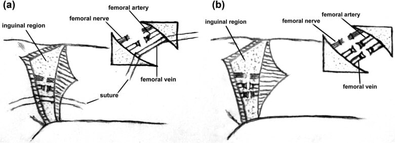
- Hip disarticulation: racquet incision based on the ASIS, femoral vessels ligated individually in the femoral triangle BEFORE deep dissection, posterior gluteus maximus flap sewn forward to the pubis/adductor origin for a weight-tolerant stump
- Forequarter amputation removes the entire upper limb with scapula and lateral clavicle; anterior (Berger) approach gives early subclavian vessel control, posterior (Littlewood) approach gives superior exposure for posteriorly based tumours
- Ligate the subclavian artery and vein separately (never en masse — risk of arteriovenous fistula) and divide the brachial plexus sharply under gentle traction with local anaesthetic infiltration
- Oncologic handling: never enter the tumour, take the biopsy tract with the specimen, orientate and ink margins with the pathologist
- Prosthetic reality must be discussed pre-operatively: Canadian-type hip disarticulation socket has very high energy cost; a forequarter prosthesis is essentially a cosmetic shoulder cap
- “The corona mortis — a retropubic anastomosis between obturator and external iliac/inferior epigastric systems — can retract into the pelvis if avulsed during obturator dissection and cause life-threatening haemorrhage
- “Pre-emptive multimodal analgesia (regional catheter, gabapentinoids, ketamine) reduces the severity of phantom limb pain; targeted muscle reinnervation at the index amputation is an emerging preventive strategy
- “Chest-wall preservation is the key intra-operative constraint in forequarter amputation — breach of the pleura converts the case to an intrathoracic emergency
In both operations the great vessels are ligated early and individually: femoral artery and vein in the femoral triangle for hip disarticulation; subclavian artery then vein for forequarter. En masse ligation risks arteriovenous fistula. Suture-ligate plus tie major arteries (double control).
Present in a substantial minority of patients. During obturator vessel ligation, an avulsed vessel retracts through the obturator foramen — control it before it disappears. Have pelvic packing and a vascular set available.
These are usually curative-intent operations for tumours failing limb salvage. Contaminating the field (tumour breach, leaving the biopsy tract) converts a curative amputation into a palliative one. Plan flaps so margins are never compromised for closure.
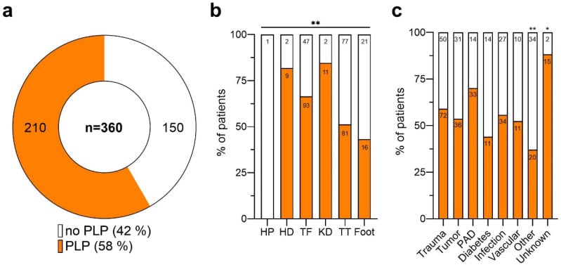
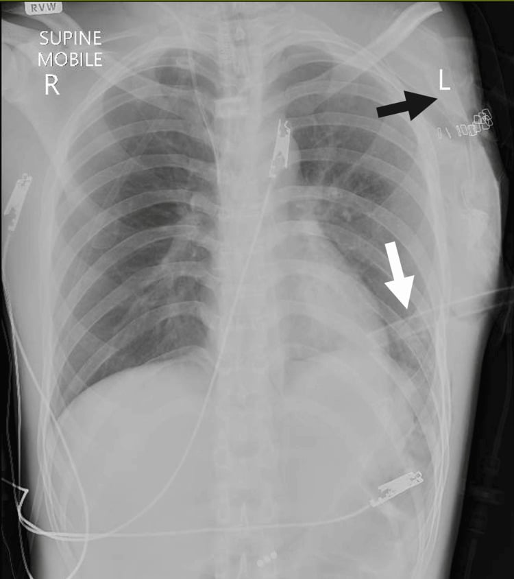
The dissection plane hugs the chest wall (serratus anterior, ribs). Warn the anaesthetist; if the pleura is opened, place a chest drain and obtain a post-operative chest radiograph. Always check for pneumothorax at the end regardless.
Indications and Decision-Making
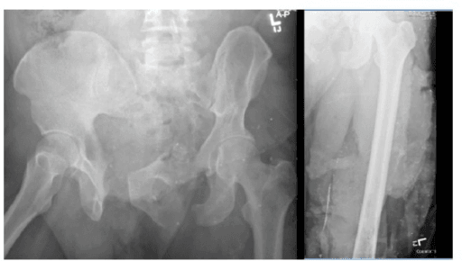
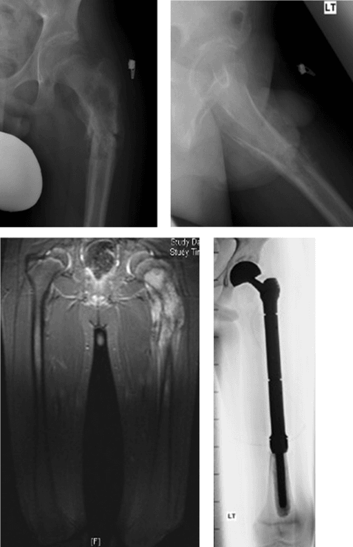
- 1Step 1 — Current staging
MRI of the whole limb, CT chest, plus PET or bone scan as indicated; histology and prior radiotherapy field reviewed
Staging must be current — never amputate on stale imaging
- 2Step 2 — Is there major neurovascular encasement?
True encasement, not mere abutment: sciatic nerve plus femoral vessels at the hip, brachial plexus encasement at the shoulder
Abutment alone, with a reconstructable vessel or preserved motor nerve, keeps salvage on the table
- 3Step 3 — Is tumour hygiene achievable?
Fungating, infected or haemorrhagic tumour where dressings, odour and bleeding cannot be controlled
Ablation justified even without cure as the goal
- 4Step 4 — Recurrence or multi-compartment disease?
Local recurrence after limb salvage, especially in a previously irradiated field; massive tumours crossing compartments with skip lesions or extensive skin involvement; pathological fracture through a chemotherapy-unresponsive sarcoma with compartmental contamination
Re-resection considered only if a durable soft-tissue envelope and functional limb remain possible
- 5Step 5 — Sepsis, trauma or ischaemia branch
Necrotising fasciitis of thigh, buttock or shoulder girdle failing serial debridement; chronic osteomyelitis in a nonfunctional, insensate or painful limb; mangled proximal limb with unreconstructable vascular injury or a dead limb with systemic toxicity; critical ischaemia extending above any possible transfemoral level
Mortality of proximal necrotising infection is high, and ischaemic cases in this frail cohort carry very high perioperative mortality
- 6Step 6 — The important negative
Metastatic disease is not an automatic contraindication
Balance systemic burden against symptom relief in explicit palliative discussion — do not amputate for a futile lesion without it
- 7Step 7 — Psychological and prosthetic preparation
Body image, phantom phenomena normalised in advance, realistic prosthetic expectations, sexual function and continence perception after hip disarticulation, return to work
Consent explicitly for haemorrhage and transfusion, wound necrosis, phantom pain, possible pleural breach in forequarter amputation, and intra-operative change of flap design
- 8Step 8 — Default
None of the above thresholds met
Proximal amputation is chosen only when it offers what salvage cannot: oncologic clearance, source control of infection, or removal of a painful, useless limb
Proximal amputation is chosen only when it offers something limb salvage cannot: oncologic clearance, source control of infection, or removal of a painful, useless limb.
Applied Anatomy
- Femoral triangle (inguinal ligament, adductor longus, sartorius): femoral nerve, artery, vein lateral to medial (NAVeL toward the lymphatics) — the site of primary vascular control
- Obturator vessels and nerve exit the obturator foramen deep to pectineus; the corona mortis connects obturator and external iliac/inferior epigastric systems over the superior pubic ramus in roughly 20 to 40 percent of patients
- Sciatic nerve emerges below piriformis with the inferior gluteal vessels; it carries a substantial arterial companion (artery to the sciatic nerve) which must be ligated separately
- Gluteus maximus with its inferior gluteal pedicle is the workhorse posterior flap — its blood supply (superior and inferior gluteal arteries from the internal iliac) is independent of the femoral system, which is why it survives femoral ligation
- Muscle detachment map: iliopsoas from lesser trochanter, abductors and short external rotators from greater trochanter, adductors and hamstrings from ischiopubic origins or divided in bulk depending on margins
Hip Disarticulation — Operative Technique
Boyd hip disarticulation — operative sequence (PIPADRAW)
The classic technique is that of Boyd. PIPADRAW sequence:
- Position: supine with the ipsilateral buttock elevated on a sandbag (or lateral/semi-lateral if a large posterior flap is needed for a posterior tumour); the whole limb, hemipelvis, flank and perineum are prepped; the perineum is excluded with an adhesive drape; urinary catheter in situ
- Imaging/equipment: cross-matched blood (at least 2 to 4 units), cell salvage where oncologically appropriate (contraindicated with tumour contamination or infection), vascular instruments, warmed fluids; review the MRI to confirm flap territories are tumour-free
- Preparation: prophylactic antibiotics; epidural or regional catheter placed for pre-emptive analgesia; mark the racquet incision with the flaps drawn on the awake or anaesthetised patient and re-checked against imaging
Corona mortis and obturator vessels (retropubic haemorrhage), femoral vessels (adequate suture-ligation — a slipped femoral artery tie is catastrophic), sciatic companion artery (persistent posterior bleeding), pudendal structures and perineum (medial dissection), bladder/spermatic cord (medial flap), and the inferior gluteal pedicle — the lifeline of the posterior flap, which must not be skeletonised or kinked at myodesis.
The gluteal flap is supplied by the internal iliac system, so it remains perfused after femoral ligation; it provides sensate, padded, muscle-backed skin able to tolerate socket end-bearing. An anteriorly based flap (quadriceps) is the salvage option when posterior tissues are involved by tumour or infection — plan this from the pre-operative MRI, not on the table.
Forequarter (Interscapulothoracic) Amputation — Operative Technique
- Hip disarticulation
- Entire lower limb through the hip joint; acetabulum and pelvis retained
- Anterior forequarter (Berger)
- Upper limb with scapula and clavicle (interscapulothoracic quarter); biopsy tract en bloc
- Posterior forequarter (Littlewood)
- Upper limb with scapula and clavicle; wider posterior chest-wall margin
- Hip disarticulation
- Supine, sandbag under the affected buttock, limb draped free
- Anterior forequarter (Berger)
- Supine or semi-lateral, shoulder elevated, limb draped free
- Posterior forequarter (Littlewood)
- Lateral, tumour side up, whole quarter prepped, limb rotated forward over the chest
- Hip disarticulation
- Femoral artery then vein ligated separately at the outset, distal to the inguinal ligament
- Anterior forequarter (Berger)
- Early — middle third of clavicle resected first to unroof the vessels; subclavian artery double-ligated and suture-ligated, then vein separately, never together
- Posterior forequarter (Littlewood)
- Late — scapula hinged off the chest wall first ('open-book'), bundle reached from behind at the apex of the axilla and first rib
- Hip disarticulation
- Sciatic nerve infiltrated with local anaesthetic, divided sharply under tension and allowed to retract; femoral nerve likewise
- Anterior forequarter (Berger)
- Brachial plexus trunks infiltrated then divided sharply proximally, retracting behind scalenus anterior
- Posterior forequarter (Littlewood)
- Plexus divided as for the anterior approach once the bundle is exposed posteriorly
- Hip disarticulation
- Massive haemorrhage from an unsecured femoral or profunda stump; ischial pressure necrosis of the flap
- Anterior forequarter (Berger)
- Phrenic nerve injury and, on the left, thoracic duct injury; pleural breach
- Posterior forequarter (Littlewood)
- Greater blood loss during muscular dissection before ligation; pleural breach during serratus and rhomboid release
- Hip disarticulation
- Anterior femoral group, adductors, then posterior flap raised from gluteus maximus for coverage
- Anterior forequarter (Berger)
- Pectoralis major off the clavicle, subclavius, pectoralis minor, latissimus dorsi, then trapezius, omohyoid, levator scapulae, rhomboids and finally serratus anterior
- Posterior forequarter (Littlewood)
- Trapezius, rhomboids, levator scapulae and serratus anterior first, then anterior musculocutaneous division and clavicular osteotomy last
- Hip disarticulation
- Posterior gluteal myocutaneous flap over the acetabulum, drained
- Anterior forequarter (Berger)
- Advance the posterior/medial skin flap over the chest-wall defect; fillet forearm free flap from the amputated limb if skin goes with the specimen
- Posterior forequarter (Littlewood)
- Same; keep dissection superficial to ribs and intercostal fascia unless en bloc chest wall resection is planned with mesh or flap reconstruction
- Hip disarticulation
- Drain, stump haemostasis, pressure care over the ischium
- Anterior forequarter (Berger)
- Saline fill plus Valsalva to detect pleural breach; chest drain if breached; mandatory post-operative chest radiograph
- Posterior forequarter (Littlewood)
- Identical pleural check and mandatory post-operative chest radiograph
- Hip disarticulation
- Canadian-type hip disarticulation prosthesis; high energy cost of gait, many become part-time or wheelchair users
- Anterior forequarter (Berger)
- Cosmetic shoulder cap and light passive prosthesis; functional prosthetic use is low, aim is contour and clothing fit
- Posterior forequarter (Littlewood)
- As for the anterior approach; loss of scapular platform means no useful powered prosthesis
- Anterior (Berger)
- Early — clavicle resected first, subclavian vessels ligated before mobilisation
- Posterior (Littlewood)
- Late — scapula mobilised first, vessels reached from behind at the end
- Anterior (Berger)
- Anteriorly based tumours; surgeons prioritising early haemorrhage control
- Posterior (Littlewood)
- Posteriorly based tumours (scapular, posterior chest-wall sarcoma); wider posterior margin
- Anterior (Berger)
- Lower once vessels controlled early
- Posterior (Littlewood)
- Potentially greater during muscular dissection before ligation
- Anterior (Berger)
- Supine/semi-lateral
- Posterior (Littlewood)
- Lateral, limb draped free and rotated forward over the chest
Many surgeons use a combined approach: anterior first stage for vascular control, then reposition or roll the patient to complete the posterior release.
The biopsy tract must be excised en bloc with the specimen in both operations. Never rupture the tumour or dissect within its reactive zone; if margins are in doubt, sacrifice the planned flap and take more tissue — closure problems are solvable, a contaminated margin is not. Orientate the specimen with sutures and ink margins with the pathologist; send separate marginal samples from any area of concern.
Complications
- Where
- Retropubic, crossing the superior pubic ramus between obturator and external iliac systems
- Mechanism
- Avulsed on retraction or blind division of the pubic attachments during hip disarticulation
- Avoid by
- Deliberate obturator dissection; identify and double ligate with suture-ligature before dividing pubic structures
- If injured
- Pack and apply pressure, gain proximal iliac control or pelvic packing, call vascular surgery
- Where
- Femoral triangle beneath the inguinal ligament, medial to the nerve
- Mechanism
- Slipped or single tie retracting proximally beneath the ligament after division
- Avoid by
- Individual double ligation with suture-ligature, divided distal to a secure proximal tie; early vascular control before flap elevation
- If injured
- Extend proximally for iliac control, retrieve the vessel, re-ligate; do not blindly clamp
- Where
- Within or alongside the sciatic nerve in the posterior thigh and buttock
- Mechanism
- Nerve divided sharply without first ligating its accompanying vessel, causing brisk delayed bleeding
- Avoid by
- Ligate the companion artery before traction neurectomy; divide the nerve sharply under traction with local anaesthetic so it retracts into muscle
- If injured
- Retrieve the retracted nerve stump, suture-ligate the vessel, reimplant the nerve into muscle
- Where
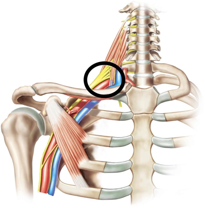
- Thoracic inlet, crossing the first rib behind the clavicle
- Mechanism
- Tie slips off the short stump after clavicular division in forequarter amputation
- Avoid by
- Obtain proximal control at the thoracic inlet first; double ligate plus transfix suture with generous stump length
- If injured
- Immediate digital pressure on the inlet, sternotomy or supraclavicular extension for proximal control
- Where
- Deep to the first and second ribs and the scalene insertions
- Mechanism
- Dissection carried deep to the ribs during scapulothoracic release, or thoracic duct injury on the left
- Avoid by
- Stay superficial to the ribs; identify the thoracic duct on the left; saline test before closure
- If injured
- Chest drain for pneumothorax; low-fat diet plus or minus duct ligation for chylous leak
- Where
- Between the scalenes, entering the axilla with the subclavian vessels
- Mechanism
- Traction division leaving long stumps in the wound bed, or nerves cut without ligating vasa nervorum
- Avoid by
- Ligate the plexus stumps, divide sharply under traction with local anaesthetic so ends retract into soft tissue; consider targeted muscle reinnervation at index surgery
- If injured
- Excise and reimplant into muscle; TMR or regenerative peripheral nerve interface for refractory neuroma pain
- Prevention
- Individual double ligation with suture-ligature; deliberate obturator dissection; early vascular control
- Management
- Pressure and packing, proximal control (pelvic packing/iliac control; thoracic inlet for subclavian), vascular surgery support
- Prevention
- Tension-free, well-vascularised flaps planned on pre-operative imaging; avoid skeletonising the inferior gluteal pedicle
- Management
- Debridement, negative-pressure dressing, secondary closure or flap (fillet spare-parts, latissimus if available)
- Prevention
- Meticulous haemostasis, obliteration of dead space by myodesis, suction drains
- Management
- Aspiration, compression; re-exploration if infected or expanding
- Prevention
- Pre-emptive regional analgesia, gabapentinoids, ketamine; sharp proximal nerve division under traction with local anaesthetic; TMR at index surgery (emerging)
- Management
- Multimodal: gabapentinoids, tricyclics, mirror therapy, graded motor imagery, desensitisation; TMR or nerve capping for refractory neuroma pain
- Prevention
- Traction neurectomy so nerve ends retract into muscle; targeted muscle reinnervation
- Management
- Excision and reimplantation into muscle, TMR or regenerative peripheral nerve interface
- Prevention
- Stay superficial to ribs; identify thoracic duct on left; saline test before closure
- Management
- Chest drain; low-fat diet plus or minus duct ligation for chyle leak
- Prevention
- Pre-operative psychology input, peer support, honest expectation-setting
- Management
- Ongoing psychological support, amputee groups, early prosthetic/rehabilitation engagement
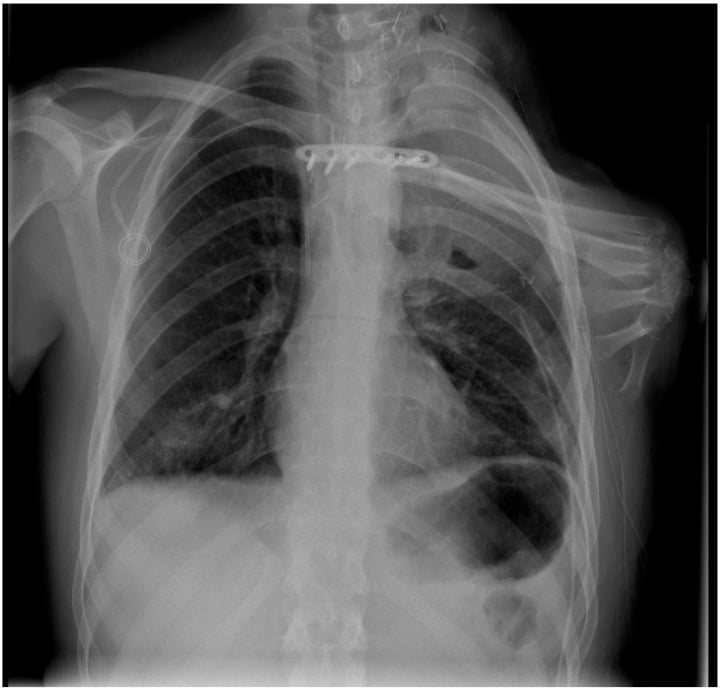
Phantom sensation (non-painful awareness of the limb — near universal, needs only reassurance), phantom pain (pain perceived in the absent limb — central, treated with gabapentinoids, mirror therapy, graded motor imagery), and residual limb/stump pain (local, peripheral — think neuroma, infection, bony prominence, poor socket fit). The management pathways differ, so name which one the patient has.
Prosthetic Reality and Rehabilitation
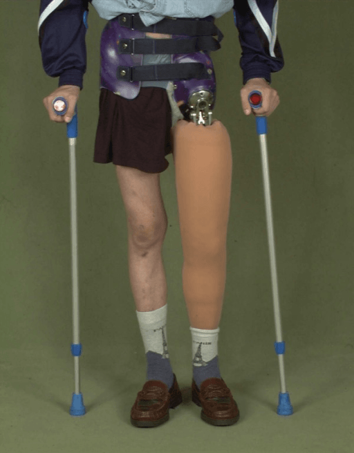
- The standard device is the Canadian-type hip disarticulation prosthesis (Colin McLaurin's design): a moulded socket embracing the hemipelvis and bearing weight on the ischium and gluteal pad, with an anteriorly placed free hip joint, a stance-stable knee and a dynamic foot; the hip joint anterior placement gives inherent alignment stability in stance
- Gait is a stiff, tilting, step-to or slow step-through pattern. The measured cost, from the only study of this level (Nowroozi 1983, 8 hip disarticulation and 10 hemipelvectomy amputees), is 80 to 125 percent more energy per metre walked than able-bodied controls — note that this is cost per distance, not per minute
- The trade patients make without being told to. Oxygen uptake per minute was no different from controls at comfortable speed; what fell was speed, to just 51 to 61 percent of normal. Pushed to walk fast, they reached only 70 to 75 percent of the controls' comfortable speed and paid 40 to 50 percent more oxygen to do it. So the patient does not feel more breathless — they simply go half as far for the same effort, and run out of reserve when hurried
- Honest counselling: many patients ultimately abandon the prosthesis in favour of elbow crutches or a wheelchair, because crutch gait is faster and less exhausting; younger, fitter patients and those with strong motivation do best
- Rehabilitation sequence: sitting balance and trunk strengthening from day one, oedema control and stump shaping, standing frame, casting for the socket once wounds are sound and volume stable, then gait training with the rehabilitation team
RAVE-CODHip disarticulation operative sequence
Hook:You RAVE about vascular control before the COD — Capsule, Open, Drape — completes the disarticulation.
SPelD-CForequarter danger structures
Hook:SPelD-C — spell out the danger list before you divide the clavicle.
Guidelines, Registries & Global Practice
- Global epidemiology: in high-resource sarcoma centres, limb salvage is achieved in well over 90 percent of extremity sarcomas, so proximal amputations are rare, concentrated operations best done in specialist units. In lower-resource settings, late presentation with fungating or massive tumours makes primary hip disarticulation and forequarter amputation proportionally far more common, and they remain essential curative operations where advanced reconstruction and radiotherapy are unavailable.
- Society guidance: sarcoma pathways from ESMO/EURACAN, NCCN and national sarcoma guidelines (including UK NICE sarcoma service guidance) converge on the same principles — management within a specialist sarcoma MDT, image-guided biopsy along a planned resectable tract, and amputation reserved for tumours where salvage cannot achieve adequate margins with a functional limb. BOA/BAPRAS ortho-plastic principles (as in open-fracture standards) apply to flap planning and combined soft-tissue decision-making in trauma indications.
- Registries: national amputee rehabilitation datasets (e.g. UK limb-fitting service data, Scandinavian amputation registers) consistently show hip disarticulation and hindquarter levels form a very small fraction of lower-limb amputations, with low sustained prosthetic use compared with transfemoral levels — evidence to quote when counselling.
- Practice variation: targeted muscle reinnervation and osseointegration expertise is concentrated in a few centres; mirror therapy and multimodal pharmacological phantom-pain management are deliverable in any resource setting and should be universal. Spare-parts (fillet flap) reconstruction is particularly valuable where free-flap microsurgery for separate donor sites is unavailable.
Controversies & Areas of Uncertainty
- Amputation versus heroic limb salvage at the extremes: with modern vascular grafting and nerve-tolerant strategies, even encased vessels can sometimes be reconstructed — but a salvaged limb that is insensate, painful and functionless serves the patient worse than a well-rehabilitated amputation. The decision remains individualised MDT judgement, not algorithm.
- Targeted muscle reinnervation at the index amputation: growing evidence supports primary TMR for reducing neuroma and phantom pain, but data at hip-disarticulation and forequarter levels are limited, target muscles are scarce, and it adds operative time in often-frail patients.
- Palliative "toilet" forequarter amputation for fungating incurable disease: strong quality-of-life rationale versus major surgery in patients with short prognosis; small series support it in selected patients, but patient selection criteria are poorly defined.
- Anterior versus posterior forequarter approach: no comparative trial exists; choice is driven by tumour location and surgeon training.
- Osseointegration has transformed transfemoral prosthetic use but has no role at disarticulation levels — whether pelvic-anchored solutions will ever be viable is speculative.
MCQ Practice Points
Q: Why must the subclavian artery and vein be ligated separately? A: En masse ligation risks arteriovenous fistula formation. The artery is doubly ligated and suture-ligated first, then the vein separately.
Q: Which flap covers a standard hip disarticulation and why does it survive femoral ligation? A: A posterior gluteus maximus myofasciocutaneous flap myodesed to the pubis/adductor origin; it is supplied by the superior and inferior gluteal arteries from the internal iliac system, independent of the ligated femoral system.
Q: What is the corona mortis and why does it matter here? A: A retropubic anastomosis between the obturator and external iliac/inferior epigastric systems, present in roughly a fifth to a third of patients; avulsion during obturator vessel dissection causes haemorrhage from a vessel that retracts into the pelvis and is difficult to control.
Q: Which forequarter approach gives early vascular control? A: The anterior Berger approach — clavicular resection first unroofs and allows early ligation of the subclavian vessels; the posterior Littlewood approach mobilises the scapula first and reaches the vessels late, but gives better exposure for posteriorly based tumours.
Q: Name three perioperative strategies to reduce phantom limb pain. A: Pre-emptive regional analgesia (epidural or perineural catheter), perioperative gabapentinoids plus or minus ketamine, and sharp traction neurectomy with local anaesthetic infiltration so nerve ends retract into muscle; targeted muscle reinnervation at the index operation is an emerging fourth option.
Q: Define the forequarter specimen. A: The entire upper limb en bloc with the scapula and the lateral portion of the clavicle (interscapulothoracic amputation), with preservation of the chest wall unless en bloc rib resection is oncologically required.
Exam Viva Scenarios
Practise clinical reasoning and management decisions out loud
“A 54-year-old presents with local recurrence of a high-grade soft-tissue sarcoma of the proximal thigh two years after wide excision and radiotherapy. MRI shows the recurrence encasing the femoral vessels and abutting the sciatic nerve, with skin tethering in the irradiated field. Staging CT chest is clear. How do you proceed?”
“During the medial dissection of a hip disarticulation for necrotising fasciitis, brisk bleeding erupts from behind the superior pubic ramus and the vessel retracts out of sight. The anaesthetist reports falling blood pressure. What is happening and what do you do?”
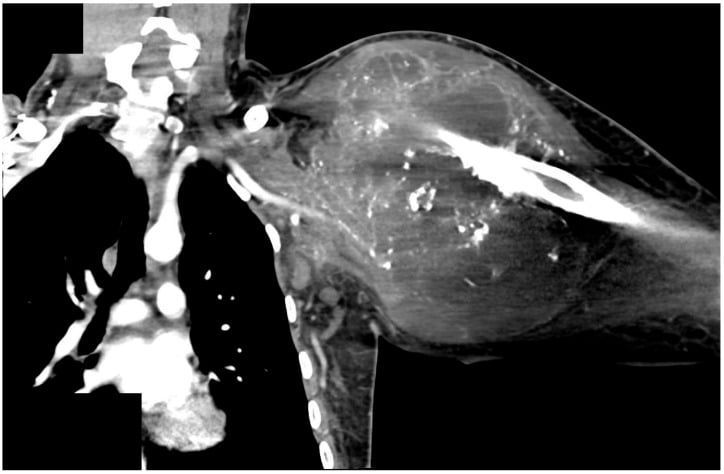
“A 47-year-old presents with a massive, fungating, malodorous recurrent sarcoma of the shoulder girdle encasing the brachial plexus and axillary vessels, with a flail painful arm. Staging shows two small lung nodules. She asks whether anything can be done.”
“A 28-year-old awaiting hip disarticulation for pelvic-girdle-sparing proximal femoral osteosarcoma asks: 'Will I walk again, and what will the leg be like?' How do you counsel him?”
Indications
- Tumour: neurovascular encasement precluding a functional limb, fungation, recurrence after failed salvage, massive multicompartment disease
- Infection: necrotising fasciitis failing debridement, chronic osteomyelitis in a nonfunctional limb
- Trauma/ischaemia: unreconstructable proximal injury or non-viable limb above transfemoral level
- Metastases do not preclude palliative toilet amputation for quality of life
Hip disarticulation key steps
- Racquet incision from ASIS; femoral artery and vein ligated individually in the femoral triangle first
- Deliberate obturator vessel ligation — beware corona mortis retraction
- Iliopsoas off lesser trochanter, abductors/rotators off greater trochanter, adductors/hamstrings off pelvis
- Capsulotomy, divide ligamentum teres, deliver head
- Gluteus maximus posterior flap myodesis to pubis/adductor origin — protect the inferior gluteal pedicle
Forequarter key steps
- Specimen: whole upper limb plus scapula plus lateral clavicle; preserve chest wall
- Anterior Berger: clavicle resected first, early subclavian control — artery then vein, separately
- Posterior Littlewood: scapular release first, best for posterior tumours
- Sharp brachial plexus division with local anaesthetic; protect phrenic nerve, pleura, thoracic duct (left)
- Saline test for pleural breach; mandatory post-op chest radiograph; consider fillet spare-parts flap for coverage
Oncologic principles
- MDT decision with current staging; excise biopsy tract and previous scars en bloc
- Never breach the tumour; sacrifice planned flaps rather than margins
- Orientate and ink the specimen with the pathologist
Complications & rehabilitation
- Haemorrhage (corona mortis, slipped ties), wound necrosis, seroma, pneumothorax/chyle leak (forequarter)
- Phantom pain in the majority — pre-emptive regional analgesia, gabapentinoids, mirror therapy, TMR emerging
- Canadian hip disarticulation prosthesis: ischial/gluteal bearing, very high energy cost, many abandon for crutches
- Forequarter prosthesis is essentially a cosmetic shoulder cap; rehabilitation targets one-handed independence and psychological adjustment
Evidence Base
The Treatment of Soft-Tissue Sarcomas of the Extremities - Prospective Randomized Evaluations of Limb-Sparing Surgery Plus Radiation Therapy Compared with Amputation, and the Role of Adjuvant Chemotherapy
- 43 adults with HIGH-GRADE extremity soft-tissue sarcoma randomised 2:1 - 27 to limb-sparing resection plus radiotherapy, 16 to amputation at or above the joint proximal to the tumour; both arms received postoperative doxorubicin, cyclophosphamide and high-dose methotrexate
- Four local recurrences in the limb-sparing arm and none after amputation (p = 0.06)
- No difference in five-year disease-free survival (71 versus 78 per cent, p = 0.75) or overall survival (83 versus 88 per cent, p = 0.99)
- On multivariate analysis the ONLY correlate of local recurrence was the final margin - positive margins recurred more often (p less than 0.0001) even with postoperative radiotherapy
- A parallel randomisation of 65 patients found adjuvant chemotherapy improved three-year continuous disease-free survival (92 versus 60 per cent) and overall survival (95 versus 74 per cent)