Massive structural allograft plus stemmed prosthesis: bone stock restoration and biological soft-tissue reattachment after segmental skeletal loss
- The defining advantage of APC over megaprosthesis is biological soft-tissue reattachment (abductors, extensor mechanism, rotator cuff) and restoration of bone stock for future revision.
- Fresh-frozen, size-matched allograft from an accredited bone bank is preferred for structural roles; high-dose irradiated grafts are mechanically weakened and should be avoided for load-bearing segments.
- The host-allograft junction is the biological Achilles heel: nonunion in roughly 10-30 percent; fixation is by compression plating across the junction, a long cemented stem bridging it, or both.
- Step-cut osteotomy increases junction contact area and rotational stability compared with a transverse cut but is technically harder to match.
- Tendon-to-tendon repair to a retained allograft tendon stump (for example host abductor tendon to allograft abductor tendon) is stronger and heals more reliably than tendon-to-bone fixation.
- Cement the stem within the allograft (its canal cannot osseointegrate and cement protects against fracture); fix in the host by press-fit or cement depending on host bone quality — never rely on bone ingrowth into allograft.
- Infection is the most devastating complication (10-15 percent in tumour series) and usually mandates removal of the entire composite.
- “If asked why choose APC in a young patient: bone stock for the revisions they will inevitably need over a lifetime, plus functional soft-tissue attachment.
- “Allograft fracture is a late complication, typically after 2 to 3 years as creeping substitution weakens unremodelled cortex — a cemented long stem spanning the graft is protective.
- “Proximal humerus and proximal tibia have the strongest functional case for APC over megaprosthesis; the proximal femur is genuinely debated.
Concept and Rationale
An allograft-prosthetic composite combines a massive structural allograft (proximal femur, proximal tibia, proximal humerus or an intercalary segment) with a conventional or long-stemmed prosthesis. The prosthesis provides the articulation; the allograft provides the bone and the soft-tissue anchor points that a megaprosthesis cannot.
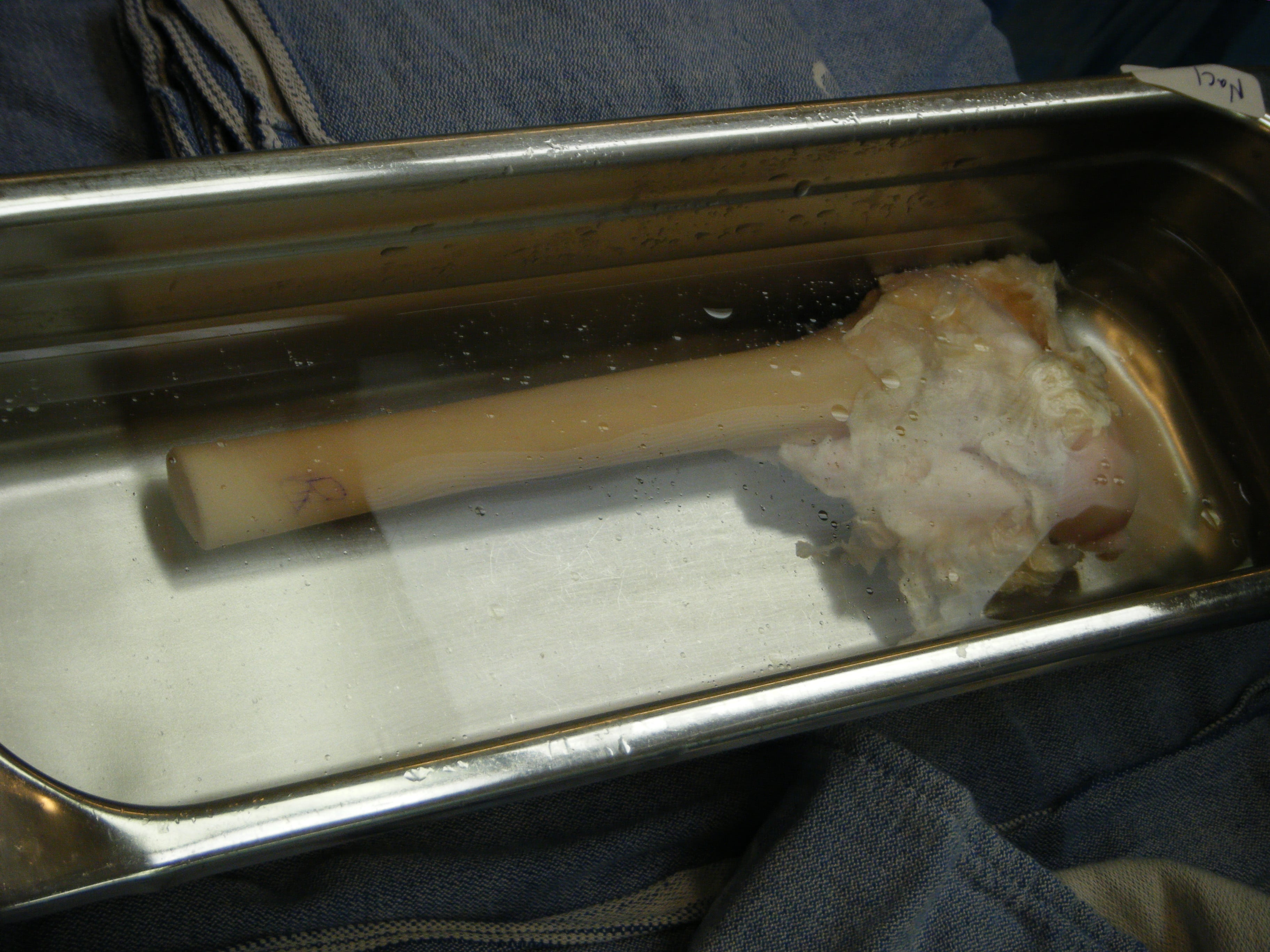
Bone stock. A failed APC usually leaves more bone than a failed megaprosthesis. That is critical in young patients, who will require future revision.
Soft-tissue attachment. This is the decisive functional advantage over a megaprosthesis, and it is site-specific:
- Proximal femur: host abductor tendon repaired to the allograft's retained abductor tendon and greater trochanter, for better gait, less Trendelenburg lurch and potentially lower dislocation risk
- Proximal tibia: host patellar tendon sutured to the allograft's retained patellar tendon, a biological extensor mechanism reconstruction that is superior to tendon-to-metal fixation
- Proximal humerus: host rotator cuff, capsule and remaining tuberosity tissue repaired to allograft cuff stumps, restoring stability and some active motion
Two philosophies in one construct. The junction and the soft tissues heal biologically; the articulation is a mechanically reliable prosthesis. That avoids the cartilage degeneration and joint instability of osteoarticular allografts, which APCs have largely superseded at the knee and shoulder.
The trade-offs. Operating time is longer, the operation depends on bone bank availability and size matching, and the junction must unite before full loading, so rehabilitation is delayed compared with a megaprosthesis. The allograft also brings its own late complications: fracture, resorption and infection of dead bone.
Indications and Patient Selection
APC buys bone stock and biological soft-tissue attachment at the cost of union time and infection risk; a megaprosthesis buys immediate function. The decision matches that trade-off to life expectancy, site and adjuvant therapy.
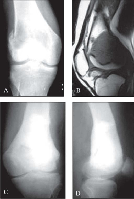
The defect. Segmental bone loss after primary sarcoma resection (osteosarcoma, chondrosarcoma, Ewing), failed arthroplasty with a Paprosky IIIB/IV femoral defect, an unreconstructable periprosthetic fracture, a failed osteoarticular allograft or a failed megaprosthesis. Confirm first that the joint cannot be reconstructed by conventional revision means and plan the resection level; only then is the patient a candidate for an APC or a megaprosthesis.
Who gets an APC. The younger and more active the patient, the stronger the case, because bone stock is preserved for the lifetime of revisions ahead. The proximal humerus and proximal tibia depend on soft-tissue reattachment (rotator cuff, extensor mechanism, abductors) for function, so favour APC where tendon-to-tendon reattachment materially changes function; the proximal femur is genuinely debated, because modern megaprostheses with dual mobility narrow the gap. A failed osteoarticular allograft with a united shell that can be retained and converted is a further indication.
Who gets a megaprosthesis instead. Each of these tips the decision away from allograft:
- Active or recent deep infection at the site. Dead allograft placed in an infected bed fails. Stage the reconstruction, eradicate sepsis and reassess; if sepsis cannot be cleared, do not use an APC
- Metastatic disease or limited life expectancy. The bone-stock advantage is never realised and no soft-tissue reconstruction is achievable in the timeframe available
- Immediate full weight-bearing required. A megaprosthesis needs no wait for union
- Age over roughly 65 to 70 years, or low functional demand
- Planned or ongoing high-dose local radiotherapy across the junction. It sharply reduces union rates. Heavy chemotherapy, by contrast, delays union but does not preclude APC; it lengthens the protected-loading period
- Inability to comply with protected loading while the host-graft junction unites, typically 6 to 12 months
- Very poor host bone or soft tissues, heavy smoking, poorly controlled diabetes, immunosuppression. All raise nonunion and infection risk; optimise what is modifiable
- No suitably size-matched allograft. Default to megaprosthesis if no matched graft exists
Allograft Selection, Banking and Preparation
Source. An accredited musculoskeletal tissue bank, with the cadaveric donor screened by serology (HIV, hepatitis B and C, HTLV, syphilis) and culture per national tissue regulations: Human Tissue Authority licensing in the UK, FDA/AATB standards in the USA, EU tissue directives elsewhere.
Preservation. Fresh-frozen at minus 80 degrees Celsius is the standard for structural grafts: freezing reduces immunogenicity while largely preserving mechanical strength. Freeze-dried grafts are brittle when rehydrated and unsuitable for major structural roles.
Irradiation. Gamma irradiation above roughly 25 kGy significantly weakens cortical bone, with a dose-dependent loss of bending and torsional strength and fatigue life. Avoid high-dose irradiated grafts for structural APC segments; low-dose or non-irradiated aseptically processed grafts are preferred.
Size matching. Match donor and host on calibrated radiographs or CT: canal diameter (to accept the stem plus a 2 mm cement mantle), cortical diameter at the planned junction, and segment length. A slightly larger allograft cortex can be telescoped or step-cut over host bone; a smaller graft is harder to salvage.
Soft-tissue retention. Request the graft with retained tendon stumps: abductor tendon and trochanter for the femur, patellar tendon for the tibia, rotator cuff insertions for the humerus. These are the anchor points for tendon-to-tendon repair.
Theatre preparation. In order:
- Thaw in warm antibiotic saline
- Take culture swabs before implantation
- Ream and lavage the allograft canal to remove marrow elements, which reduces the immunogenic load and prepares the canal for cement
- Fashion the osteotomy on the back table against trial components
Host-Allograft Junction: Osteotomy and Fixation
Preparing the junction. The size-matched allograft is trimmed to restore length and version, and the cut surfaces are cleaned of marrow and debris. The junction is packed circumferentially with autogenous iliac crest or reamer aspirate graft. Compression, autograft and rotational control across the junction are what deliver union.
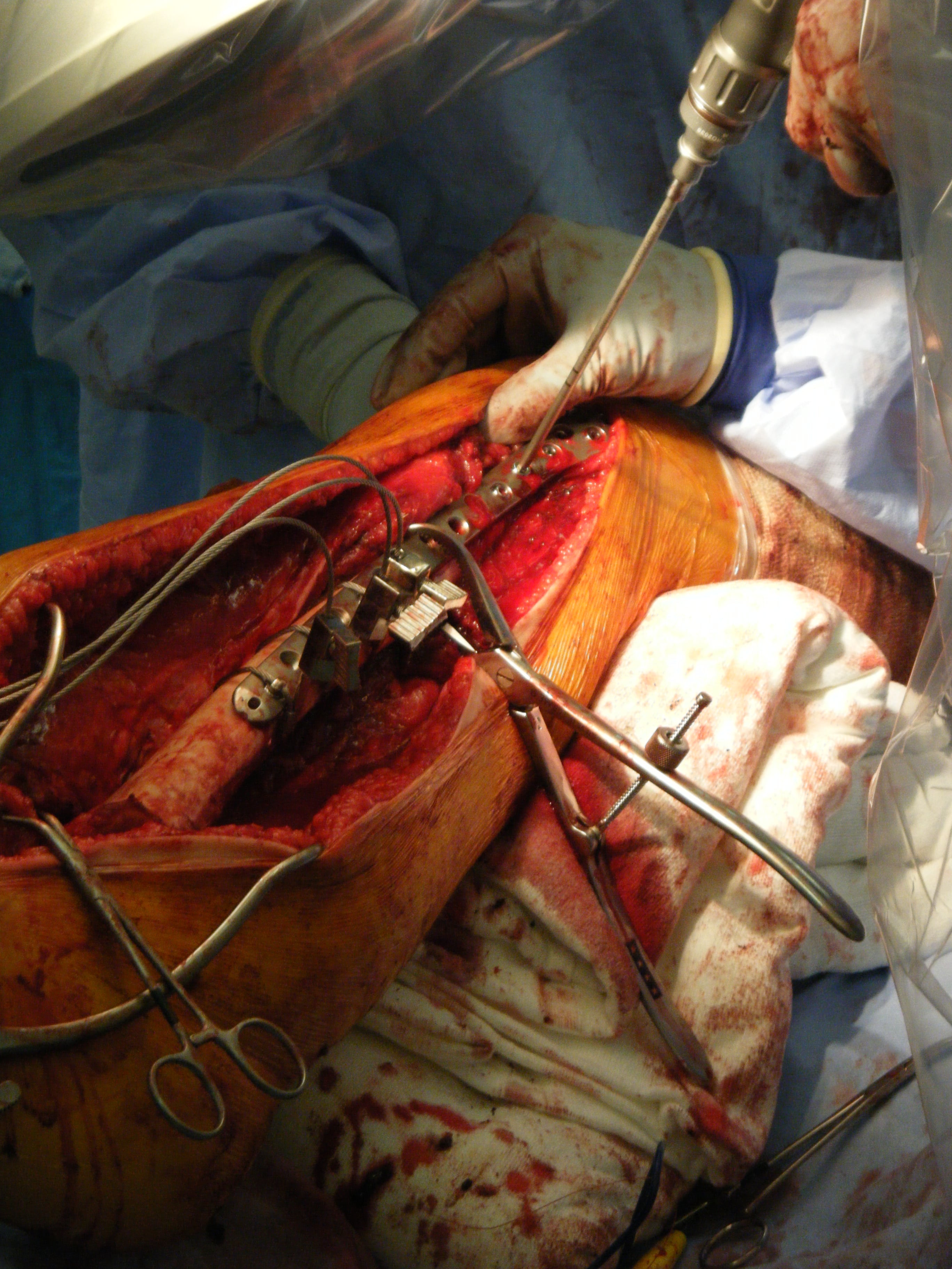
The cut. Transverse or step-cut. The trade is contact area and rotational control against ease of matching and of applying compression:
- Transverse osteotomy
- Smaller, single plane
- Step-cut osteotomy
- Larger, interdigitating surfaces
- Transverse osteotomy
- Poor without supplementary fixation
- Step-cut osteotomy
- Intrinsically better
- Transverse osteotomy
- Simple, easy length adjustment
- Step-cut osteotomy
- Demanding to match cuts precisely; gaps if imperfect
- Transverse osteotomy
- Excellent with a compression plate perpendicular to the cut
- Step-cut osteotomy
- Compression harder to apply uniformly
- Transverse osteotomy
- Most common; combined with plate and/or long stem
- Step-cut osteotomy
- Where rotational control matters and cuts can be templated
The plate. A long lateral plate with bicortical screws in both host and allograft, applied perpendicular to a transverse cut, delivers excellent interfragmentary compression and neutralises the torsion a stem controls poorly.
The bridging stem. A long cemented stem crosses from the prosthesis through the entire allograft into host bone, so the junction is splinted and the graft protected against fracture. Cement is confined to the allograft canal, and to the host if that is the chosen host fixation.
The allograft canal is dead bone: it cannot osseointegrate, so ingrowth surfaces are wasted inside it. Cementing the stem within the allograft gives immediate rigid fixation, fills the canal (reducing hoop-stress fracture risk) and allows the stem to act as an internal splint. Fixation in the host segment follows normal arthroplasty principles, press-fit if bone quality permits and cemented if not, and some surgeons deliberately leave the host portion uncemented press-fit to ease future revision.
Both. A long bridging stem plus a junction plate is biomechanically the most stable construct, and increasingly the default for proximal femoral and tibial allograft-prosthetic composites in the lower limb.
Cement interposed between host and allograft cut surfaces is a classic cause of nonunion. Pressurise cement only within the canal and remove any extrusion at the osteotomy.
Operative Technique by Site
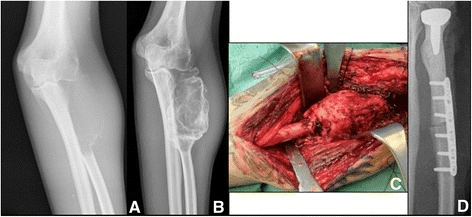
Set-up. Lateral decubitus on a radiolucent table with the whole limb and iliac crest prepped, and an image intensifier available. Have the templated, size-matched fresh-frozen proximal femoral allograft with its abductor tendon, a long-stem revision hip system with cemented and uncemented options, cables and a long lateral plate. Cell salvage is used where oncologically appropriate, which excludes tumour cases.
Graft preparation. Thaw, culture, ream and back-table trial of the stem and cement mantle as above; the osteotomy, transverse or step-cut, is fashioned against the measured resection.
Approach and dissection. Extended posterolateral or lateral. In tumour cases the biopsy tract is excised en bloc and the approach is dictated by oncological margins; the resection is extra-osseous with a cuff of normal tissue, preserving as long a host abductor tendon stump as the margins allow. In revision cases the failed implant is extracted, the bed debrided, and frozen sections and cultures taken if infection is suspected.
At risk. The sciatic nerve (posterior retraction, and limb lengthening beyond roughly 4 cm), the femoral vessels anteromedially during tumour dissection, and the superior gluteal neurovascular bundle.
Reconstruction. Cement the long stem into the allograft on the back table or in situ, reduce allograft to host with the chosen osteotomy, then compress and fix the junction (plate and/or press-fit or cemented stem in the host) and autograft it. Trial and implant the acetabular side; a dual-mobility or larger head is strongly considered.
Soft tissue. The host abductor tendon is repaired tendon-to-tendon to the allograft abductor stump with heavy non-absorbable sutures (Krackow), supplemented by trochanteric cables if osseous, followed by capsular repair.
Aftercare. Layered closure over a drain, abduction precautions, and toe-touch to partial weight-bearing for 6 to 12 weeks, progressing as junction union appears radiographically. Abductor strengthening is deferred 6 weeks.
Pitfalls. Leg-length and version errors (mark rotation before the osteotomy), cement at the junction, an undersized allograft cortex, and dislocation from deficient abductor tension.
Complications: Rates, Prevention and Management
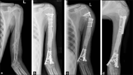
Junction nonunion. Seen at the osteotomy interface at any site in 10 to 30 percent, declared at 9 to 12 months. The mechanisms are inadequate compression, a gap at the cut, cement interposed at the junction, and irradiated or devitalised graft ends; the risk factors are a transverse osteotomy without compression, poor host bone contact, chemotherapy, an irradiated graft and inadequate fixation. Prevention is the junction construct described above, with a non-irradiated graft. An established nonunion is treated by autogenous bone grafting plus revision compression plating: take down the fibrous tissue, remove any interposed cement, refresh the cut surfaces and upgrade to a longer or dual-plate compression construct with generous autograft. Union is usually achievable.
Deep infection. 10 to 15 percent in tumour series, early or late, and highest at the proximal tibia. The drivers are a large avascular graft mass, a long operating time and a thin soft-tissue envelope, and the allograft is dead bone that antibiotics cannot sterilise. Prevention is gastrocnemius flap coverage at the tibia, antibiotic-loaded cement, meticulous soft-tissue handling and a short operating time. Debridement rarely salvages the graft: established deep infection almost always requires removal of allograft and prosthesis and staged reconstruction, described under revision below.
Allograft fracture. Roughly 5 to 20 percent, typically beyond 2 years, mid-graft or at a stress riser. The mechanisms are a graft that never fully revascularises, screw and drill holes acting as stress risers, and an unsupported graft segment. A long cemented stem spanning the whole graft is protective; minimise unfilled drill holes and unicortical hardware clusters in the graft, and select a graft of adequate cortical thickness and diameter match. With the stem intact and the fracture minimally displaced, plate and graft; segmental failure means revision to a new APC (young patient, allograft available) or conversion to megaprosthesis.
Allograft resorption. Late and progressive, in a minority. Micromotion and soft-tissue stripping devitalise the graft surface, so prevention is rigid fixation and avoiding circumferential stripping of the graft periosteal envelope. Observe if mechanically stable; graft or revise once cortical integrity is threatened.
Instability and dislocation. Proximal femoral APCs dislocate at a meaningful rate despite abductor repair, through loss of abductor tension and the capsular envelope after resection, or component malversion. Tendon-to-tendon abductor repair to the graft trochanter, capsular repair, dual mobility or large heads, and correct combined version are the defences. Closed reduction and address the cause; recurrent instability is revised to a constrained or dual-mobility bearing.
Extensor lag and cuff failure. The site-specific functional failures of the proximal tibia and proximal humerus: a patellar tendon or cuff attached to the graft heals by tenodesis and stretches out. Prevention is the tibial repair under tension in full extension with 6 weeks of protection, and a reverse design at the humerus. Salvage is revision tendon reconstruction, an allograft extensor mechanism, or acceptance of the deficit.
APC versus Megaprosthesis by Site
Proximal humerus. The deltoid and axillary nerve must be intact. A hemiarthroplasty megaprosthesis behaves as a spacer with poor active elevation, and osteoarticular allograft alone has been largely abandoned here. In Abdeen's series the failure modes were prosthetic loosening (3 of 36) and delayed union at the osteosynthesis site (4 of 36); dislocation was rare, one in 36; construct survival was 88% at 10 years, and all patients had mild or no pain and normal hand function. The case for APC is strong, but counsel on function by what must be resected: deltoid loss predicts reduced motion and extra-articular resection predicts a lower MSTS score.
Proximal tibia. Extensor lag, infection and wound breakdown bite before junctional nonunion at this site. Tendon-to-metal fixation with mesh or tube augments gives inferior extensor function, so the functional case for APC is strong where the soft tissues allow; the thin envelope means flap coverage is required either way.
Proximal femur. The construct is a long-stem hip prosthesis cemented into the allograft, with a dual-mobility or constrained head where the abductors are deficient, and the failure modes are dislocation, junctional nonunion (10% in Farid's APC arm, all of which united after grafting) and trochanteric escape if the abductor repair fails. The case against megaprosthesis is not survivorship: Kaplan-Meier implant survival was identical at 86% at 10 years for both. The difference is abductor power, 4.6/5 with APC against 2.8/5 with endoprosthesis, and MSTS 82% against 70%. Decide on abductor function and life expectancy, not on survival, and note the caveat in the source: APC follow-up was a median 76 months against 146 for endoprostheses, so the 10-year APC figure is the less mature of the two.
Revision of the Failed APC
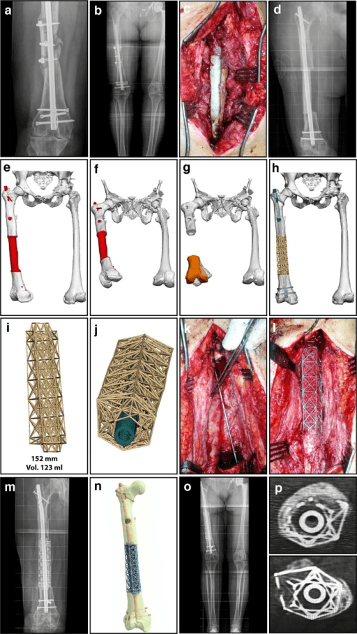
Aseptic loosening or stem failure with a united junction. The united allograft has become part of the host skeleton, so the prosthetic side alone is revised, with a longer stem through the retained allograft shell. This scenario is the payoff of the APC philosophy: bone stock has been banked.
Junction nonunion with an intact implant. Compression plating and autografting as described under complications; exchange to a longer bridging stem if the existing stem is short or loose.
Infection. Staged: remove prosthesis and allograft (dead bone cannot be sterilised), antibiotic spacer and targeted systemic antibiotics, then reconstruct with a megaprosthesis, a new APC in selected cases after confirmed eradication, or arthrodesis. Amputation is the salvage for uncontrolled sepsis or recalcitrant infection.
Failed osteoarticular allograft converting to APC. If the original allograft-host junction has united, resurface through the allograft with a stemmed prosthesis, a well-described salvage that preserves the united segment.
Union Biology and Postoperative Course
Massive allografts unite to host at the junction like a fracture, but the graft itself revascularises only superficially and slowly, with creeping substitution limited to the outer millimetres over years. The bulk remains dead scaffold indefinitely, hence the late fracture risk and the rationale for a protective intramedullary stem.
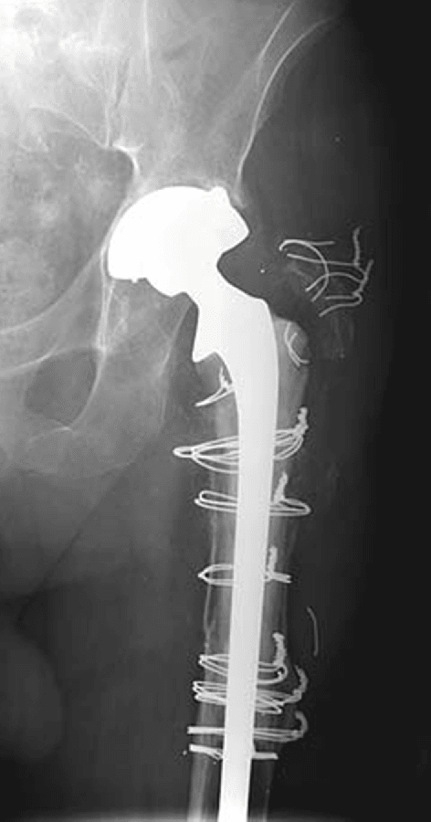
Toe-touch or partial weight-bearing (lower limb); extensor, abductor or cuff repair protected. Wound surveillance, because early infection declares here.
Radiographs for callus and maintained fixation; graded loading if the junction shows bridging; begin protected strengthening of the repaired muscle groups.
Most junctions unite between 6 and 12 months; chemotherapy prolongs this. CT if union is uncertain at 9 to 12 months. Full weight-bearing once bridging is seen on at least three cortices.
Annual radiographs for allograft resorption, fracture and hardware failure and, in tumour patients, local recurrence within oncological follow-up.
Guidelines, Registries & Global Practice
- Global epidemiology and practice variation: APC use tracks the availability of accredited musculoskeletal tissue banks. Well-resourced centres in North America, Europe, Australasia, Japan, Brazil and Argentina (which pioneered large bone-banking programmes) use APCs routinely; in regions without bone banks, megaprostheses, extracorporeally irradiated or pasteurised/liquid-nitrogen-treated autografts (recycled autograft techniques popular in parts of Asia) substitute for allograft.
- Tissue regulation: allograft procurement and processing are governed nationally — Human Tissue Authority licensing in the UK, FDA regulations and American Association of Tissue Banks standards in the USA, EU Tissues and Cells Directives across Europe, Therapeutic Goods Administration frameworks in Australia. Surgeons must know their bank is accredited and grafts serologically screened.
- Society guidance: no society issues an APC-specific guideline; principles are embedded in musculoskeletal oncology consensus practice (ISOLS — International Society of Limb Salvage — meetings and classification of reconstruction failure by Henderson and colleagues, which categorises failures into soft tissue, aseptic loosening, structural, infection and tumour progression and is the standard reporting framework for APC series). AO principles govern junction fixation. BOA/BOOS guidance in the UK directs sarcoma reconstruction to specialist centres, where bone banking is concentrated.
- Registry evidence: national arthroplasty registries (NJR, AJRR, AOANJRR, SHAR, Norwegian, NZJR) capture megaprosthetic and some composite revision constructs only coarsely; APC evidence therefore rests on institutional series and ISOLS multicentre reports rather than registry data — a recognised evidence gap.
- Resource-setting adaptation: where allograft is unavailable, alternatives include megaprosthesis, recycled tumour-bearing autograft (irradiated, pasteurised or frozen) combined with a prosthesis — an "autograft-prosthetic composite" applying identical junction and soft-tissue principles.
Related pages: Limb Salvage Surgery Principles sets the margins and the resection this reconstruction has to fill, and Osteosarcoma, Ewing Sarcoma, Chondrosarcoma and Giant Cell Tumour of Bone are the diagnoses that generate the defect - chemotherapy timing in the first two is what makes wound healing and union so much harder here than in a non-oncological reconstruction. Megaprosthesis in Non-Oncologic Salvage is the alternative construct at every site discussed above, and Metastatic Bone Disease is the setting in which it wins outright, because a construct that depends on union is the wrong operation for a patient who may not live to unite it. Periprosthetic Joint Infection behaves differently here - the allograft is dead bone and becomes a sequestrum, so eradication means removing the construct rather than exchanging a liner. Masquelet Induced Membrane Technique and Femoral Impaction Bone Grafting are the biological alternatives for segmental and cavitary loss respectively, Revision THA and Revision TKA Bone Loss: Cones and Sleeves cover the non-tumour route to the same problem, and TKA Extensor Mechanism Failure with Extensor Mechanism Ruptures explain why tendon-to-tendon repair at the proximal tibia is worth the wait for union. Hip Disarticulation and Forequarter Amputation is the salvage when the composite fails irretrievably.
Controversies & Areas of Uncertainty
- Proximal femur: APC or megaprosthesis? Comparable survivorship in most comparative series; dual-mobility megaprostheses have eroded the dislocation argument for APC, while APC retains the bone-stock argument in the young. No randomised evidence exists or is likely.
- Stem fixation in the host: fully cemented (immediate stability, easier in poor bone) versus press-fit host fixation (easier future revision, biological fixation) — practice varies by centre with no comparative superiority shown.
- Plate, stem, or both at the junction: combined constructs are biomechanically stiffest but add soft-tissue stripping and hardware; some series report equivalent union with a well-fitted compressed junction and long stem alone.
- Step-cut versus transverse osteotomy: theoretical advantages of step-cut are not consistently reflected in union rates; precision of contact and compression may matter more than geometry.
- Reverse shoulder APC: increasingly favoured over anatomical/hemiarthroplasty APC in the proximal humerus, but long-term data on glenoid survival in young tumour patients are limited.
- Role of APC in the era of custom 3D-printed implants: additive-manufactured endoprostheses with porous collars and soft-tissue attachment augments may replicate some APC advantages without allograft-specific risks; comparative data are immature.
MCQ Practice Points
Q: What is the principal functional advantage of an APC over a megaprosthesis at the proximal tibia? A: Biological reattachment of the host patellar tendon to the retained allograft patellar tendon (tendon-to-tendon), giving superior extensor mechanism function compared with tendon-to-metal fixation — plus restoration of bone stock. The equivalent advantage at the proximal femur is abductor reattachment to the allograft trochanter, worth nearly two grades of abductor power.
Q: What is the reported host-allograft junction nonunion rate, and what is first-line treatment? A: Approximately 10-30 percent; treated with autogenous bone grafting and revision compression plating, with attention to removing interposed cement or fibrous tissue. In the largest proximal femoral comparative series every junction eventually united after grafting.
Q: Why are high-dose irradiated allografts avoided in APC? A: Gamma irradiation at sterilising doses (around and above 25 kGy) causes dose-dependent loss of bending strength, toughness and fatigue life of cortical bone, predisposing the structural graft to fracture. Lyophilised, irradiated cortical allograft recovered only 12% of normal torsional strength experimentally, against 64% for deep-frozen.
Q: How should the stem be fixed within the allograft and why? A: Cemented — the allograft is avascular and cannot osseointegrate; cement provides immediate rigid fixation, fills the canal reducing fracture risk, and the stem splints the graft. Host-side fixation is press-fit or cemented according to host bone quality.
Q: When do massive allograft fractures typically occur and why? A: Late — commonly beyond 2 to 3 years — because revascularisation is superficial and creeping substitution weakens unremodelled cortex. Deep-frozen cortical allograft recovers only about two-thirds of normal torsional strength even experimentally, so a long cemented stem spanning the graft is protective.
Q: Why does deep infection of an APC usually require removal of the allograft? A: The allograft is dead, avascular bone that antibiotics cannot penetrate or sterilise; it behaves as a large sequestrum, so eradication requires staged removal of the composite before reconstruction with megaprosthesis, new APC, arthrodesis, or amputation as salvage.
Exam Viva Scenarios
Practise clinical reasoning and management decisions out loud
“A 19-year-old with a proximal tibial osteosarcoma has completed neoadjuvant chemotherapy. Planned resection removes 14 cm of proximal tibia including the patellar tendon insertion. How will you reconstruct, and why?”
“A 34-year-old, 11 months after proximal femoral APC for chondrosarcoma, has persistent thigh pain on loading. Radiographs show a lucent transverse junction with no bridging callus; the cemented long stem crossing the junction appears well fixed. Inflammatory markers are normal. Manage this.”
“A 52-year-old has a loose cemented femoral stem with Paprosky IIIB/IV bone loss and a non-functional greater trochanter after two prior revisions. Infection workup is negative. Discuss your reconstructive options and how you would decide.”
“Your bone bank offers two proximal femoral allografts for a planned APC: one fresh-frozen and non-irradiated, one freeze-dried and gamma-irradiated at 25 kGy. Which do you choose and how do you prepare it?”
Concept and indications
- APC = massive structural allograft plus stemmed prosthesis: restores bone stock AND soft-tissue attachment
- Best case: young patient, tumour resection or failed arthroplasty with massive segmental loss
- Site-specific attachments: abductors (femur), patellar tendon (tibia), rotator cuff/tuberosities (humerus)
- Avoid in: active infection, limited life expectancy/metastases, irradiated bed, no size-matched graft
Graft and technique
- Fresh-frozen, size-matched, non-irradiated, accredited bank, retained tendon stumps
- Cement stem within allograft; press-fit or cement in host per bone quality
- Junction: step-cut or compressed transverse; plate and/or long bridging stem; autograft; NO cement at osteotomy
- Tendon-to-tendon repair to allograft stumps is strongest; gastrocnemius flap for proximal tibia
Complications and rates
- Junction nonunion 10-30 percent — graft and re-compress
- Deep infection 10-15 percent — usually remove entire composite, staged reconstruction
- Allograft fracture — late (beyond 2 years); protected by long cemented stem
- Instability (proximal femoral APC) — dual mobility/large heads, abductor and capsular repair
Evidence and decisions
- Mankin's MGH series: most allograft failures within 3 years; survivors durable
- Proximal humerus and proximal tibia: strongest case for APC over megaprosthesis
- Proximal femur: genuine equipoise — APC for young (bone stock), megaprosthesis for older/metastatic
- United junction pays off at revision: prosthetic side revised through retained allograft bone stock
- Henderson/ISOLS failure classification: soft tissue, aseptic loosening, structural, infection, tumour progression
Evidence Base
Long-term outcomes of massive allograft reconstruction
- Over 870 massive frozen cadaveric allografts implanted over 24 years, most from the authors' own institutional bone bank
- Only TWO variables predicted outcome: tumour STAGE (Enneking 2 or 3 did worse than 0 or 1) and the TYPE of graft - not age, site or fixation
- Infection 10% (concentrated in the first year) and fracture 19% (risk elevated through the third year)
- After year three the grafts stabilise: roughly 75% are retained and considered successful beyond 20 years
- Allograft ARTHRODESES did considerably worse than osteoarticular, intercalary and allograft-plus-prosthesis reconstructions
- Osteoarthritis appears at about 6 years in osteoarticular grafts; 16% of femoral and tibial graft patients later needed joint replacement
Proximal femoral APC versus megaprosthesis
- 52 endoprostheses versus 20 APCs for proximal femoral tumour resection, 1974-2002
- Kaplan-Meier implant survivorship IDENTICAL at 86% for both groups at 10 years
- Hip abductor strength markedly better with APC: 4.6 of 5 versus 2.8 of 5 for endoprosthesis
- MSTS score 82% (APC) versus 70% (endoprosthesis); nonunion was the commonest APC complication (10%), aseptic loosening the commonest endoprosthetic one (10%)
- All host-allograft junctions eventually healed after bone grafting
Proximal humeral reconstruction options
- 36 consecutive proximal humeral APCs at one institution over 16 years; mean follow-up 5 years in survivors
- Construct survival 88% at 10 years with revision as the endpoint; 3 failures from progressive prosthetic loosening
- Only ONE glenohumeral dislocation in the whole series; all patients had mild or no pain and normal hand function
- The measured determinants of function were DELTOID preservation (range of motion) and INTRA-articular resection (MSTS score) - not cuff repair
- Four patients needed additional bone grafting for delayed union at the osteosynthesis site