When the Humeral Diaphysis Fails to Unite
- Humeral shaft nonunion is failure of the diaphysis to unite (no progressive healing, typically by around 6 months); the humeral shaft is relatively prone to nonunion with certain risk factors - TRANSVERSE fracture pattern, fracture DISTRACTION/gapping, proximal-third location, inadequate immobilisation, open fracture, infection, and patient factors (smoking, NSAIDs, obesity, diabetes, poor bone quality).
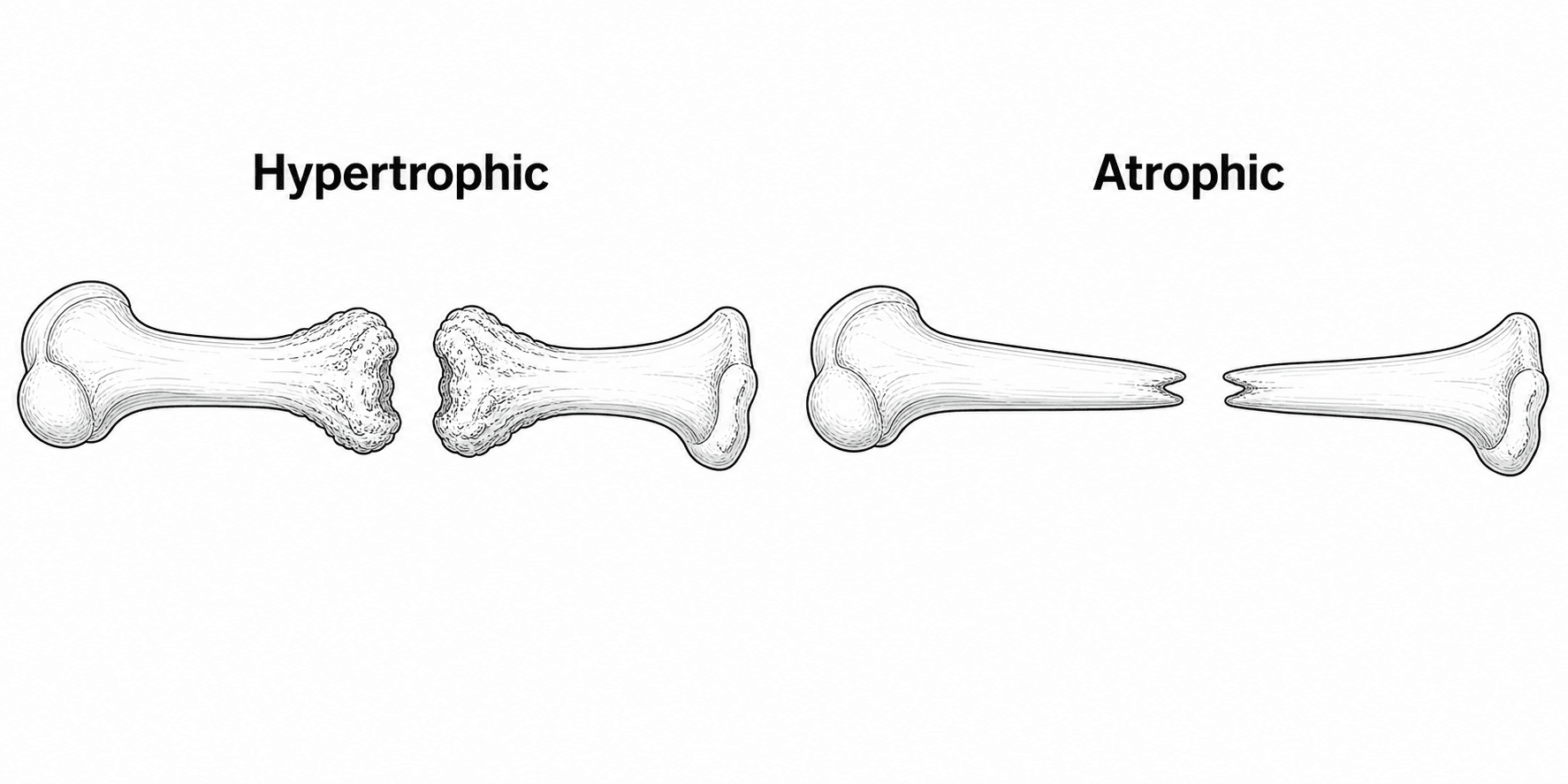
- Classify the nonunion: HYPERTROPHIC (abundant 'elephant-foot' callus that fails to bridge - a MECHANICAL/stability problem, with viable bone) versus ATROPHIC (tapered, sclerotic, avascular ends with a gap and little callus - a BIOLOGICAL problem) versus INFECTED.
- Always EXCLUDE INFECTION before treating (history, inflammatory markers, prior surgery), and document RADIAL NERVE function pre-operatively - the nerve is intimately related to the shaft and at risk both from the nonunion and during surgery.
- The GOLD-STANDARD treatment of aseptic humeral shaft nonunion is OPEN reduction with COMPRESSION PLATING (dynamic compression/locking plate) plus AUTOGENOUS (iliac crest) BONE GRAFT - this achieves UNION in the large majority (around 90-100% in series), restoring shoulder and elbow function.
- Unlike the femur and tibia, EXCHANGE NAILING is UNRELIABLE for humeral shaft nonunion; a failed nail is best converted to COMPRESSION PLATE FIXATION WITH BONE GRAFT rather than a larger nail.
- Atrophic and gap nonunions need the BIOLOGY addressed (autograft, and biological adjuncts such as BMP where appropriate); infected nonunions require infection ERADICATION (debridement, culture-directed antibiotics, sometimes staged reconstruction) alongside stabilisation - following the 'diamond concept' of stability + biology + a viable host.
- “Hypertrophic = stability problem (give compression); atrophic = biology problem (give compression PLUS bone graft).
- “In the HUMERUS, exchange nailing does NOT reliably achieve union - convert to compression plate + autograft.
- “Always exclude infection and check/protect the radial nerve before and during surgery.
For diaphyseal nonunion after nailing, exchange nailing (a larger reamed nail) is often effective - it adds stability and reams in biology.
Exchange nailing is UNRELIABLE in the humerus. A failed humeral nail (or a nonunion) is best treated by conversion to COMPRESSION PLATING with autogenous bone graft, which is the gold standard.
Risk Factors & Patterns
Most humeral shaft fractures heal with non-operative (functional bracing) or operative care, but nonunion occurs with identifiable risks:
- Fracture-related: a transverse fracture pattern, distraction/gapping (e.g. over-distraction in a brace or by gravity, soft-tissue interposition), proximal-third fractures, comminution/bone loss, open fractures and infection.
- Treatment-related: inadequate immobilisation/instability, and (with surgery) inadequate fixation.
- Patient-related: smoking, NSAIDs, diabetes, obesity, poor bone quality and other comorbidities. Recognising these helps both prevention and the management plan.
Classification & Assessment
- Hypertrophic - exuberant callus ('elephant-foot') but no bridging. The biology is intact; the problem is mechanical (insufficient stability). Providing rigid compression alone usually achieves union.
- Atrophic - tapered, avascular, sclerotic bone ends with a gap and little/no callus. The problem is biological (and often mechanical too); treatment must add bone graft / biology to stability.
- Infected (septic) nonunion - must be recognised and the infection eradicated as part of the plan.
- Radiographs (+/- CT) to confirm nonunion, characterise it (hyper/atrophic), and assess any implant/bone loss
- EXCLUDE INFECTION - history (wound problems, prior surgery), CRP/ESR/FBC, and intra-operative cultures where suspicion exists
- Document RADIAL NERVE function (and other neurovascular status) pre-operatively
- Optimise patient factors (stop smoking, review NSAIDs, correct metabolic/endocrine causes)
Successful union needs mechanical stability, osteogenic cells, an osteoconductive scaffold, osteoinductive signals (growth factors) and an adequate vascular/host environment - the framework guiding what each nonunion needs.
Management
The gold-standard treatment of aseptic humeral shaft nonunion is open reduction and internal fixation with a COMPRESSION PLATE (dynamic compression or locking plate, achieving direct compression across the nonunion) supplemented with AUTOGENOUS (iliac crest) BONE GRAFT - especially for atrophic/gap nonunions. This combination addresses both stability and biology and achieves union in the large majority of cases (around 90-100% in large series), with restoration of shoulder and elbow function. The nonunion site is freshened/decorticated, any sclerotic/avascular bone and interposed tissue removed, and the canal reopened.
The biology is fine - provide rigid stability and compression (compression plating). Bone graft is often not essential but may be added. Union is highly reliable once stability is restored.
The radial nerve spirals around the humeral shaft and is intimately related to the mid/distal diaphysis (and to the nonunion and any callus). Document its function pre-operatively, identify and protect it intra-operatively (it may be encased in callus/scar at the nonunion), and counsel the patient about the risk of (usually transient) radial-nerve palsy, which is the commonest neurological complication of humeral nonunion surgery.
Evidence & Key Studies
Treatment of nonunion of humeral shaft fracture with dynamic compression plate and cancellous bone graft
- In 105 humeral shaft nonunions (67 atrophic, 20 hypertrophic) treated with a dynamic compression plate plus cancellous bone graft, ALL united, at an average of 16 weeks (range 10-26).
- Complications were limited to 4 temporary radial-nerve palsies and 3 wound infections; shoulder and elbow function were satisfactory at follow-up.
- Compression plating with supplemental cancellous bone graft is a reliable, effective treatment for humeral shaft nonunion.
Repair of humeral shaft nonunion with plate and screw fixation and iliac crest bone graft
- Describes the technique for humeral shaft nonunion repair: nonunion-site preparation, direct compression of the fracture using plate osteosynthesis, and iliac crest bone graft harvest/utilisation.
- Reinforces compression plating with autogenous bone graft as the management approach for humeral shaft nonunion.
- Emphasises freshening the nonunion and achieving direct compression across the site.
According to PubMed, the high union rates and complication profile of compression plating plus cancellous bone graft come from the cited Hsu series, and the technique (nonunion preparation, direct compression, iliac-crest autograft) from the cited Egol-group description. The hypertrophic/atrophic classification, the diamond concept, and the unreliability of exchange nailing in the humerus are standard, well-established teaching. (See also our Nonunion Management, Malunion/Delayed Union and Humeral Shaft Fracture material.)
Clinical Decision Scenarios
Practise clinical reasoning and management decisions out loud
“A patient has a humeral shaft nonunion 8 months after a transverse mid-shaft fracture treated in a brace. How do you classify and assess it, and what is the gold-standard treatment?”
“How does your management differ for a hypertrophic versus an atrophic versus an infected humeral nonunion, and why is the humerus different from the femur?”
Mnemonics & Memory Aids
UNITE
Hook:To UNITE a humeral nonunion: type it, protect the nerve, exclude infection, compression-plate + graft, don't re-nail.
DIAMOND
Hook:The DIAMOND concept applied: stability + biology + viable host - and plate, don't re-nail, the humerus.
Risk factors
- Transverse pattern, distraction/gapping, proximal-third, open, infection, comminution
- Inadequate immobilisation/instability
- Smoking, NSAIDs, diabetes, obesity, poor bone quality
Classify & assess
- Hypertrophic (elephant-foot callus = stability problem) vs atrophic (tapered/avascular = biology) vs infected
- EXCLUDE infection (CRP/ESR, cultures); document RADIAL NERVE
- Diamond concept: stability + cells + scaffold + signals + viable host
Treatment
- Gold standard (aseptic): compression plate + autogenous iliac-crest bone graft (~90-100% union)
- Hypertrophic: compression often suffices; atrophic/gap: add graft (+/- BMP/structural/transport)
- Infected: eradicate infection (staged) + stabilise + graft
Key pitfalls
- Exchange nailing UNRELIABLE in the humerus - convert to compression plate + graft
- Radial nerve at risk (commonest neuro complication) - protect it
- Don't reconstruct over unrecognised infection