A lesion centred on the shaft — age, permeation and periosteal reaction do most of the work
- The diaphysis is the shaft between the metaphyseal flares; a lesion is diaphyseal only if its epicentre lies there — measure the centre, not the edges.
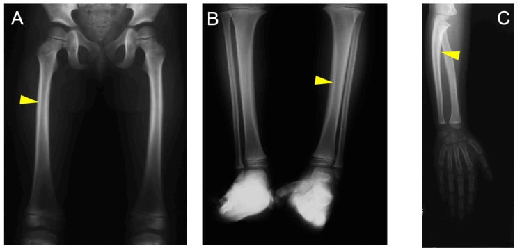
- In a child, the diaphyseal aggressive lesion differential is Ewing sarcoma, osteomyelitis, Langerhans cell histiocytosis and leukaemia/lymphoma until proven otherwise.
- Over 40 years, a destructive shaft lesion is metastasis or myeloma until proven otherwise — never biopsy before staging CT chest/abdomen/pelvis and serum electrophoresis.
- A well-defined lucency with thick sclerotic rim, ground-glass matrix or a central nidus is benign and often needs no biopsy at all.
- Endosteal scalloping greater than two-thirds of cortical thickness, cortical breach or a soft-tissue mass upgrade any lesion to aggressive.
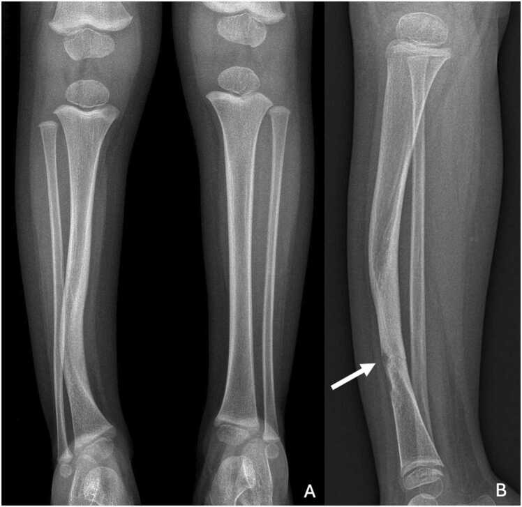
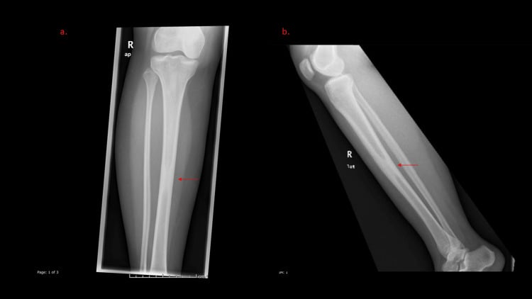
- Adamantinoma and osteofibrous dysplasia both sit in the anterior tibial cortex — the two must be distinguished histologically, not radiographically.
- “Say the epicentre, the zone of transition, the matrix, the periosteal reaction and the soft-tissue component — in that order — before you offer a diagnosis.
- “Lamellated (onion-skin) periosteum is not specific for Ewing; osteomyelitis and LCH both produce it.
- “A sequestrum can appear in osteomyelitis, LCH, lymphoma and desmoplastic fibroma — it is not pathognomonic of infection.
- “Bone infarct has a serpiginous sclerotic margin; enchondroma has ring-and-arc calcification. Both are 'chondroid-looking' shaft lesions and are commonly confused.
Most osteosarcomas and chondrosarcomas that reach the shaft started in the metaphysis. Localise the epicentre. A metadiaphyseal epicentre in a 15-year-old reopens osteosarcoma, which you had excluded.
A permeative femoral shaft lesion in a 65-year-old is metastasis or myeloma far more often than primary sarcoma. Staging CT, myeloma screen and PSA/breast examination first — you may find the primary and spare a biopsy, or you may find it is solitary and needs a sarcoma-centre biopsy tract.
Description is not enough. Comment on cortical involvement, lesion length and pain on weight-bearing. Mirels score of 9 or more, or axial cortical destruction greater than 30 mm, means prophylactic fixation.
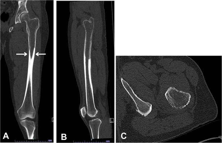
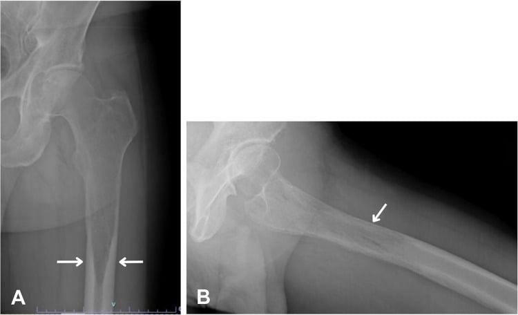
A longitudinal or oblique cortical lucency with fusiform periosteal new bone in a runner or recruit is a stress fracture. Aggressive appearance on radiograph and marrow oedema on MRI mimic malignancy — the linear low-signal fracture line is the discriminator.
Recognising the Pattern
Definition. A diaphyseal lesion has its epicentre in the tubular shaft, between the distal and proximal metaphyseal flares — that is, in the segment where the cortex is thickest and the medullary canal narrowest. Confirm by drawing a line through the centre of the abnormality on both orthogonal views; if that centre lies within the flare or within 1–2 cm of the physeal scar, call it metadiaphyseal and change your differential.
Confirming it is real. Two views, always. A "lesion" seen on one projection only is usually a nutrient foramen (an oblique lucent channel with sclerotic margins running in a predictable direction — away from the knee, towards the elbow), an overlying skin lesion, a plaster or dressing artefact, or Mach effect at a cortical margin. Confirm continuity of the cortex on both views before you commit.
The words to use out loud.
"This is an AP and lateral radiograph of the left femur in a skeletally immature patient. There is a lesion centred on the mid-diaphysis, measuring approximately 6 cm in craniocaudal extent. It is intramedullary with a permeative pattern of bone destruction and a wide zone of transition. There is lamellated and spiculated periosteal reaction along the lateral cortex, cortical breach medially, and an associated soft-tissue mass. There is no visible matrix mineralisation. The appearances are aggressive. My primary diagnosis is Ewing sarcoma; the principal differential is osteomyelitis and Langerhans cell histiocytosis."
Mimics — the false positives.
- Nutrient canal: oblique lucency crossing the cortex, sclerotic walls, no medullary component.
- Cortical desmos / periosteal fibrous cortical defect: posteromedial distal femur, scalloped surface lesion, universally in children, no follow-up needed.
- Stress fracture: cortical thickening and a linear lucency; the fusiform periosteal reaction is symmetrical and the patient has activity-related pain.
- Bone island (enostosis): dense, small, brush borders blending with trabeculae, cold on bone scan.
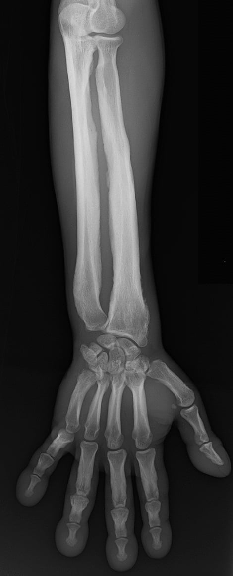
- Longitudinal split / normal cortical striation in elderly osteoporotic bone or in hyperparathyroidism (intracortical tunnelling).
- Old healed osteomyelitis or fracture callus: sclerosis with cortical thickening but no medullary destruction and no interval change on old films.
Always get the old films. An unchanged lesion over two years is benign; this saves more biopsies than any single imaging test.
Next Investigation
FEMALEFEMALE — lucent diaphyseal lesions of childhood
Hook:A long-established mnemonic for the lucent diaphyseal differential. Add osteomyelitis mentally to every line — it mimics all of them.
The Differential
- Typical age / setting
- 5–25 yrs; femoral/tibial shaft, pelvis, ribs; fever, raised ESR/CRP mimicking infection
- Discriminating feature
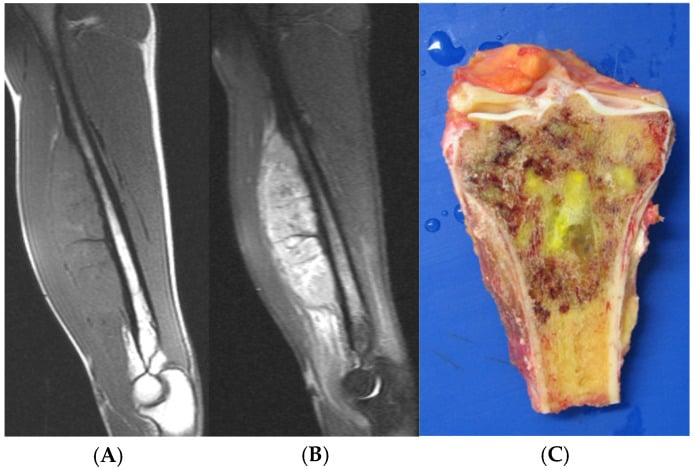
- Permeative destruction with a soft-tissue mass disproportionately large for the amount of bone destroyed; lamellated periosteum
- What confirms it
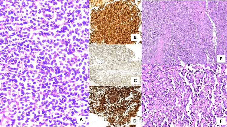
- MRI whole bone plus core biopsy — small round blue cells, CD99 membranous, EWSR1 rearrangement
- Typical age / setting
- Over 40 yrs; lung, breast, renal, thyroid, prostate; femoral and humeral shaft
- Discriminating feature
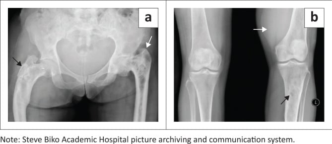
- Multiple lesions; lytic with cortical destruction but usually NO periosteal reaction and no soft-tissue mass; renal/thyroid deposits are expansile and hypervascular
- What confirms it
- Staging CT chest/abdomen/pelvis, bone scan or FDG-PET, biopsy of the most accessible lesion
- Typical age / setting
- Over 50 yrs; humerus, femur, skull, spine; anaemia, raised ESR, renal impairment
- Discriminating feature
- Endosteal scalloping with multiple discrete punched-out lucencies and no reactive sclerosis; bone scan often negative
- What confirms it
- Serum/urine electrophoresis and free light chains, whole-body MRI or low-dose CT, marrow aspirate
- Typical age / setting
- Any age; children commonly; immunosuppression, diabetes, sickle cell
- Discriminating feature
- Sequestrum within an involucrum; Brodie abscess shows the 'penumbra sign' — T1 high-signal granulation rim around a low-signal cavity
- What confirms it
- MRI with contrast, aspirate/biopsy for culture before antibiotics; CRP and blood cultures
- Typical age / setting
- Typically 40–70 yrs but any age; femur most common
- Discriminating feature
- Extensive permeative marrow infiltration with a large soft-tissue mass yet relatively preserved cortex on radiograph; patient looks better than the film
- What confirms it
- MRI shows marrow replacement out of proportion to radiograph; biopsy plus immunophenotyping, staging PET-CT
- Typical age / setting
- 10–25 yrs; classically metaphyseal, 2–11% purely diaphyseal
- Discriminating feature
- Cloud-like osteoid matrix; Codman triangle and sunburst spiculation; raised ALP
- What confirms it
- MRI whole bone, CT chest, biopsy in sarcoma unit along the planned resection tract
- Typical age / setting
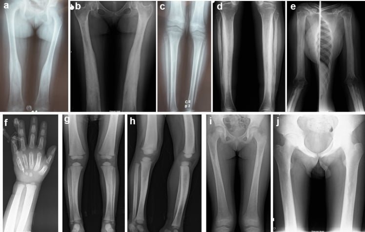
- Childhood to 30 yrs; femur ('shepherd's crook'), tibia, ribs, skull; McCune–Albright with café-au-lait and precocious puberty
- Discriminating feature
- Ground-glass matrix, long lesion in a long bone, no periosteal reaction, thinned but intact endosteally scalloped cortex
- What confirms it
- Radiograph alone is usually diagnostic; GNAS mutation if biopsied
- Typical age / setting
- 5–15 yrs; proximal humerus and femur, migrates into diaphysis with growth
- Discriminating feature
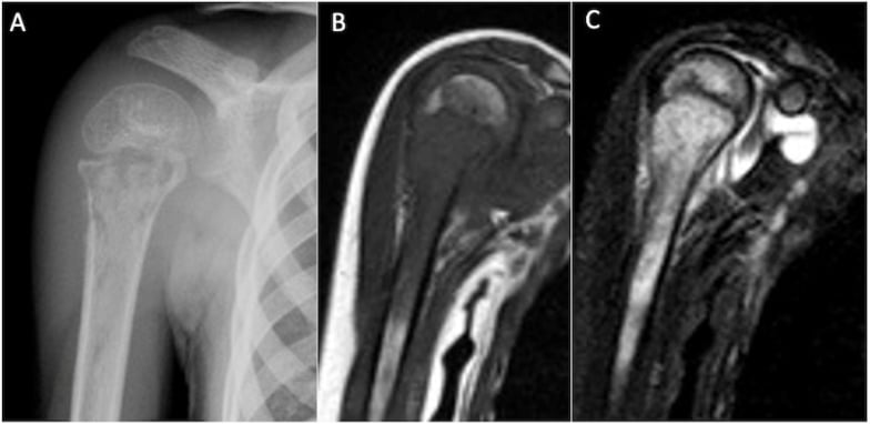
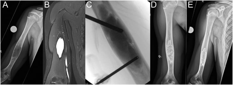
- Central, well-defined, fluid-density, cortex thinned not expanded beyond the physeal width; 'fallen fragment' sign after fracture
- What confirms it
- Radiograph; MRI fluid signal without solid enhancing component
- Typical age / setting
- 20–50 yrs; hands and feet most common, also femoral/humeral shaft; Ollier and Maffucci if multiple
- Discriminating feature
- Ring-and-arc / popcorn chondroid calcification with endosteal scalloping less than two-thirds of cortex and no periostitis
- What confirms it
- Radiograph; MRI if painful — deep scalloping, cortical breach or a soft-tissue mass suggests chondrosarcoma
- Typical age / setting
- Adults; steroids, alcohol, sickle cell, caisson disease, SLE
- Discriminating feature
- Serpiginous, sharply defined sclerotic 'smoke-up-a-chimney' margin with lucent centre; no endosteal scalloping
- What confirms it
- MRI double-line sign; no further action unless pain or a malignant transformation (rare MFH/UPS) is suspected
- Typical age / setting
- 1–15 yrs; skull, femur, ribs, vertebra plana
- Discriminating feature
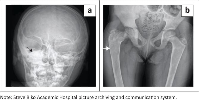
- Can look like anything; classically a lytic diaphyseal lesion with laminated periosteum, a 'hole within a hole' skull lesion, and marked pain but a well child
- What confirms it
- Skeletal survey, biopsy — Birbeck granules, CD1a and langerin positive
- Typical age / setting
- 10–25 yrs, male predominance; femoral and tibial shaft
- Discriminating feature
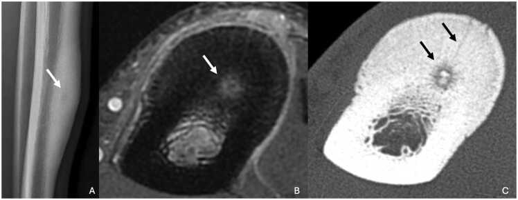
- Night pain relieved dramatically by NSAIDs; lucent nidus less than 1.5 cm with dense fusiform reactive cortical sclerosis
- What confirms it
- Thin-slice CT shows the nidus; CT-guided radiofrequency ablation is diagnostic and therapeutic
- Typical age / setting
- 20–40 yrs; anterior tibial diaphysis, occasionally fibula
- Discriminating feature
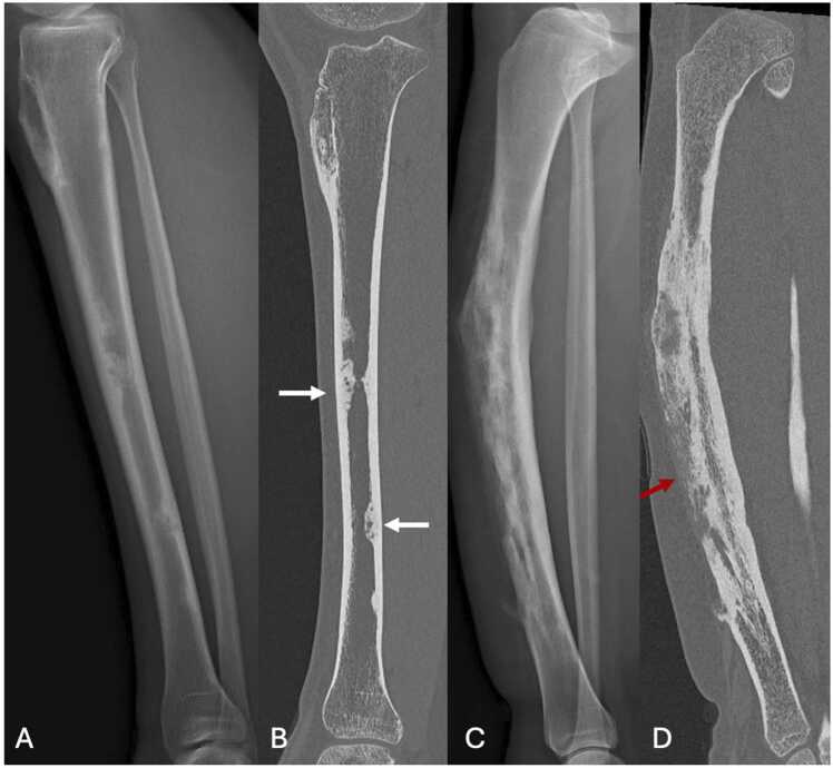
- Multiple lucencies in the anterior tibial cortex with a 'soap-bubble' appearance and anterior bowing; slow, painless, sometimes decades of history
- What confirms it
- MRI to define marrow extent; open biopsy — epithelial nests in fibrous stroma, cytokeratin positive; needs wide resection
- Typical age / setting
- Under 10 yrs; anterior tibial cortex
- Discriminating feature
- Radiographically identical to adamantinoma but intracortical, in a much younger child, and self-limiting after skeletal maturity
- What confirms it
- Observation with serial radiographs; biopsy only if enlarging — keratin-positive cells would raise adamantinoma
- Typical age / setting
- 4–14 yrs; clavicle, tibia, distal femur; associated palmoplantar pustulosis
- Discriminating feature
- Multifocal, symmetrical, sterile lesions with sclerosis and hyperostosis; relapsing course; cultures negative
- What confirms it
- Whole-body MRI showing multifocal marrow oedema; diagnosis of exclusion after biopsy excludes infection and malignancy
Narrowing It Down
- 1Step 1 — How old is the patient?
Under 5 yrs: metastatic neuroblastoma, leukaemia, osteomyelitis, LCH. 5–25 yrs: Ewing, osteosarcoma, osteomyelitis, LCH, simple cyst, fibrous dysplasia, osteoid osteoma. 25–40 yrs: fibrous dysplasia, enchondroma, adamantinoma, giant cell tumour (if it reaches the shaft from the metaphysis), infection. Over 40 yrs: metastasis, myeloma, lymphoma, chondrosarcoma, infarct, Paget disease. Age alone eliminates roughly half the list.
Age alone eliminates roughly half the list - say it first
- 2Step 2 — Solitary or multiple?
Multiple lucent lesions in an adult means metastasis, myeloma, or (rarely) brown tumours of hyperparathyroidism. Multiple in a child means LCH, fibrous dysplasia (polyostotic), enchondromatosis, or CRMO. A truly solitary aggressive lesion in a young patient shifts weight decisively towards primary sarcoma.
Multiple redirects to staging and biochemistry; solitary points at sarcoma
- 3Step 3 — Where in the cross-section: medullary, cortical or surface?
Intramedullary: Ewing, myeloma, metastasis, infarct, cyst, enchondroma. Intracortical: osteoid osteoma, osteofibrous dysplasia, adamantinoma, stress fracture, intracortical abscess. Surface/juxtacortical: periosteal osteosarcoma, periosteal chondroma, osteochondroma. This is the step candidates skip and it discriminates powerfully.
Intracortical and anterior tibial is a different disease from intramedullary
- 4Step 4 — What is the matrix?
Ground-glass equals fibrous dysplasia. Ring-and-arc or popcorn equals chondroid — enchondroma or chondrosarcoma. Cloud-like, amorphous density equals osteoid — osteosarcoma. Serpiginous sclerotic rim equals infarct. No matrix keeps the round-cell and metastatic differential fully open.
Matrix names the tissue; absent matrix excludes nothing
- 5Step 5 — How aggressive? Zone of transition, cortex, periosteum
Narrow zone with sclerotic rim (Lodwick IA) is benign — observe. Wide zone with permeative (Lodwick III) or moth-eaten (II) destruction is aggressive. Interrupted periosteal reaction — Codman triangle, sunburst, lamellated — and a soft-tissue mass mean malignancy or infection. Solid, thick, uninterrupted periosteum means a slow process.
Grade I is 94 per cent benign and grade III 81 per cent malignant; grade IC decides nothing
- 6Step 6 — Systemic features and bloods
Fever, raised CRP and ESR occur in BOTH Ewing and osteomyelitis — they do not discriminate. Weight loss, anaemia, hypercalcaemia and raised ESR over 40 yrs suggest myeloma. Raised ALP suggests osteosarcoma or Paget. Night pain abolished by aspirin means osteoid osteoma. Take a full history of previous malignancy, smoking, and breast/prostate examination.
The bloods cannot separate Ewing from infection - only tissue can
- 7Step 7 — Decide the pathway
Benign, characteristic (fibrous dysplasia, infarct, cyst, cortical desmoid): reassure, discharge or interval radiograph. Indeterminate: MRI whole bone plus old films. Aggressive: MRI whole bone, CT chest, and referral to a specialist sarcoma centre for biopsy — do not biopsy locally.
Aggressive means refer BEFORE biopsy, never after
MCQ Practice Points
Q: Which single radiographic feature best predicts the biological aggressiveness of a bone lesion?
A: The zone of transition. A narrow, sharply defined transition with a sclerotic rim indicates slow growth; a wide, imperceptible transition with permeative or moth-eaten destruction indicates rapid growth. It outperforms size, matrix and location as a predictor and underpins the Lodwick grading system.
Q: A 9-year-old has a lytic diaphyseal femoral lesion with laminated periosteal reaction and severe local pain, but is systemically well with normal inflammatory markers. What is the likely diagnosis?
A: Langerhans cell histiocytosis. It is the great mimic and reproduces both the Ewing and the osteomyelitis appearance. Diagnosis is by biopsy showing CD1a- and langerin-positive cells with Birbeck granules on electron microscopy. Solitary lesions frequently heal spontaneously or after intralesional steroid.
Q: Which sign on MRI distinguishes a Brodie abscess from a small round-cell tumour?
A: The penumbra sign — a rim of granulation tissue that is relatively high signal on unenhanced T1 surrounding the low-signal abscess cavity, with rim enhancement after gadolinium. It is reasonably specific for subacute osteomyelitis, though not universally present. Note the limit of this and every other imaging sign here: Durbin's two infants show that neither the clinical picture nor routine bloods separate Ewing from infection, so a negative penumbra sign is not permission to skip the biopsy.
Q: What distinguishes an enchondroma from a low-grade central chondrosarcoma in a long-bone shaft?
A: Endosteal scalloping deeper than two-thirds of cortical thickness, lesion length greater than 5–6 cm, cortical thickening or breach, periosteal reaction, a soft-tissue mass, and — critically — pain not attributable to another cause, plus interval growth on serial imaging. Histology alone is unreliable; the radiological and clinical picture drives the diagnosis.
Q: Why must an aggressive diaphyseal lesion never be nailed before diagnosis?
A: Intramedullary reaming disseminates tumour along the entire canal and through the entry portal, converting a localised, resectable sarcoma into a whole-bone contamination that usually mandates amputation. Stage and biopsy first; fix only once metastatic disease is confirmed or the sarcoma team has planned the tract.
Exam Viva Scenarios
Practise clinical reasoning and management decisions out loud
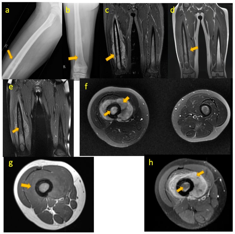
“You are shown this radiograph of the right femur in a 13-year-old boy with six weeks of thigh pain, a low-grade fever and an ESR of 68. There is a permeative mid-diaphyseal lesion with lamellated periosteal reaction and a soft-tissue mass.”
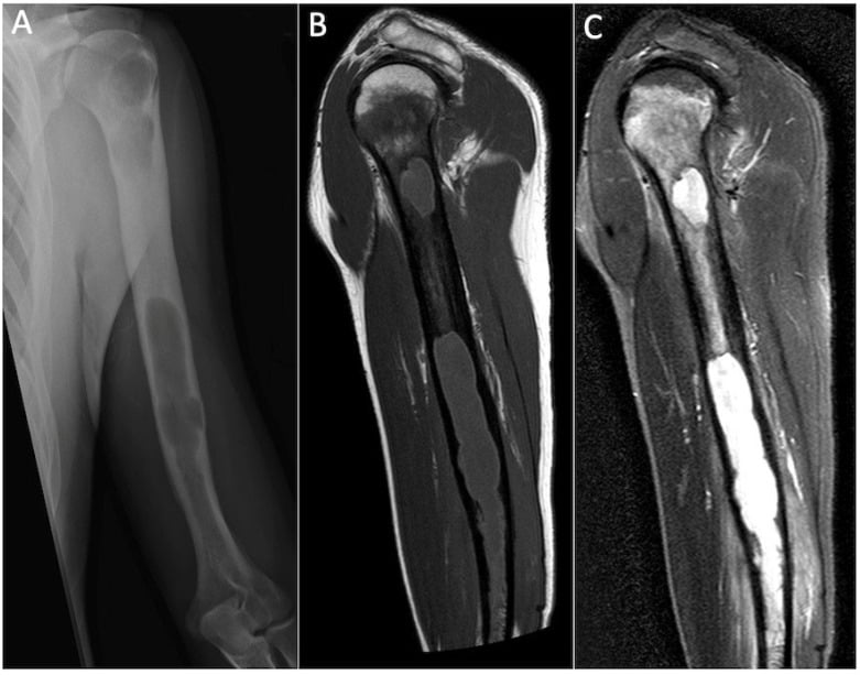
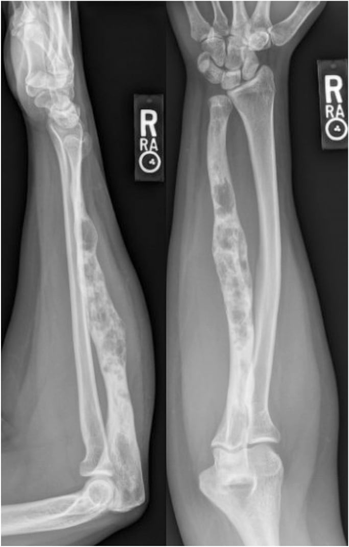
“You are shown this radiograph of the left humerus in a 68-year-old woman with two months of arm pain, now unable to lift a kettle. There is a lytic mid-diaphyseal lesion with cortical thinning over 4 cm and endosteal scalloping.”
“You are shown this lateral tibial radiograph of a 28-year-old man with a two-year history of a painless anterior shin swelling and mild anterior bowing. There are multiple lucencies in the anterior tibial cortex with intervening sclerosis, giving a soap-bubble appearance.”
Describe in this order
- Patient age and bone
- Epicentre: diaphysis, metadiaphysis; medullary, cortical or surface
- Zone of transition and Lodwick grade
- Matrix: ground-glass, chondroid, osteoid, none
- Periosteal reaction: solid, lamellated, spiculated, Codman
- Cortex and soft-tissue mass
- Then commit to a diagnosis and two differentials
Age shortcuts
- Under 5: neuroblastoma metastasis, leukaemia, infection, LCH
- 5–25: Ewing, osteosarcoma, LCH, osteomyelitis, simple cyst, osteoid osteoma
- 25–40: fibrous dysplasia, enchondroma, adamantinoma, infection
- Over 40: metastasis, myeloma, lymphoma, chondrosarcoma, infarct, Paget
Signature discriminators
- Ground-glass, long lesion in a long bone: fibrous dysplasia
- Serpiginous sclerotic margin: bone infarct
- Ring-and-arc calcification: chondroid
- Nidus less than 1.5 cm plus night pain relieved by NSAIDs: osteoid osteoma
- Anterior tibial cortex, soap-bubble, adult: adamantinoma
- Sequestrum plus penumbra sign: Brodie abscess
- Big soft-tissue mass, little bone destruction, child: Ewing
- Punched-out lucencies, no sclerosis, over 50: myeloma
Do not do
- Do not biopsy an adult shaft lesion before staging CT and electrophoresis
- Do not biopsy or nail outside the treating sarcoma unit
- Do not give antibiotics before an aspirate
- Do not call a one-view finding a lesion
- Do not discharge a lytic femoral lesion without assessing fracture risk
Next test
- Old films first — unchanged over two years equals benign
- MRI whole bone with contrast for indeterminate or aggressive lesions
- Thin-slice CT for matrix, sequestrum and nidus
- CT chest plus PET or bone scan for staging
- No imaging at all for NOF, cortical desmoid, bone island, classic infarct
Evidence Base
Adamantinoma of Long Bones - A Clinicopathologic Study of 85 Cases
- 70 of 85 were in the TIBIA (11 also involving the fibula); the remainder were 6 femur, 3 ulna, 2 humerus, 2 fibula, 1 radius and 1 arising in the soft tissue anterior to the tibia.
- Most presented with pain and swelling, aged 10 to 30 years.
- 26 (31 per cent) recurred locally, 13 (15 per cent) developed LUNG metastasis and 6 (7 per cent) lymph node metastasis.
- NINE of the 13 with lung metastasis had preceding local recurrence - local failure comes first and is the warning.
- Risk factors for recurrence or metastasis: male sex, pain, symptoms of LESS than 5 years' duration, and initial treatment by biopsy, curettage, excision or resection. The only histological risk factor was LACK of squamous differentiation.
- 41 patients were alive without disease from 1 month to 47 YEARS after treatment; 11 died of disease and 7 were alive with metastases.
Ewing's Sarcoma Masquerading as Osteomyelitis
- Two infants in whom Ewing sarcoma was initially diagnosed and treated as osteomyelitis - a tibia at 16 months and a finger phalanx at 7 months.
- States the reason the error happens: the presentations may be similar AND routine laboratory evaluation may not distinguish the two.
- In both cases the correct diagnosis was delayed by the initial misdiagnosis.
Primary Lymphoma of Bone - Outcome and Role of Surgery
- 21 consecutive patients aged 19 to 82; commonest site the spine (6), then ilium, femur and mandible (3 each).
- Surgery was performed in only 4 of 21 (19 per cent); the rest had chemotherapy, 17 of them with radiotherapy as well.
- Overall survival 74.2 per cent at five years. The ONLY significant positive prognostic factor was complete remission after chemotherapy.
- Six patients presented with a pathological fracture.
- Concludes that surgery should aim to restore function and relieve pain while MINIMISING DELAY to chemotherapy, and that where feasible it is best postponed until after chemotherapy.